. 2020 Jun 8;21(6):2285-2297.
doi: 10.1021/acs.biomac.0c00229. Epub 2020 Apr 23.

Role of Sheet-Edge Interactions in β-sheet Self-Assembling Peptide Hydrogels

Affiliations

Role of Sheet-Edge Interactions in β-sheet Self-Assembling Peptide Hydrogels

Jacek K Wychowaniec et al. Biomacromolecules. .

Abstract

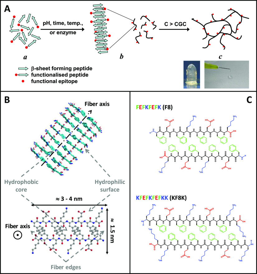
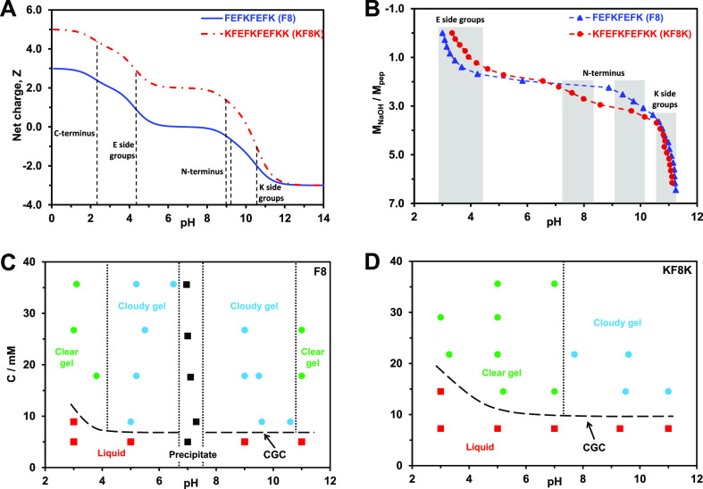
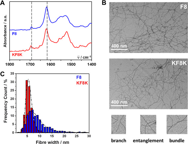
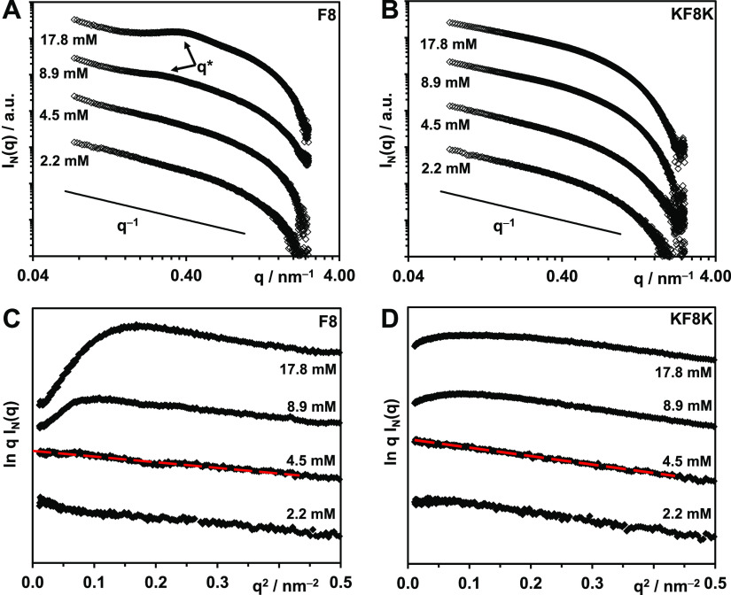
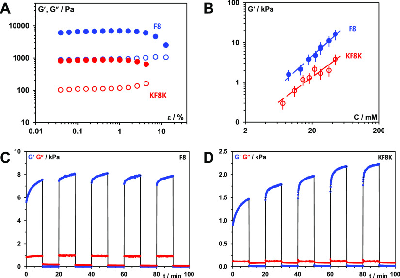
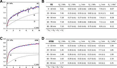
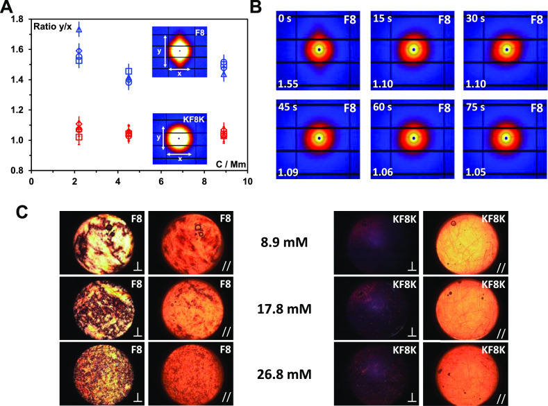
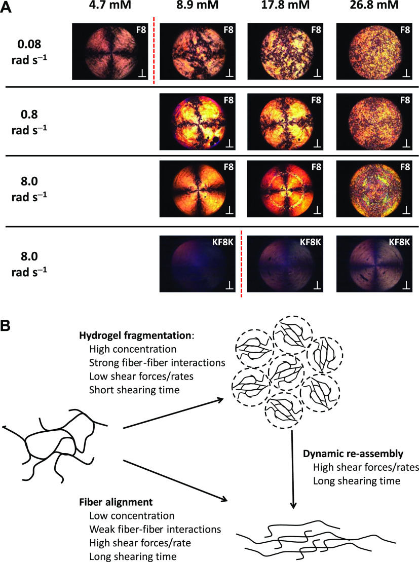
Hydrogels' hydrated fibrillar nature makes them the material of choice for the design and engineering of 3D scaffolds for cell culture, tissue engineering, and drug-delivery applications. One particular class of hydrogels which has been the focus of significant research is self-assembling peptide hydrogels. In the present work, we were interested in exploring how fiber-fiber edge interactions affect the self-assembly and gelation properties of amphipathic peptides. For this purpose, we investigated two β-sheet-forming peptides, FEFKFEFK (F8) and KFEFKFEFKK (KF8K), the latter one having the fiber edges covered by lysine residues. Our results showed that the addition of the two lysine residues did not affect the ability of the peptides to form β-sheet-rich fibers, provided that the overall charge carried by the two peptides was kept constant. However, it did significantly reduce edge-driven hydrophobic fiber-fiber associative interactions, resulting in reduced tendency for KF8K fibers to associate/aggregate laterally and form large fiber bundles and consequently network cross-links. This effect resulted in the formation of hydrogels with lower moduli but faster dynamics. As a result, KF8K fibers could be aligned only under high shear and at high concentration while F8 hydrogel fibers were found to align readily at low shear and low concentration. In addition, F8 hydrogels were found to fragment at high concentration because of the high aggregation state stabilizing the fiber bundles, resulting in fiber breakage rather than disentanglement and alignment.


Conflict of interest statement

The authors declare no competing financial interest.

Figures

Figure 1
Figure 1
Schematic representation of (A) the self-assembly and gelation pathway of β-sheet forming peptides and (B) the fibers formed (side and top views). The peptide illustrated here is FEFKFEFK (F8) (F: phenylalanine, E: glutamic acid, K: lysine). (C) Chemical structures of F8 and KF8K (KFEFKFEFKK) peptides presented in a schematic antiparallel β-sheet conformation (top view).
Figure 2
Figure 2
(A) Theoretical charge carried by each peptide vs pH (dotted lines indicate the theoretical pKa of the different ionic groups present on the peptides); (B) molar ratio of added NaOH to peptide vs pH (shadowed regions indicate the protonation/deprotonation transition regions of the different ionic groups); (C,D) concentration vs pH phase diagrams describing the samples’ physical appearance/state.
Figure 3
Figure 3
(A) FTIR–ATR normalized spectra obtained for F8 and KF8K hydrogels prepared at a concentration of 26.8 mM (dotted lines indicate the position of the two bands characteristic of adoption by peptides of β-sheet conformations); (B) TEM images obtained for F8 and KF8K diluted hydrogels and bottom characteristic examples of topological features observed across the fibrillar networks; (C) measured fiber and fiber bundle width distributions and lognormal fits obtained (fitting parameters: F8: μ = 8.4 ± 0.1, σ = 3.5 ± 0.1 and KF8K: μ = 5.8 ± 0.1, σ = 1.3 ± 0.1).
Figure 4
Figure 4
(A,B) Double logarithmic plots of SAXS patterns obtained for F8 and KF8K samples prepared at different concentrations in In(q) vs q representation. (C,D) SAXS patterns of F8 and KF8K samples plotted for low q in a ln q I(q) vs q2 representation (red lines show the linear fits used to calculate the fiber cross-section radii of gyration, Rσ).
Figure 5
Figure 5
(A) Storage (G′—closed symbols) and loss (G″—open symbols) shear moduli vs strain (ε) curves obtained at 1 Hz frequency for F8 and KF8K hydrogels prepared at 26.8 mM concentration. (B) Storage modulus (G′) vs peptide concentration (C) plots obtained for F8 and KF8K hydrogels measured at 0.2% strain and 1 Hz frequency. (C,D) Shear thinning and recovery behavior of F8 and KF8K hydrogels prepared at 26.8 mM peptide concentration. Experiments were performed at 1 Hz and shear strain of 0.2 and 100% were applied alternatingly in 10 min intervals.
Figure 6
Figure 6
(A,C) Shear storage modulus (G′) vs time (t) curves obtained for the 60–70 min recovery cycle (4th recovery cycle, Figure 5C,D); black dotted lines: the two dynamic recovery processes curves derived from eq 8: ① “fast” process, ② “slow” process (curves have been shifted upwards by G0 for ease of visualization); red line: curve’s fit obtained using eq 8. (B,D) fitting parameters obtained by fitting each recovery cycle curves using eq 8 for F8 and KF8K hydrogels prepared at 26.8 mM peptide concentration. The standard fitting errors are shown in brackets. All fits had R-square values of 0.98 or higher.
Figure 7
Figure 7
(A) Degree of peptide fiber alignment taken as the ratio of the meridian (y) over the equatorial axis (x) lengths of the 2D SAXS pattern observed vs sample concentration. Flow rates: 80 (△), 90 (◊), 120 (○), 150 mL s–1 (□). Inset shows the 2D scattering patterns obtained for the F8 and KF8K sample prepared at 8.9 mM; (B) 2D scattering patterns obtained for the F8 8.9 mM sample after stopping the flow: time elapsed from stopping the flow is shown in the top left corner and the degree of fiber alignment is shown in the bottom left corner; (C) PLIs of the sample taken after loading the samples in the SIPLI rheometer (⊥ indicates that the polarizer and analyzer are oriented at 90° and // indicates that the polarizer and analyzer are oriented at 0°).
Figure 8
Figure 8
(A) PLIs of the F8 and KF8K peptides at different concentrations after 120 s shearing (⊥ indicates that the polarizer and analyzer are oriented at 90°). The red dotted line indicates the CGC of the peptides. (B) Schematic representation of the different morphological transformations taking place when shearing peptides are assembled in fibers.

References

    1. Sun J. E. P.; Stewart B.; Litan A.; Lee S. J.; Schneider J. P.; Langhans S. A.; Pochan D. J. Sustained release of active chemotherapeutics from injectable- solid - hairpin peptide hydrogel. Biomater. Sci. 2016, 4, 839–848. 10.1039/c5bm00538h. - DOI - PMC - PubMed
    1. Yamada Y.; Chowdhury A.; Schneider J. P.; Stetler-Stevenson W. G. Macromolecule- network electrostatics controlling delivery of the biotherapeutic cell modulator TIMP- 2. Biomacromolecules 2018, 19, 1285.10.1021/acs.biomac.8b00107. - DOI - PMC - PubMed
    1. Mujeeb A.; Miller A. F.; Saiani A.; Gough J. E. Self-assembled octapeptide scaffolds for in vitro chondrocyte culture. Acta Biomater. 2013, 9, 4609–4617. 10.1016/j.actbio.2012.08.044. - DOI - PubMed
    1. Qi Y.; Min H.; Mujeeb A.; Zhang Y.; Han X.; Zhao X.; Anderson G. J.; Zhao Y.; Nie G. Injectable hexapeptide hydrogel for localized chemotherapy prevents breast cancer recurrence. ACS Appl. Mater. Interfaces 2018, 10, 6972–6981. 10.1021/acsami.7b19258. - DOI - PubMed
    1. Tang J. D.; Mura C.; Lampe K. J. Stimuli-responsive, pentapeptide, nanofiber hydrogel for tissue engineering. J. Am. Chem. Soc. 2019, 141, 4886–4899. 10.1021/jacs.8b13363. - DOI - PubMed

Publication types