Case Reports
. 2020 May 5;153(6):725-733.
doi: 10.1093/ajcp/aqaa062.

COVID-19 Autopsies, Oklahoma, USA

Affiliations
Case Reports

COVID-19 Autopsies, Oklahoma, USA

Lisa M Barton et al. Am J Clin Pathol. .

Erratum in

  • Corrigendum.
    [No authors listed] [No authors listed] Am J Clin Pathol. 2020 May 5;153(6):852. doi: 10.1093/ajcp/aqaa070. Am J Clin Pathol. 2020. PMID: 32369558 Free PMC article. No abstract available.

Abstract

Objectives: To report the methods and findings of two complete autopsies of severe acute respiratory syndrome coronavirus 2 (SARS-CoV-2) positive individuals who died in Oklahoma (United States) in March 2020.

Methods: Complete postmortem examinations were performed according to standard procedures in a negative-pressure autopsy suite/isolation room using personal protective equipment, including N95 masks, eye protection, and gowns. The diagnosis of coronavirus disease 2019 (COVID-19) was confirmed by real-time reverse transcriptase polymerase chain reaction testing on postmortem swabs.

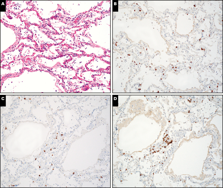
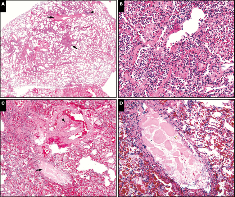
Results: A 77-year-old obese man with a history of hypertension, splenectomy, and 6 days of fever and chills died while being transported for medical care. He tested positive for SARS-CoV-2 on postmortem nasopharyngeal and lung parenchymal swabs. Autopsy revealed diffuse alveolar damage and chronic inflammation and edema in the bronchial mucosa. A 42-year-old obese man with a history of myotonic dystrophy developed abdominal pain followed by fever, shortness of breath, and cough. Postmortem nasopharyngeal swab was positive for SARS-CoV-2; lung parenchymal swabs were negative. Autopsy showed acute bronchopneumonia with evidence of aspiration. Neither autopsy revealed viral inclusions, mucus plugging in airways, eosinophils, or myocarditis.

Conclusions: SARS-CoV-2 testing can be performed at autopsy. Autopsy findings such as diffuse alveolar damage and airway inflammation reflect true virus-related pathology; other findings represent superimposed or unrelated processes.

Keywords: Acute lung injury; Autopsy; COVID-19; Coronavirus; Diffuse alveolar damage; Pulmonary pathology; SARS-CoV-2.


Figures

Image 1
Image 1
A pathology technician in complete autopsy attire. Behind her is an example of the reverse airflow tables at the Oklahoma Office of the Chief Medical Examiner autopsy stations. The main autopsy suite is equipped with 8 identical stations. Suspected coronavirus disease 2019 (COVID-19) autopsies are performed in an isolation room equipped with two identical stations.
Image 2
Image 2
Postmortem anterior-posterior chest radiographs. A, Case 1. Diffuse, dense bilateral airspace consolidations (complete “whiteout”). Multiple air bronchograms are present (arrows). The autopsy in this case showed diffuse alveolar damage. B, Case 2. Diffuse airspace opacities in both lungs, less consolidative in comparison to part A. Multiple bilateral air bronchograms are highlighted (arrows). The left lung is asymmetrically slightly more consolidated compared to the right. An endotracheal tube is shown with its tip above the level of the clavicular heads in the cervical trachea (white arrow). There is marked gastric distension with air (asterisk). The large opaque circular artifact on the right chest represents the grommet of the sealed body bag, and the small opaque circular artifacts represent buttons on clothing. Autopsy revealed acute bronchopneumonia.
Image 3
Image 3
Case 1. Microscopic findings in the lungs of a 77-year-old man who died of coronavirus disease 2019 (COVID-19). A, The airways are patent, with no evidence of mucus plugging. The upper arrow points to a patent bronchiole. The structure marked by the lower arrow is a patent bronchus. Neither airway shows evidence of mucus plugging. The pale appearance of the thickened bronchial mucosa is caused by mucosal edema. B, Diffuse alveolar damage in the acute stage. Note hyaline membranes (arrow). C, Chronic inflammation in the mucosa of an airway (arrow). The inflammatory cells are mainly lymphocytes. D, Patchy interstitial chronic inflammation. This image is taken from one of the few areas where interstitial inflammation was obvious even at low magnification. In most areas, the inflammatory infiltrate was very sparse or absent.
Image 4
Image 4
Immunohistochemistry in case 1. A, Diffuse alveolar damage with minimal, patchy chronic inflammation (H&E, x200). T-lymphocytes are highlighted by immunohistochemical stains for CD3 (B), CD4 (C), and CD8 (D).
Image 5
Image 5
Antemortem chest computed tomographic scan from case 2, showing bilateral ground-glass opacities. Bilateral consolidations were present elsewhere (not shown).
Image 6
Image 6
Pathologic findings in the lungs in case 2. A, Low magnification view of a small pulmonary artery branch (upper arrow) and its partner bronchiole (arrowhead). The area indicated by the lower arrow is shown at higher magnification in B. B, The airspaces are filled by a mix of neutrophils and histiocytes (acute bronchopneumonia). C, Low magnification view of another area. The arrowhead points to a small pulmonary artery. The arrow indicates its partner airway, which contains a foreign particle. The particle is shown at higher magnification in D. D, The foreign particle is a piece of aspirated vegetable matter.

Comment in

References

    1. World Health Organization. Coronavirus disease (COVID-2019) situation reports https://www.who.int/emergencies/diseases/novel-coronavirus-2019/situatio.... Accessed April 3, 2020.
    1. Guarner J. Three emerging coronaviruses in two decades. Am J Clin Pathol. 2020;153:420-421. - PMC - PubMed
    1. Huang C, Wang Y, Li X, et al. . Clinical features of patients infected with 2019 novel coronavirus in Wuhan, China. Lancet. 2020;396:497-506. - PMC - PubMed
    1. Mukhopadhyay S, Philip AT, Stoppacher R. Pathologic findings in novel influenza A (H1N1) virus (“Swine Flu”) infection: contrasting clinical manifestations and lung pathology in two fatal cases. Am J Clin Pathol. 2010;133:380-387. - PMC - PubMed
    1. Mukhopadhyay S, Mehrad M, Dammert P, et al. . Lung biopsy findings in severe pulmonary illness associated with E-cigarette use (vaping). Am J Clin Pathol. 2020;153:30-39. - PubMed

Publication types

MeSH terms