Practice Guideline
. 2020 Jul;158(1):106-116.
doi: 10.1016/j.chest.2020.04.003. Epub 2020 Apr 7.

The Role of Chest Imaging in Patient Management During the COVID-19 Pandemic: A Multinational Consensus Statement From the Fleischner Society

Affiliations
Practice Guideline

The Role of Chest Imaging in Patient Management During the COVID-19 Pandemic: A Multinational Consensus Statement From the Fleischner Society

Geoffrey D Rubin et al. Chest. 2020 Jul.

Abstract

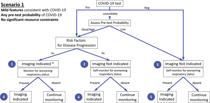
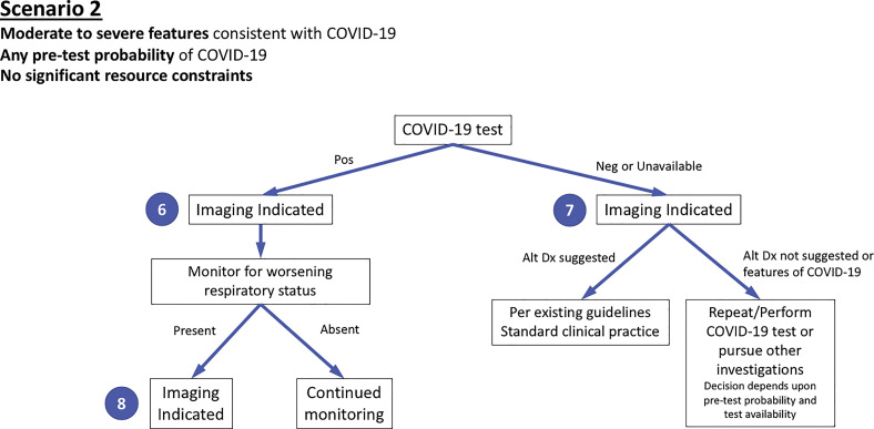
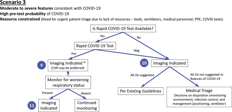
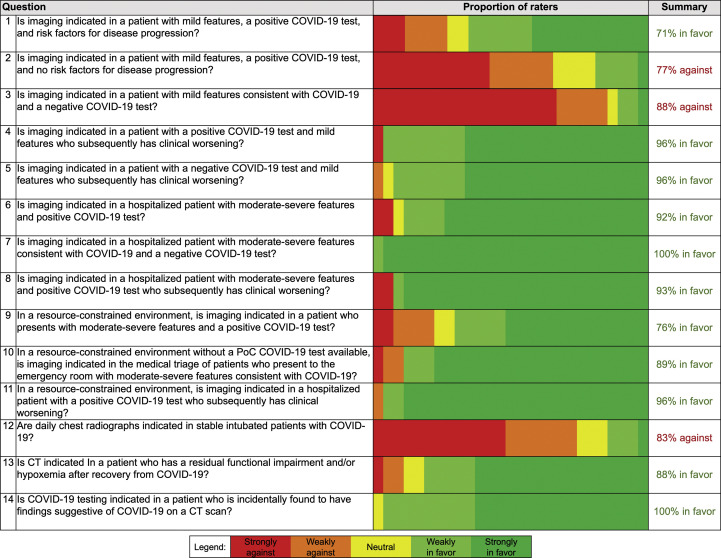
With more than 900,000 confirmed cases worldwide and nearly 50,000 deaths during the first 3 months of 2020, the coronavirus disease 2019 (COVID-19) pandemic has emerged as an unprecedented health care crisis. The spread of COVID-19 has been heterogeneous, resulting in some regions having sporadic transmission and relatively few hospitalized patients with COVID-19 and others having community transmission that has led to overwhelming numbers of severe cases. For these regions, health care delivery has been disrupted and compromised by critical resource constraints in diagnostic testing, hospital beds, ventilators, and health care workers who have fallen ill to the virus exacerbated by shortages of personal protective equipment. Although mild cases mimic common upper respiratory viral infections, respiratory dysfunction becomes the principal source of morbidity and mortality as the disease advances. Thoracic imaging with chest radiography and CT are key tools for pulmonary disease diagnosis and management, but their role in the management of COVID-19 has not been considered within the multivariable context of the severity of respiratory disease, pretest probability, risk factors for disease progression, and critical resource constraints. To address this deficit, a multidisciplinary panel comprised principally of radiologists and pulmonologists from 10 countries with experience managing patients with COVID-19 across a spectrum of health care environments evaluated the utility of imaging within three scenarios representing varying risk factors, community conditions, and resource constraints. Fourteen key questions, corresponding to 11 decision points within the three scenarios and three additional clinical situations, were rated by the panel based on the anticipated value of the information that thoracic imaging would be expected to provide. The results were aggregated, resulting in five main and three additional recommendations intended to guide medical practitioners in the use of chest radiography and CT in the management of COVID-19.


Figures

Figure 1
Figure 1
Diagram illustrates the first of three clinical scenarios presented to the panel with final recommendations. Mild features refer to absence of significant pulmonary dysfunction or damage. Pretest probability is based on background prevalence of disease and may be further modified by individual’s exposure risk. The absence of resource constraints corresponds to sufficient availability of personnel, personal protective equipment, coronavirus disease 2019 (COVID-19) testing, hospital beds, and/or ventilators with the need to rapidly triage patients. Numbers in blue circles indicate key questions referenced in the text and presented in Figure 4. Contextual detail and considerations for imaging with chest radiography versus CT are presented in the text. Although not covered by this scenario and not shown in the figure, in the presence of substantial resources constraints, there is no role for imaging of patients with mild features of COVID-19. ∗ = Clinical judgment should dictate the use of imaging through consideration of patient risk factors and local resources. Mod = moderate; Neg = negative; Pos = positive.
Figure 2
Figure 2
Diagram illustrates the second of three clinical scenarios presented to the panel with final recommendations. Moderate-to-severe features refer to evidence of significant pulmonary dysfunction or damage. Pretest probability is based on background prevalence of disease and may be further modified by individual’s exposure risk. The absence of resource constraints corresponds to sufficient availability of personnel, personal protective equipment, coronavirus disease 2019 (COVID-19) testing, hospital beds, and/or ventilators with the need to rapidly triage patients. Numbers in blue circles indicate key questions referenced in the text and presented in Figure 4. Contextual detail and considerations for imaging with chest radiography versus CT are presented in the text. Alt Dx = alternate diagnosis; Neg = negative; Pos = positive.
Figure 3
Figure 3
Diagram illustrates the third of three clinical scenarios presented to the panel with final recommendations. Moderate-to-severe features refer to evidence of significant pulmonary dysfunction or damage. High pretest probability is based on high background prevalence of disease associated with community transmission. Rapid coronavirus disease 2019 (COVID-19) test is a point-of-care test with a turnaround time of less than 1 hour. Numbers in blue circles indicate key questions referenced in the text and presented in Figure 4. Contextual detail and considerations for imaging with chest radiography (CXR) versus CT are presented in the text. ∗ = Lower priority if severely resource constrained, relative to question 10 or 11. Alt Dx = alternate diagnosis; Neg = negative; Pos = positive; PPE = personal protection equipment.
Figure 4
Figure 4
Panel members (n = 27) developed 14 key questions used to support creation of common scenarios and recommendations related to the use of chest imaging in patients with features of coronavirus disease 2019 (COVID-19). The proportion of panel member votes for each question is presented on a 5-point scale as well as a summary column that shows the total percentage of committee members who voted for or against imaging for each key question, excluding those members who were neutral or who abstained (one panel member abstained for questions 1 and 2). Numbers in left column correspond to question numbers in text and Figure 1, Figure 2, Figure 3. PoC = point of care.

References

    1. World Health Organization Director-General’s opening remarks at the media briefing on COVID-19 - March 11, 2020. https://www.who.int/dg/speeches/detail/who-director-general-s-opening-re...
    1. World Health Organization Coronavirus disease (COVID-19) situation dashboard. https://experience.arcgis.com/experience/685d0ace521648f8a5beeeee1b9125cd
    1. Johns Hopkins University Coronavirus COVID-19 global cases by the Center for Systems Science and Engineering (CSSE) https://gisanddata.maps.arcgis.com/apps/opsdashboard/index.html#/bda7594...
    1. Ai T, Yang Z, Hou H, et al. Correlation of chest CT and RT-PCR testing in coronavirus disease 2019 (COVID-19) in China: a report of 1014 cases [published online ahead of print February 26, 2020]. Radiology. . - DOI - PMC - PubMed
    1. Li Y, Yao L, Li J, et al. Stability issues of RT-PCR testing of SARS-CoV-2 for hospitalized patients clinically diagnosed with COVID-19 [published online ahead of print March 26, 2020]. J Med Virol. . - DOI - PMC - PubMed

Publication types

MeSH terms