. 2020 Jul 15:215:116800.
doi: 10.1016/j.neuroimage.2020.116800. Epub 2020 Apr 8.

Towards HCP-Style macaque connectomes: 24-Channel 3T multi-array coil, MRI sequences and preprocessing

Affiliations

Towards HCP-Style macaque connectomes: 24-Channel 3T multi-array coil, MRI sequences and preprocessing

Joonas A Autio et al. Neuroimage. .

Abstract

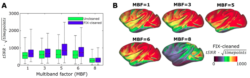
Macaque monkeys are an important animal model where invasive investigations can lead to a better understanding of the cortical organization of primates including humans. However, the tools and methods for noninvasive image acquisition (e.g. MRI RF coils and pulse sequence protocols) and image data preprocessing have lagged behind those developed for humans. To resolve the structural and functional characteristics of the smaller macaque brain, high spatial, temporal, and angular resolutions combined with high signal-to-noise ratio are required to ensure good image quality. To address these challenges, we developed a macaque 24-channel receive coil for 3-T MRI with parallel imaging capabilities. This coil enables adaptation of the Human Connectome Project (HCP) image acquisition protocols to the in-vivo macaque brain. In addition, we adapted HCP preprocessing methods to the macaque brain, including spatial minimal preprocessing of structural, functional MRI (fMRI), and diffusion MRI (dMRI). The coil provides the necessary high signal-to-noise ratio and high efficiency in data acquisition, allowing four- and five-fold accelerations for dMRI and fMRI. Automated FreeSurfer segmentation of cortex, reconstruction of cortical surface, removal of artefacts and nuisance signals in fMRI, and distortion correction of dMRI all performed well, and the overall quality of basic neurobiological measures was comparable with those for the HCP. Analyses of functional connectivity in fMRI revealed high sensitivity as compared with those from publicly shared datasets. Tractography-based connectivity estimates correlated with tracer connectivity similarly to that achieved using ex-vivo dMRI. The resulting HCP-style in vivo macaque MRI data show considerable promise for analyzing cortical architecture and functional and structural connectivity using advanced methods that have previously only been available in studies of the human brain.

Keywords: Cortex; Diffusion; Human connectome project; Macaque; Parallel imaging; Primate; Resting-state.


Conflict of interest statement

The other authors have no conflicts of interest to declare.

Figures

Figure 1
Figure 1
The design and development of macaque 24-channel receive-only coil. (A) Design of coil geometry and (B) element locations. (C) Outlook of element alignment on a 3D print. (D) Coil with final element arrangements. (D) Schematic of a coil element circuit. (E) Coil circuitry. (F) Coil outlook with animal holder attached to the gantry of the MRI scanner. (G) Macaque head phantom.
Figure 2
Figure 2
Macaque 24-channel coil performance and geometry. (A) Noise correlation matrix. (B) Coil element arrangement and labeling flattened into a 2D representation. (C) Inverse geometry (1/g)-factor maps using gradient echo imaging with generalized autocalibrating partially parallel acquisitions (GRAPPA) reduction factors (R=2, 3 and 4) in LR-direction used for diffusion MRI (see later). (D) The boxplot shows 1/g-factor with respect to reduction factor. While geometric distortions are small with acceleration factor of 2 (1/g=1.03±0.07), further reduction yields large signal degradations. Geometric distortions were evaluated using a phantom whose contour was matched to the average macaque brain.
Figure 3
Figure 3
Data quality assessment of structural and functional MRI. Axial slices acquired with 500 μm isotropic resolution (A) T1-weighted MPRAGE and (B) T2-weighted SPACE. Flip-angle (C) axial and (D) surface maps. The values indicate the difference between experimental and nominal flip-angle (90°) in units of degree. B0 (E) axial and (F) surface field-maps. Unit radian per second. White and black lines (in E and G, respectively) outline the pial surface. Temporal signal-to-noise ratio (tSNR) (G) axial and (H) surface maps of FIX-cleaned fMRI. The tSNR map was acquired using multiband 2D-EPI sequence (TR=0.755s, TE=30ms, MBF=5, isotropic resolution=1.25mm). Data at https://balsa.wustl.edu/Z44X3
Figure 4
Figure 4
Cortical surface mapping of three widely studied macaque monkeys. Japanese rhesus (Macaca fuscata, N=1), rhesus (Macaca mulatta, N=1), and crab-eating monkey (Macaca fascicularis, N=1) and average maps across the species (N=12; N=4 for each species). (A, B, C, D) Pial surface. (E, F, G, H) Curvature and (I, J, K, L) bias-corrected myelin maps shown on very inflated cortical surface. Cortical thickness (M, N, O, P) maps and (Q, R, S, T) histograms. Data at https://balsa.wustl.edu/VjjZV
Figure 5
Figure 5
Optimization of fMRI multiband acceleration. (A) Relationship between temporal signal-to-noise ratio (tSNR) multiplied by a square-root of acquired time-points and multiband factor (MBF). Acquisition times are matched in the data points (each scan 10 minutes, N=1). The boxplot shows distributions of tSNR in the greyordinates (a total of 26k) for FIX-uncleaned (green) and FIX-cleaned data (blue). (B) Cortical surface presentation of FIX cleaned tSNR × sqrt (#timepoints) vs multiband factor. Note that MBF=5 produces the highest tSNR.
Figure 6
Figure 6
Classification of resting-state fMRI variance and their relative contributions of the total variance in macaque (N=20) and the human connectome project (HCP, N=20). The variances were computed using a development version of the Resting State Stats HCP pipeline. Abbreviations: struct noise=structured noise (scanner artefacts and nuisance signals etc.), BOLD=’neural’ blood oxygen level dependent signal, MGT=FIX-cleaned mean greyordinate timeseries.
Figure 7
Figure 7
Representative macaque resting-state functional connectivity in a single subject. (A) An example resting-state network (RSN) obtained in spatial ICA, which shows positive connectivity over posterior parietal cortex (areas 7A, DP, LIP), precuneus (areas 23, 31), temporo-occipital areas (MST, PGa) and prefrontal cortex (areas 46d, 8b, as defined in M132 atlas). Timeseries and frequency of this component (lower panels) exhibited pronounced hemodynamic-like low-frequency oscillations. (B) Exemplar functional connectivity seeded from a single greyordinate in the area 7A (white circle). Spatial distribution of connectivity resembled to that of the component in (A), as well as timeseries and frequency of the seed signal (lower panels). Data was from two 51-min fMRI scans (subject N=1), preprocessed for correction of motion, distortion, inhomogeneity, and denoising with multi-run FIX. The dense timeseries was further reduced in random noise by Wishart filter and used for seed-based dense connectivity (Pearson’s correlation). Other components classified into signal or noise, and dense connectivity seeded from other vertices can be interactively viewed using Workbench using data at https://balsa.wustl.edu/3ggwG
Figure 8
Figure 8
Comparison of dMRI quality measures between macaque and the HCP (blue and red bars, respectively; N=10). Plots show whole brain SNR (A) and CNR across b-values 1000, 2000 and 3000 (B), as well as three-crossing fiber ratio (C) and dispersion uncertainties (in degree) of 1st, 2nd and 3rd fiber orientations in the white matter voxels (D). Overall, the quality measures were comparable across the studies.
Figure 9
Figure 9
Representative diffusion magnetic resonance imaging (dMRI) applications. Parcellated cortical surface distributions of mean diffusivity (MD) (A) and fractional anisotropy (FA) (B) calculated in diffusion tensor model, and neurite density index (NDI) and (C) orientation dispersion index (ODI) (D) calculated in NODDI (see main text; N=6). (E) Parcellated diffusion tractography (N=1, ID=A18031601) seed from left premotor area, F5 (blue color) and (F) the quantitative ground-truth derived from retrograde tracer injected into F5. Note the correspondence between tractography and tracer connectivities (see main text for details). Data at https://balsa.wustl.edu/zppXg

References

    1. Andersson JLR, Skare S, Ashburner J. How to correct susceptibility distortions in spin-echo echo-planar images: application to diffusion tensor imaging. NeuroImage. 2003;20:870–888. doi: 10.1016/S1053-8119(03)00336-7. - DOI - PubMed
    1. Andersson JLR, Sotiropoulos SN. An integrated approach to correction for off-resonance effects and subject movement in diffusion MR imaging. NeuroImage. 2016;125:1063–1078. - PMC - PubMed
    1. Andersson JLR, Sotiropoulos SN. Non-parametric representation and prediction of single- and multi-shell diffusion-weighted MRI data using Gaussian processes. NeuroImage. 2015;122:166–176. doi: 10.1016/j.neuroimage.2015.07.067. - DOI - PMC - PubMed
    1. Azevedo FAC, Carvalho LRB, Grinberg LT, Farfel JM, Ferretti REL, Leite REP, Filho WJ, Lent R, Herculano-Houzel S. Equal numbers of neuronal and nonneuronal cells make the human brain an isometrically scaled-up primate brain. J Comp Neurol. 2009;513:532–541. doi: 10.1002/cne.21974. - DOI - PubMed
    1. Bastiani M, Cottaar M, Fitzgibbon SP, Suri S, Alfaro-Almagro F, Sotiropoulos SN, Jbabdi S, Andersson JLR. Automated quality control for within and between studies diffusion MRI data using a non-parametric framework for movement and distortion correction. NeuroImage. 2019;184:801–812. doi: 10.1016/j.neuroimage.2018.09.073. - DOI - PMC - PubMed

Publication types

MeSH terms