Review
. 2020 Apr 8;9(4):910.
doi: 10.3390/cells9040910.

A G-Protein Coupled Receptor and Macular Degeneration

Affiliations
Review

A G-Protein Coupled Receptor and Macular Degeneration

Anna G Figueroa et al. Cells. .

Abstract

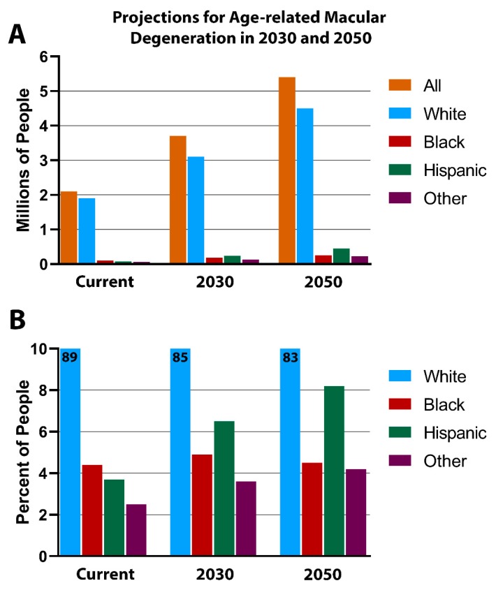
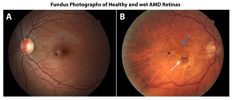
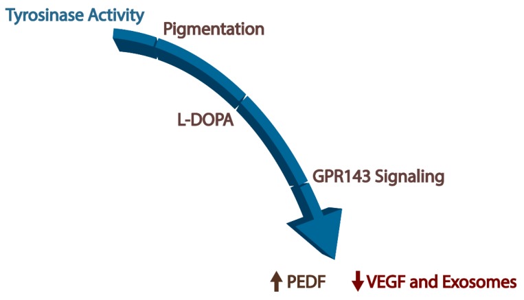
Age-related macular degeneration (AMD) is a leading cause of irreversible blindness in the world. The risk of AMD increases with age and is most common among the white population. Here, we discuss the convergence of factors related to race, pigmentation, and susceptibility to AMD, where the primary defect occurs in retinal support cells, the retinal pigment epithelium (RPE). We explore whether the observed racial bias in AMD incidence is related to innate differences in the basal level of pigmentation between races, and whether the pigmentation pathway activity in the RPE might protect from retinal degeneration. More specifically, we explore whether the downstream signaling activity of GPR143, a G-protein coupled receptor in the pigmentation pathway, might underly the racial bias of AMD and be a target to prevent the disease. Lastly, we summarize the past findings of a large retrospective study that investigated the relationship between the stimulation of GPR143 with L-DOPA, the pigmentation pathway, and AMD, to potentially help develop new ways to prevent or treat AMD. The reader of this review will come to understand the racial bias of AMD, which is related to the function of the RPE.

Keywords: AMD; G-protein; GPR143; L-DOPA; OA1; RPE; macular degeneration; melanin.


Conflict of interest statement

The authors declare no conflict of interest.

Figures

Figure 1
Figure 1
One of the many prominent features of the retinal pigment epithelium (RPE) is the production and accumulation of melanin granules. Representative phase-contrast micrograph of the cell morphology of human RPE cells from a 78-year-old healthy female donor. The primary cells were cultured with Dulbecco’s Modified Eagle Medium (DMEM) + 5% FBS. Total magnification 200×, scale bar = 25 µm.
Figure 2
Figure 2
Representative images of possible mechanisms of intercellular communication by exosomes; exosome uptake mediated by endocytosis (A) or exosome fusion at the target cell’s plasma membrane (B). Paired phase-contrast and fluorescence micrographs of bovine ex-vivo RPE challenged with apical in situ bovine RPE exosomes. Exosomes were labeled with CF555 membrane protein dye to illustrate internalized exosomes taken up by the target unpigmented RPE cells from the tapetum lucidum region of the bovine eye (A) or lipophilic octadecyl rhodamine B, R18, membrane dye to illustrate sites of exosome fusion along the plasma membrane of pigmented RPE cells neighboring the tapetum lucidum (B). Total magnification = 630×.
Figure 3
Figure 3
Projections for age-related macular degeneration. The white population will continue to account for most age-related macular degeneration (AMD)-related cases, as the disease incidence level is expected to more than double by 2050, from 2 million to 5.4 million people (A). The percentage of people affected with AMD by race is illustrated in (B). Races with the highest levels of pigmentation have the lowest incidence of AMD, which is possibly due to the stimulation of GPR143 by L-DOPA. Image adapted from the National Eye Institute’s AMD Data Tables, last updated 17 July 2019.
Figure 4
Figure 4
A healthy retina can undergo severe morphological changes due to wet age-related macular degeneration (AMD). Color photograph of a heavily pigmented fundus of a healthy volunteer (A) and the lightly pigmented fundus of a patient with wet AMD (B), which illustrates drusen (blue arrow) and retinal hemorrhage (white arrow). The black asterisk denotes a circular image artifact. (Images courtesy of Robert W. Snyder, MD, PhD).
Figure 5
Figure 5
Schematic diagram of the path from tyrosinase activity in the retinal pigment epithelium to increased pigment epithelium-derived factor (PEDF) secretion, decreased secretion of vascular endothelial growth factor (VEGF), and decreased release of exosomes. Together, these downstream effectors of GPR143 signaling by L-DOPA are likely to offer protection of the retina during aging.

References

    1. Strauss O. The retinal pigment epithelium in visual function. Physiol. Rev. 2005;85:845–881. doi: 10.1152/physrev.00021.2004. - DOI - PubMed
    1. Fronk A.H., Vargis E. Methods for culturing retinal pigment epithelial cells: A review of current protocols and future recommendations. J. Tissue Eng. 2016;7 doi: 10.1177/2041731416650838. - DOI - PMC - PubMed
    1. McKay B.S., Burke J.M. Separation of phenotypically distinct subpopulations of cultured human retinal pigment epithelial cells. Exp. Cell Res. 1994;213:85–92. doi: 10.1006/excr.1994.1176. - DOI - PubMed
    1. Schmidt S.Y., Peisch R.D. Melanin concentration in normal human retinal pigment epithelium. Investig. Ophthalmol. Vis. Sci. 1986;27:1063–1067. - PubMed
    1. Weiter J.J., Delori F.C., Wing G.L., Fitch K.A. Retinal pigment epithelial lipofuscin and melanin and choroidal melanin in human eyes. Invest. Ophthalmol. Vis. Sci. 1986;27:145–152. - PubMed

Publication types

MeSH terms