Review
. 2020 Apr 8;12(4):911.
doi: 10.3390/cancers12040911.

MicroRNAs as Emerging Regulators of Signaling in the Tumor Microenvironment

Affiliations
Review

MicroRNAs as Emerging Regulators of Signaling in the Tumor Microenvironment

Shahzad Nawaz Syed et al. Cancers (Basel). .

Abstract

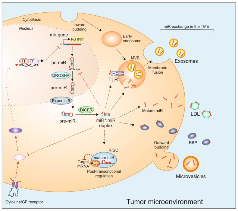
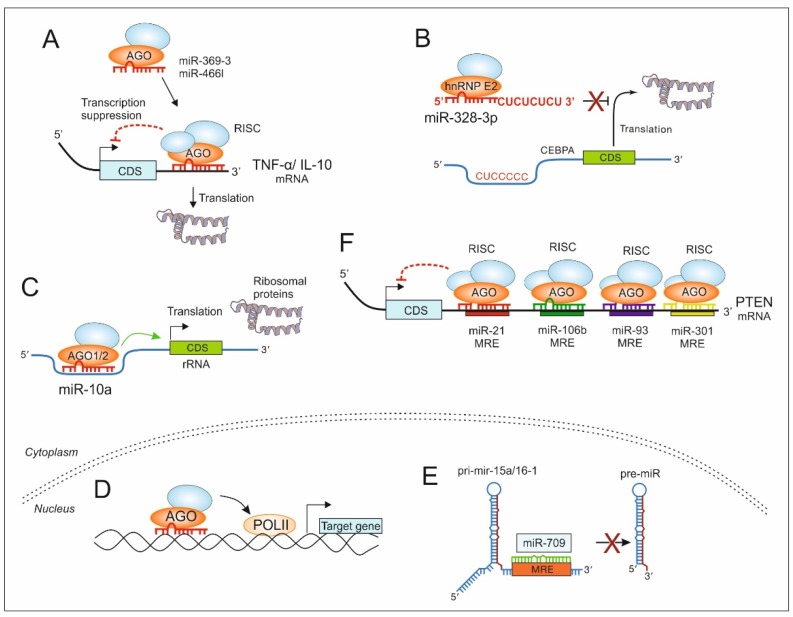
A myriad of signaling molecules in a heuristic network of the tumor microenvironment (TME) pose a challenge and an opportunity for novel therapeutic target identification in human cancers. MicroRNAs (miRs), due to their ability to affect signaling pathways at various levels, take a prominent space in the quest of novel cancer therapeutics. The role of miRs in cancer initiation, progression, as well as in chemoresistance, is being increasingly investigated. The canonical function of miRs is to target mRNAs for post-transcriptional gene silencing, which has a great implication in first-order regulation of signaling pathways. However, several reports suggest that miRs also perform non-canonical functions, partly due to their characteristic non-coding small RNA nature. Examples emerge when they act as ligands for toll-like receptors or perform second-order functions, e.g., to regulate protein translation and interactions. This review is a compendium of recent advancements in understanding the role of miRs in cancer signaling and focuses on the role of miRs as novel regulators of the signaling pathway in the TME.

Keywords: RNA therapeutics; breast cancer; cancer; carcinoma; inflammation; microRNA.


Conflict of interest statement

The authors declare no conflict of interest.

Figures

Figure 1
Figure 1
MiR genesis, exchange, and inter-cellular transfer. MiRs are transcribed by RNA polymerase II (Pol II) and III into primary-miRs (pri-miR), which are further processed by the Drosha complex. The resultant precursor-miR (pre-miR) is exported to the cytoplasm by exportin 5 and cleaved by Dicer to form a double-stranded miR–miR duplex. One of the strands is degraded and the mature miR strand is loaded to the Argonaute protein (AGO) complex, which together with other proteins forms the RNA-induced silencing complex (RISC). RISC complex binds, in a sequence complementary manner to the 3′-untranslated region (UTR) of mRNA targets to provoke post-transcriptional gene suppression. MiRs can be exported from donor cells into the tumor microenvironment (TME), where they can also repress target gene expression in recipient cells. Several carriers such as extracellular vesicles (exosomes, microvesicles, apoptotic bodies), RNA-binding proteins (RBP), or low-density lipoproteins (LDL) aid miR transport and exchange. Intracellularly, miRs can be loaded into multivesicular bodies (MVB), which are formed by the plasma membrane inward budding. The fusion of the MVBs with the plasma membrane provokes the release of exosomal miRs into the extraluminal space. Apart from canonical mRNA targets, inter- and intra-cellular targets of miRs include TLRs in the endosome. MiRs also interfere with cytokine and growth factor receptor signaling cascades by targeting intermediary protein expression. In turn, cell surface receptor signaling can also regulate miR biogenesis. GF: growth factor, TF: transcription factor, TLR: toll-like receptor.
Figure 2
Figure 2
MiRs as regulators of signaling in the TME. The canonical function of miRs includes binding to 3′ UTR of target mRNA to provoke post-transcriptional gene suppression either by decreasing the mRNA stability or decreasing translation (A). However, non-canonical functions include miRs that increase translation by binding to the 3′ UTR that masks the binding site of an RNA-binding protein that would otherwise induce mRNA degradation [78,79] (A). Increased translation can also be achieved by miR-328 by binding to the translation inhibitor heterogeneous ribonuclear protein E2 (hnRNP E2) in a seed sequence-independent manner that prevents and/or displaces CCAAT/enhancer-binding protein alpha (CEBPA) mRNA binding and rescues CEBPA mRNA translation [80] (B). Furthermore, miR-10a has been shown to bind the 5′ UTR of ribosomal protein mRNAs and to enhance their translation (C) [81]. MiRs also activate transcription either by recruiting transcription-activating factors to the complementary elements in gene promoters (D) [82,83] or by acting as a scaffold for transcription-regulating RNA [84]. MiRs can also regulate expression of its peer in the nucleus as in case of miR-709 that directly binds to a 19-nt MRE on pri-miR-15a/16-1 and blocks its processing into pre-miR-15a/16-1, thereby suppressing miR-15a/16-1 maturation (E) [65]. Target mRNA can be regulated by multiple miRs by targeting respective MRE in the 3′ UTR of e.g., PTEN, which can be targeted by miR-21, miR-106b, miR-93, and miR-301 (F) [85,86,87,88,89].

References

    1. Odes E.J., Randolph-Quinney P.S., Steyn M., Throckmorton Z., Smilg J.S., Zipfel B., Augustine T.N., Beer F.d., Hoffman J.W., Franklin R.D., et al. Earliest hominin cancer: 1.7-million-year-old osteosarcoma from Swartkrans Cave, South Africa. S. Afr. J. Sci. 2016;112 doi: 10.17159/sajs.2016/20150471. - DOI
    1. Randolph-Quinney P.S., Williams S.A., Steyn M., Meyer M.R., Smilg J.S., Churchill S.E., Odes E.J., Augustine T., Tafforeau P., Berger L.R. Osteogenic tumour in Australopithecus sediba: Earliest hominin evidence for neoplastic disease. S. Afr. J. Sci. 2016;112 doi: 10.17159/sajs.2016/20150470. - DOI
    1. Dagogo-Jack I., Shaw A.T. Tumour heterogeneity and resistance to cancer therapies. Nat. Rev. Clin. Oncol. 2018;15:81–94. doi: 10.1038/nrclinonc.2017.166. - DOI - PubMed
    1. Lim B., Woodward W.A., Wang X., Reuben J.M., Ueno N.T. Inflammatory breast cancer biology: The tumour microenvironment is key. Nat. Rev. Cancer. 2018;18:485–499. doi: 10.1038/s41568-018-0010-y. - DOI - PubMed
    1. Yang L., Li A., Lei Q., Zhang Y. Tumor-intrinsic signaling pathways: Key roles in the regulation of the immunosuppressive tumor microenvironment. J. Hematol. Oncol. 2019;12:125. doi: 10.1186/s13045-019-0804-8. - DOI - PMC - PubMed

LinkOut - more resources