The AMPK-PP2A axis in insect fat body is activated by 20-hydroxyecdysone to antagonize insulin/IGF signaling and restrict growth rate
- PMID: 32277029
- PMCID: PMC7196814
- DOI: 10.1073/pnas.2000963117
The AMPK-PP2A axis in insect fat body is activated by 20-hydroxyecdysone to antagonize insulin/IGF signaling and restrict growth rate
Abstract
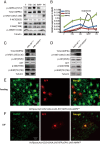
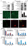
In insects, 20-hydroxyecdysone (20E) limits the growth period by triggering developmental transitions; 20E also modulates the growth rate by antagonizing insulin/insulin-like growth factor signaling (IIS). Previous work has shown that 20E cross-talks with IIS, but the underlying molecular mechanisms are not fully understood. Here we found that, in both the silkworm Bombyx mori and the fruit fly Drosophila melanogaster, 20E antagonized IIS through the AMP-activated protein kinase (AMPK)-protein phosphatase 2A (PP2A) axis in the fat body and suppressed the growth rate. During Bombyx larval molt or Drosophila pupariation, high levels of 20E activate AMPK, a molecular sensor that maintains energy homeostasis in the insect fat body. In turn, AMPK activates PP2A, which further dephosphorylates insulin receptor and protein kinase B (AKT), thus inhibiting IIS. Activation of the AMPK-PP2A axis and inhibition of IIS in the Drosophila fat body reduced food consumption, resulting in the restriction of growth rate and body weight. Overall, our study revealed an important mechanism by which 20E antagonizes IIS in the insect fat body to restrict the larval growth rate, thereby expanding our understanding of the comprehensive regulatory mechanisms of final body size in animals.
Keywords: 20-hydroxyecdysone; AMPK-PP2A axis; fat body; growth rate; insulin/IGF signaling.
Conflict of interest statement
The authors declare no competing interest.
Figures


Similar articles
-
Transcriptional regulation of the insulin signaling pathway genes by starvation and 20-hydroxyecdysone in the Bombyx fat body.J Insect Physiol. 2010 Oct;56(10):1436-44. doi: 10.1016/j.jinsphys.2010.02.011. Epub 2010 Mar 9. J Insect Physiol. 2010. PMID: 20197069
-
Insulin and 20-hydroxyecdysone action in Bombyx mori: Glycogen content and expression pattern of insulin and ecdysone receptors in fat body.Gen Comp Endocrinol. 2017 Jan 15;241:108-117. doi: 10.1016/j.ygcen.2016.06.022. Epub 2016 Jun 15. Gen Comp Endocrinol. 2017. PMID: 27317549
-
Developmental regulation of glycolysis by 20-hydroxyecdysone and juvenile hormone in fat body tissues of the silkworm, Bombyx mori.J Mol Cell Biol. 2010 Oct;2(5):255-63. doi: 10.1093/jmcb/mjq020. Epub 2010 Aug 20. J Mol Cell Biol. 2010. PMID: 20729248
-
Insulin/IGF signaling in Drosophila and other insects: factors that regulate production, release and post-release action of the insulin-like peptides.Cell Mol Life Sci. 2016 Jan;73(2):271-90. doi: 10.1007/s00018-015-2063-3. Epub 2015 Oct 15. Cell Mol Life Sci. 2016. PMID: 26472340 Free PMC article. Review.
-
The role of insulin signalling in the endocrine stress response in Drosophila melanogaster: A mini-review.Gen Comp Endocrinol. 2018 Mar 1;258:134-139. doi: 10.1016/j.ygcen.2017.05.019. Epub 2017 May 26. Gen Comp Endocrinol. 2018. PMID: 28554733 Review.
Cited by
-
Effects of Starvation on the Levels of Triglycerides, Diacylglycerol, and Activity of Lipase in Male and Female Drosophila Melanogaster.J Lipids. 2021 Mar 25;2021:5583114. doi: 10.1155/2021/5583114. eCollection 2021. J Lipids. 2021. PMID: 33833879 Free PMC article.
-
Conceptual framework for the insect metamorphosis from larvae to pupae by transcriptomic profiling, a case study of Helicoverpa armigera (Lepidoptera: Noctuidae).BMC Genomics. 2022 Aug 13;23(1):591. doi: 10.1186/s12864-022-08807-y. BMC Genomics. 2022. PMID: 35963998 Free PMC article.
-
Transcriptome analysis unveils the mechanisms of lipid metabolism response to grayanotoxin I stress in Spodoptera litura.PeerJ. 2023 Dec 6;11:e16238. doi: 10.7717/peerj.16238. eCollection 2023. PeerJ. 2023. PMID: 38077416 Free PMC article.
-
A plant virus manipulates the long-winged morph of insect vectors.Proc Natl Acad Sci U S A. 2024 Jan 16;121(3):e2315341121. doi: 10.1073/pnas.2315341121. Epub 2024 Jan 8. Proc Natl Acad Sci U S A. 2024. PMID: 38190519 Free PMC article.
-
Mmp-induced fat body cell dissociation promotes pupal development and moderately averts pupal diapause by activating lipid metabolism.Proc Natl Acad Sci U S A. 2023 Jan 3;120(1):e2215214120. doi: 10.1073/pnas.2215214120. Epub 2022 Dec 27. Proc Natl Acad Sci U S A. 2023. PMID: 36574695 Free PMC article.
References
-
- Conlon I., Raff M., Size control in animal development. Cell 96, 235–244 (1999). - PubMed
-
- Gokhale R. H., Shingleton A. W., Size control: The developmental physiology of body and organ size regulation. Wiley Interdiscip. Rev. Dev. Biol. 4, 335–356 (2015). - PubMed
-
- Nijhout H. F., The control of body size in insects. Dev. Biol. 261, 1–9 (2003). - PubMed
-
- Nijhout H. F., Callier V., Developmental mechanisms of body size and wing-body scaling in insects. Annu. Rev. Entomol. 60, 141–156 (2015). - PubMed
Publication types
MeSH terms
Substances
LinkOut - more resources
Full Text Sources
Other Literature Sources
Molecular Biology Databases

