Review
. 2020 May 15;295(20):7138-7153.
doi: 10.1074/jbc.REV120.008286. Epub 2020 Apr 10.

The many lives of type IA topoisomerases

Affiliations
Review

The many lives of type IA topoisomerases

Anna H Bizard et al. J Biol Chem. .

Abstract

The double-helical structure of genomic DNA is both elegant and functional in that it serves both to protect vulnerable DNA bases and to facilitate DNA replication and compaction. However, these design advantages come at the cost of having to evolve and maintain a cellular machinery that can manipulate a long polymeric molecule that readily becomes topologically entangled whenever it has to be opened for translation, replication, or repair. If such a machinery fails to eliminate detrimental topological entanglements, utilization of the information stored in the DNA double helix is compromised. As a consequence, the use of B-form DNA as the carrier of genetic information must have co-evolved with a means to manipulate its complex topology. This duty is performed by DNA topoisomerases, which therefore are, unsurprisingly, ubiquitous in all kingdoms of life. In this review, we focus on how DNA topoisomerases catalyze their impressive range of DNA-conjuring tricks, with a particular emphasis on DNA topoisomerase III (TOP3). Once thought to be the most unremarkable of topoisomerases, the many lives of these type IA topoisomerases are now being progressively revealed. This research interest is driven by a realization that their substrate versatility and their ability to engage in intimate collaborations with translocases and other DNA-processing enzymes are far more extensive and impressive than was thought hitherto. This, coupled with the recent associations of TOP3s with developmental and neurological pathologies in humans, is clearly making us reconsider their undeserved reputation as being unexceptional enzymes.

Keywords: BLM; DNA replication; DNA supercoiling; DNA topology; DNA transcription; PICH; TOP3A; TOP3B; chromosome segregation; chromosomes; protein translocation; reverse gyrase; translocases.


Conflict of interest statement

The authors declare that they have no conflicts of interest with the contents of this article

Figures

Figure 1.
Figure 1.
Topological constraints associated with DNA metabolism in vivo. A, twin supercoiling domain model. When a translocating machinery is not allowed to rotate around the DNA axis (arrowhead with black circle), it introduces overwinding (positive supercoiling; +ve SC) in front of and underwinding (negative supercoiling; −ve SC) behind the translocase. B, in the context of transcription, the overwinding accumulated ahead of the RNA polymerase prevents strand opening and can ultimately block transcription elongation. Underwinding generated behind the polymerase can promote strand opening and lead to the stabilization of R-loops and other secondary structures. C, during DNA replication, and when fork rotation is prevented, the degree of entanglement between the newly replicated DNA molecules is limited, but overwinding ahead of the fork can prevent replisome progression. D, during DNA replication, overwinding of the template can be limited by fork rotation, but this leads to formation of precatenanes behind the fork, which represent an obstacle during segregation.
Figure 2.
Figure 2.
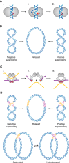
Mechanism of action and catalytic activities of Type IB and Type II topoisomerases. A, i, Type IB topoisomerases bind to dsDNA. ii, a transesterification reaction leads to the formation of a single strand break with the 3′ terminus covalently associated with the active tyrosine. Torsional stress in the substrate is dissipated by free rotation of the 5′ end of the nick. iii, a second transesterification reaction religates the nick and frees the enzyme from its covalent interaction. Open red circle, catalytic tyrosine; closed red circle, catalytic tyrosine engaged in a covalent DNA intermediate. B, Type IB topoisomerases efficiently relax both positive (+ve SC) and negative (−ve SC) supercoils. C, i, Type II topoisomerases are dimeric enzymes that possess two catalytic tyrosine residues (open red circles). ii, transesterification reactions lead to the introduction of a transient double strand break into a dsDNA molecule (G-segment; purple). After cleavage, each 5′ end of the DNA break is covalently associated with one of the tyrosines (closed red circles). Conformational changes in the protein brings the ends of the broken G-segment apart, enabling another dsDNA molecule (T-segment; yellow) to pass though the gate. iii, a second set of transesterification reactions religate the break and free the enzyme from its covalent interaction with the DNA. D, when the G-segments (purple) and T-segments (yellow) are located on the same molecule, Type II topoisomerase activity leads to the relaxation of negative and positive supercoils (top). When the G- and T-segments are located in trans, Type II topoisomerases can modify the degree of catenation between two dsDNA molecules (bottom).
Figure 3.
Figure 3.
Structure and catalytic cycle of Type IA topoisomerases. A, the structure of the core topoisomerase domain resembles a toroidal clamp in which the topo-fold subdomain II forms an arc. The catalytic site is reconstituted at the base of this arc, by the association of residues from the topoisomerase-primase (TOPRIM) subdomain I and the two catabolite activator (CAP-Y and CAP) subdomains III and IV. The deep ssDNA (and ssRNA) binding groove (G-segment–binding groove) is formed between subdomains I and IV. B, a, Type IA topoisomerases bind to single-stranded segments of DNA via the G-segment–binding groove that directs the G-segment in line with the catalytic tyrosine (i, open red circle). b, a transesterification reaction leads to the formation of a single strand break with the 5′ termini covalently associated with the catalytic tyrosine (ii, closed red circle). The 3′ end of the nick forms a tight association with the G-segment–binding groove. c, protein conformational changes enable the opening of a gate via the separation of the CAP domains and their associated DNA termini (iii). d, another nucleic acid segment (here the single-strand DNA complementary to the G-segment) is passed though the gate toward the cavity of the enzyme (iv). e, after this transport, the closure of the gate reconstitutes the catalytic cycle (v). f, a second transesterification reaction reseals the nick and frees the enzyme from its covalent interaction with the substrate (vi). g, full dissociation is enabled by opening of the gate.
Figure 4.
Figure 4.
Range of substrates for Type IA topoisomerases as a function of the nature of the G- and T- segments.
Figure 5.
Figure 5.
Functions of the RecQ helicase/Type IA topoisomerase “dissolvasome”. A, the dissolvasome is a multienzyme complex that combines the helicase activity of a RecQ family member and the topoisomerase activity of a TOP3 topoisomerase. RecQ helicases act as ssDNA translocases that rotate around the DNA axis (arrowhead with open black circle), such that the unpaired strands remain topologically entangled. In a covalently closed DNA molecule, the full dissociation of two paired strands requires the rupture of the hydrogen bounds (catalyzed by a helicase) and the dissipation of the topological entanglements resulting from the double-helical structure of DNA (catalyzed by a topoisomerase). B, synergistic cooperation between the helicase and topoisomerase activities of the dissolvasome enables the resolution of complex intermolecular entanglements, such as double Holliday junctions, late replication intermediates, and dsDNA catenanes.
Figure 6.
Figure 6.
Positive supercoiling activity of the PICH-TOP3A complex. PICH is a dsDNA translocase that extrudes DNA loops. Because it is prevented from rotating around the DNA axis (arrowhead with black circle), its translocation is associated with the redistribution of DNA torsional stress. This leads to an accumulation of negative (−ve SC) and positive (+ve SC) supercoils within and outside of the extruded loop, respectively. TOP3 relaxes the highly negatively supercoiled loop, which leads to an accumulation of net positive supercoiling in the substrate.
Figure 7.
Figure 7.
Domain organization of Type IA topoisomerases and their obligatory subunits. A, all Type IA topoisomerases share a highly conserved catalytic domain (blue) and sometimes an additional C-terminal extension (CTD), which contains multiple zinc finger motifs (black boxes) involved in protein-DNA and protein-protein interactions. In addition to putative zinc finger motifs, the C-terminal domain of HsTOP3B also exhibits RGG box motifs (green) that can be methylated and mediate interactions with RNA and the Tudor domain of TDRD3. An alternative start codon leads to the addition of a mitochondrial targeting sequence (MTS) to the TOP3A polypeptide, such that TOP3A encodes both nuclear and mitochondrial isoforms. In eukaryotes, TOP3 forms heterodimers with members of the RMI family. RMI members are characterized by a conserved association between a DUF1767/OB-fold domain (DUF-OB). In humans, the nuclear isoforms of TOP3A and TOP3B interact with their own cognate RMI protein, RMI1 and TDRD3, respectively. The RMI1 CTD exhibits a second OB-fold domain and mediates interactions with other proteins, including RMI2. TDRD3 CTD is characterized by the presence of multiple protein-protein interaction motifs including a ubiquitin-associating domain (UBA) and a Tudor domain. The Tudor domain of TDRD3 mediates interactions with methylated proteins, including histones, RNA polymerase, and TOP3B, and with the fragile X mental retardation protein. Ec, E. coli; Sc, S. cerevisiae; Hs, Homo sapiens. B, RMI1 and TDRD3 interact with the arc of TOP3A and TOP3B, respectively. RMI1 inserts a loop into the cavity of TOP3A, which restricts its size. A similar insertion loop is present in TDRD3, but this does not appear to significantly reduce the size of the TOP3B cavity.

References

    1. Liu L. F., and Wang J. C. (1987) Supercoiling of the DNA template during transcription. Proc. Natl. Acad. Sci. U.S.A. 84, 7024–7027 10.1073/pnas.84.20.7024 - DOI - PMC - PubMed
    1. Ma J., and Wang M. D. (2016) DNA supercoiling during transcription. Biophys. Rev. 8, 75–87 10.1007/s12551-016-0215-9 - DOI - PMC - PubMed
    1. Kouzine F., Gupta A., Baranello L., Wojtowicz D., Ben-Aissa K., Liu J., Przytycka T. M., and Levens D. (2013) Transcription-dependent dynamic supercoiling is a short-range genomic force. Nat. Struct. Mol. Biol. 20, 396–403 10.1038/nsmb.2517 - DOI - PMC - PubMed
    1. Teves S. S., and Henikoff S. (2014) Transcription-generated torsional stress destabilizes nucleosomes. Nat. Struct. Mol. Biol. 21, 88–94 10.1038/nsmb.2723 - DOI - PMC - PubMed
    1. Naughton C., Avlonitis N., Corless S., Prendergast J. G., Mati I. K., Eijk P. P., Cockroft S. L., Bradley M., Ylstra B., and Gilbert N. (2013) Transcription forms and remodels supercoiling domains unfolding large-scale chromatin structures. Nat. Struct. Mol. Biol. 20, 387–395 10.1038/nsmb.2509 - DOI - PMC - PubMed

Publication types

MeSH terms