

Comment
doi: 10.1016/j.jinf.2020.03.033.
Epub 2020 Apr 8.
Clinical and CT imaging features of 2019 novel coronavirus disease (COVID-19)
Affiliations
- PMID: 32277968
- PMCID: PMC7194958
- DOI: 10.1016/j.jinf.2020.03.033
Item in Clipboard
Comment
Clinical and CT imaging features of 2019 novel coronavirus disease (COVID-19)
J Infect.
2020 Jul.
No abstract available
Conflict of interest statement
Declaration of Competing Interest The authors declare that they have no competing interests.
Figures
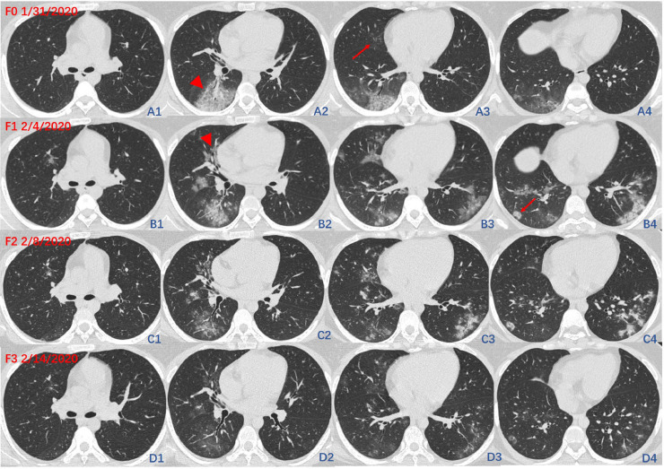
The initial CT images (F0) and three times of follow-up CT images (F1–F3) of P1. F0 showed patchy-like pure GGO located in the subpleural regions of the right middle lobe (F0, A3, arrow) and the right lower lobe, accompanied by crazy paving sign (F0, A2, arrowhead). Follow-up 1(F1, B1–B4): CT images showed diseases progression. The lesions manifested as coexisted nodular-like (F1, B4, arrow) and patchy-like lesions as well as peribronchial (F1, B2, arrowhead), central and subpleural distribution. The lesions are migratory manifested as the absorption of the primary lesions and the emergence of new lesions. CT images of Follow-up 2 (F2, C1–C4) and Follow-up 3 (F3, D1–D4) showed lesion absorption.
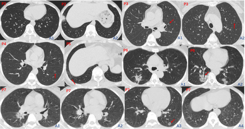
The initial CT images of P2-P7. CT images of P2 (Fig. 2, P2, A1-A2), P5 (Fig. 2, P5), P6 (Fig. 2, P6, A1-A2) and P7 (Fig. 2, P7, A1–A4) showed subpleural lesions, a nodular-like lesion with pseudocavitary sign (Fig. 2, P7, A3, arrow) and mild bronchiectasis (Fig. 2, P6, A2, arrow) were also observed within the lesion. CT images of P3 (Fig. 2, P3, A1-A2) and P4 (Fig. 2, P4) showed round nodular-like GGO lesions (P3 and P4, arrow) located in the central area of the lung.
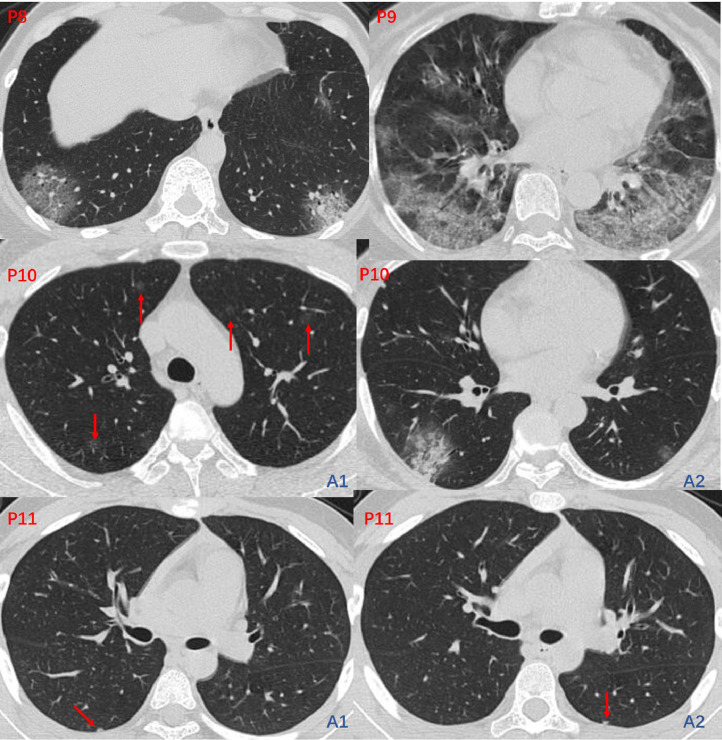
The initial CT images of P8-P11. CT images of P8 (Fig. 3, P8) and P9 (Fig. 3, P9) showed bilateral subpleural lesions with crazy paving sign. CT images of P10 (Fig. 3, P10, A1-A2) showed bilateral multiple lesions, some of them were pure GGO located in the central region of the lung. CT images of P11 (Fig. 3, P11, A1-A2) showed bilateral subpleural small nodular-like lesions.
Comment on
-
Emergence of a novel coronavirus causing respiratory illness from Wuhan, China.J Infect. 2020 Mar;80(3):350-371. doi: 10.1016/j.jinf.2020.01.014. Epub 2020 Jan 28. J Infect. 2020. PMID: 32001309 Free PMC article. No abstract available.
References
-
- Song F., Shi N., Shan F. Emerging Coronavirus 2019-nCoV Pneumonia. Radiology. 2020 doi: 10.1148/radiol.2020200274. - DOI
Publication types
MeSH terms
LinkOut - more resources
Full Text Sources
