. 2021 Jun:190:72-79.
doi: 10.1016/j.ymeth.2020.04.003. Epub 2020 Apr 8.

Investigating lymphangiogenesis in a sacrificially bioprinted volumetric model of breast tumor tissue

Affiliations

Investigating lymphangiogenesis in a sacrificially bioprinted volumetric model of breast tumor tissue

Tingting Liu et al. Methods. 2021 Jun.

Abstract

Lymphatic vessels, as a means to metastasize, are frequently recruited by tumor tissues during their progression. However, reliable in vitro models to dissect the intricate crosstalk between lymphatic vessels and tumors are still in urgent demand. Here, we describe a tissue-engineering method based on sacrificial bioprinting, to develop an enabling model of the human breast tumor with embedded multiscale lymphatic vessels, which is compatible with existing microscopy to examine the processes of lymphatic vessel sprouting and breast tumor cell migration in a physiologically relevant volumetric microenvironment. This platform will potentially help shed light on the complex biology of the tumor microenvironment, tumor lymphangiogenesis, lymphatic metastasis, as well as tumor anti-lymphangiogenic therapy in the future. We further anticipate wide adoption of the method to the production of various tissues and their models with incorporation of lymphatics vessels towards relevant applications.

Keywords: Angiogenesis; Bioprinting; Breast tumor; Lymphatic vessels; Sacrificial; Tumor microenvironment.


Figures

FIG. 1.
FIG. 1.
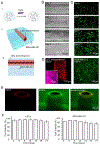
Construction of sacrificially bioprinted hydrogel-based tissue models containing embedded microchannels. (A) The sacrificial bioprinting procedure of the LV-BC model: i) preparation of a PDMS mold; ii) dispersing a layer of GelMA solution (half of the PDMS mold) followed by iii) semi-crosslinking by UV for 10 s; iv) bioprinting the agarose microfiber on top of the semi-crosslinked GelMA base in the center; v) dispensing the GelMA solution containing MDA-MB-231 breast tumor cells to fill the rest of the mold and vi) further UV crosslinking for 40 s; vii) removing the microfiber to create the microchannel; viii) seeding LECs in the microchannel; and ix) subsequent culture. (B) Photographs showing i-ii) the different designs of molds and iii-v) the prefusion of microchannels with different configurations or diameters in the GelMA constructs.
FIG. 2.
FIG. 2.
Characterizations of the sacrificially bioprinted LV-BC model. (A) Schematics showing the arrangement of the LECs and the MDA-MB-231 breast tumor cells in the model. (B) Bright-field time-lapse top-view micrographs showing the proliferation of LECs on the surface of the microchannel. (C) Fluorescence time-lapse top-view micrographs showing the proliferation of MDA-MB-231 breast tumor cells in the surrounding GelMA matrix. (D) Confocal reconstruction images showing the LECs (left) and MDA-MB-231 breast tumor cells at day 8 of culture. The inset is a high-magnification image showing the CD31 expressions, where CD31 was stained in purple and nuclei in blue. (E) Cross-section reconstruction views of LECs in the microchannel and MDA-MB231 breast tumor cells in matrix at day 8 of culture. (F) Viability quantifications of LECs and MDA-MB-231 breast tumor cells in the model for up to 21 days of culture.
FIG. 3.
FIG. 3.
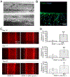
Breast tumor cells induced lymphangiogenesis in the LV-BC model, which was further promoted by VEGF-C stimulation. (A) Optical micrographs showing the migration of LECs from the microchannel inward out. (B) F-actin staining and CD31 staining of the LECs revealing the sprouting at 20 day. (C) Comparisons of LEC sprouting (CD31 staining) from the microchannels in the absence and presence of VEGF-C (250 ng mL−1) at days 14, 17, and 20 of culture. (D) Quantification of the migration distances of LECs from the microchannels under different conditions.
FIG. 4.
FIG. 4.
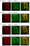
The interactions of LECs and the MDA-MB-231 breast tumor cells in the LV-BC model. (A, B) Confocal reconstruction images showing crosstalk of the cells at day 17 of culture in the (A) absence and (B) presence of VEGF-C. (C, D) Confocal reconstruction images showing crosstalk of the cells at day 20 of culture in the (C) absence and (D) presence of VEGF-C. LECs were stained with CD31 (red) and MDA-MB-231 breast tumor cells with EGFR (green).

References

    1. Siegel RL, Miller KD, and Jemal A, CA Cancer J Clin, 70(1) (2020) 7–30. - PubMed
    1. Fidler IJ and Ellis LM, Cell, 79(2) (1994) 185–188. - PubMed
    1. Lambert AW, Pattabiraman DR, and Weinberg RA, Cell, 168(4) (2017) 670–691. - PMC - PubMed
    1. Hanahan D and Weinberg RA, Cell, 144(5) (2011) 646–674. - PubMed
    1. Stacker SA, Williams SP, Karnezis T, Shayan R, Fox SB, and Achen MG, Nat Rev Cancer, 14(3) (2014) 159–172. - PubMed

Publication types