

Comparison of beamformer implementations for MEG source localization
- PMID: 32278091
- PMCID: PMC7322560
- DOI: 10.1016/j.neuroimage.2020.116797
Comparison of beamformer implementations for MEG source localization
Abstract
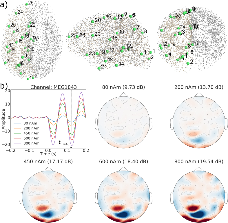
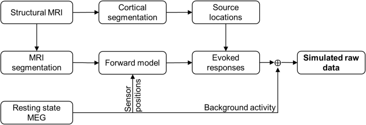
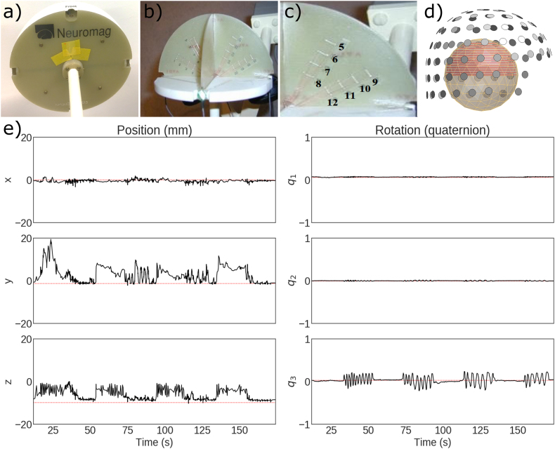
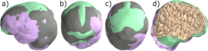
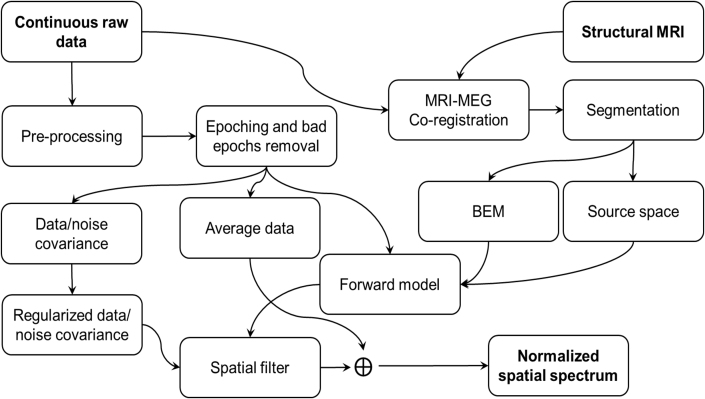
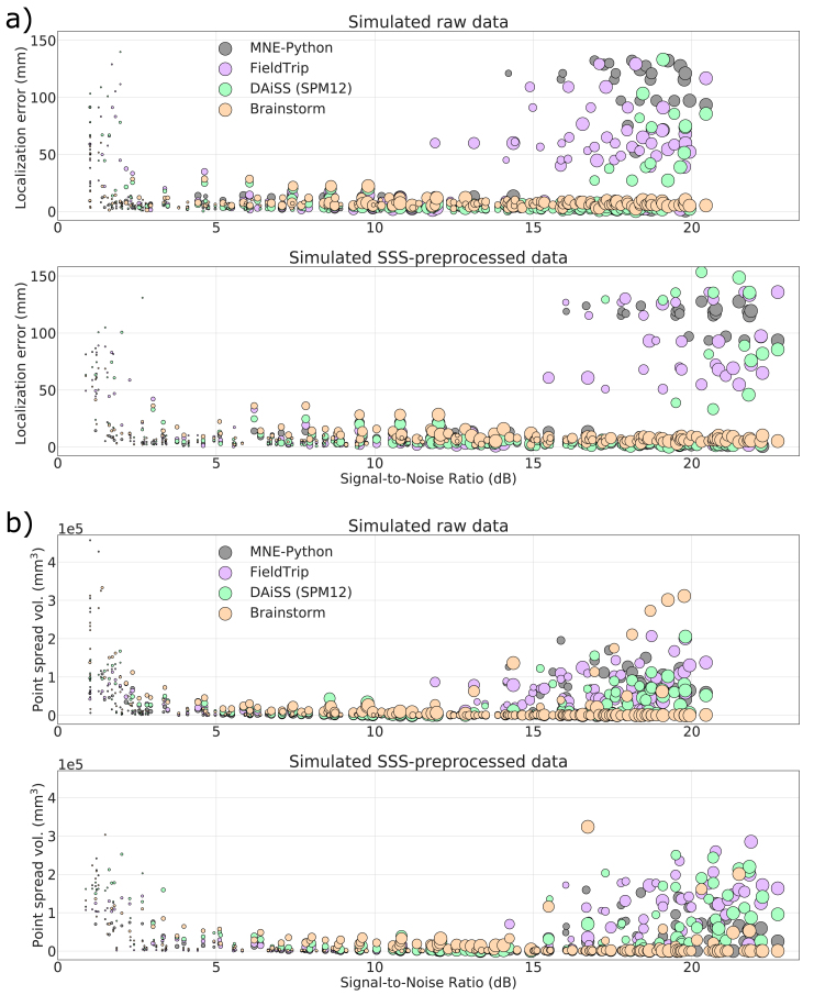
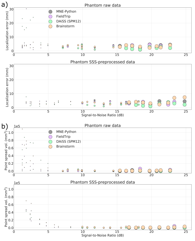
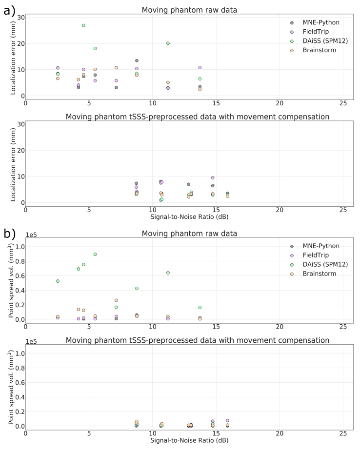
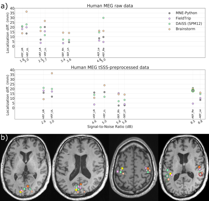
Beamformers are applied for estimating spatiotemporal characteristics of neuronal sources underlying measured MEG/EEG signals. Several MEG analysis toolboxes include an implementation of a linearly constrained minimum-variance (LCMV) beamformer. However, differences in implementations and in their results complicate the selection and application of beamformers and may hinder their wider adoption in research and clinical use. Additionally, combinations of different MEG sensor types (such as magnetometers and planar gradiometers) and application of preprocessing methods for interference suppression, such as signal space separation (SSS), can affect the results in different ways for different implementations. So far, a systematic evaluation of the different implementations has not been performed. Here, we compared the localization performance of the LCMV beamformer pipelines in four widely used open-source toolboxes (MNE-Python, FieldTrip, DAiSS (SPM12), and Brainstorm) using datasets both with and without SSS interference suppression. We analyzed MEG data that were i) simulated, ii) recorded from a static and moving phantom, and iii) recorded from a healthy volunteer receiving auditory, visual, and somatosensory stimulation. We also investigated the effects of SSS and the combination of the magnetometer and gradiometer signals. We quantified how localization error and point-spread volume vary with the signal-to-noise ratio (SNR) in all four toolboxes. When applied carefully to MEG data with a typical SNR (3-15 dB), all four toolboxes localized the sources reliably; however, they differed in their sensitivity to preprocessing parameters. As expected, localizations were highly unreliable at very low SNR, but we found high localization error also at very high SNRs for the first three toolboxes while Brainstorm showed greater robustness but with lower spatial resolution. We also found that the SNR improvement offered by SSS led to more accurate localization.
Keywords: Beamformers; EEG; LCMV; MEG; Open-source analysis toolboxes; Source modeling.
Copyright © 2020 The Authors. Published by Elsevier Inc. All rights reserved.
Figures
References
-
- Bagic A.I., Knowlton R.C., Rose D.F., Ebersole J.S., ACMEGS Clinical Practice Guideline (CPG) Committee American clinical magnetoencephalography society clinical practice guideline 1: recording and analysis of spontaneous cerebral activity. J. Clin. Neurophysiol. 2011;28(4):348–354. doi: 10.1097/WNP.0b013e3182272fed. - DOI - PubMed
-
- Burgess R.C., Funke M.E., Bowyer S.M., Lewine J.D., Kirsch H.E., Bagić A.I., ACMEGS Clinical Practice Guideline (CPG) Committee American Clinical Magnetoencephalography Society Clinical Practice Guideline 2: presurgical functional brain mapping using magnetic evoked fields. J. Clin. Neurophysiol. 2011;28(4):355–361. doi: 10.1097/WNP.0b013e3182272ffe. - DOI - PMC - PubMed
Publication types
MeSH terms
LinkOut - more resources
Full Text Sources
Research Materials
Miscellaneous
