Review
. 2020 Nov:79:100861.
doi: 10.1016/j.preteyeres.2020.100861. Epub 2020 Apr 9.

Clinical spectrum, genetic complexity and therapeutic approaches for retinal disease caused by ABCA4 mutations

Affiliations
Review

Clinical spectrum, genetic complexity and therapeutic approaches for retinal disease caused by ABCA4 mutations

Frans P M Cremers et al. Prog Retin Eye Res. 2020 Nov.

Abstract

The ABCA4 protein (then called a "rim protein") was first identified in 1978 in the rims and incisures of rod photoreceptors. The corresponding gene, ABCA4, was cloned in 1997, and variants were identified as the cause of autosomal recessive Stargardt disease (STGD1). Over the next two decades, variation in ABCA4 has been attributed to phenotypes other than the classically defined STGD1 or fundus flavimaculatus, ranging from early onset and fast progressing cone-rod dystrophy and retinitis pigmentosa-like phenotypes to very late onset cases of mostly mild disease sometimes resembling, and confused with, age-related macular degeneration. Similarly, analysis of the ABCA4 locus uncovered a trove of genetic information, including >1200 disease-causing mutations of varying severity, and of all types - missense, nonsense, small deletions/insertions, and splicing affecting variants, of which many are located deep-intronic. Altogether, this has greatly expanded our understanding of complexity not only of the diseases caused by ABCA4 mutations, but of all Mendelian diseases in general. This review provides an in depth assessment of the cumulative knowledge of ABCA4-associated retinopathy - clinical manifestations, genetic complexity, pathophysiology as well as current and proposed therapeutic approaches.

Keywords: ABCA4-associated retinopathy; Allelic heterogeneity; Autofluorescence; Hypomorphic variant; Penetrance; Phenocopies; Pseudoexon; Splice defects; Stargardt disease; Structural variant; Therapy.


Conflict of interest statement

Declaration of competing interest

The authors declare that they have no known competing financial interests or personal relationships that could have appeared to influence the work reported in this paper.

Figures

Fig. 1.
Fig. 1.
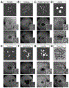
Stages of atrophy progression in ABCA4-associated retinopathy. Fundus photographs with corresponding short wavelength autofluorescence (SW-AF) images and foveal spectral domain-optical coherence tomography (SD-OCT) scans depicting the progressive stages of macular atrophy in ABCA4-associated retinopathy. (A) Early lesions exhibit a mottled appearance on funduscopy and (B) diffusely decreased autofluorescence on SW-AF imaging. (C) An apparent loss of the photoreceptor-attributable ellipsoid zone (EZ) band and appearance of hyper-reflective debris can be observed by SD-OCT within the lesion at this stage. (D) Lesions in the chorioretinal atrophy stage exhibit the canonical beaten-bronze appearance, are well-delineated and enable visibility of underlying choroidal vessels. (E) This stage is also uniquely characterized by a homogeneous and complete loss of autofluorescence; (F) A marked thinning of the retinal pigment epithelium (RPE) layer resulting in an increased transmission of the SD-OCT signal (F, inset) is typically present at this stage. (G, H) Continued progression of atrophy extends across the macula and posteriorly, sequentially involving the choriocapillaris, Sattler and Haller layers of the choroid (I, inset). (J, K) The end-stage of widespread degeneration results in a complete loss of outer retinal and choroidal layers (L) resulting in a visibility of the underlying sclera. The discernible edge of the atrophic lesion and its corresponding position on SD-OCT are denoted by yellow arrows (For interpretation of the references to color in this figure legend, the reader is referred to the Web version of this article).
Fig. 2.
Fig. 2.
Morphological spectrum of fundus flecks in ABCA4-associated retinopathy. Lipofuscin-laden flecks deposited across the fundus exhibit a yellow appearance on fundoscopy and an intense autofluorescent signal on short wavelength autofluorescence (SW-AF) imaging. The collective spatio-temporal pattern of flecks and their individual morphology (A–G) vary across disease stage and genotypic trajectories. Areas of resorbing flecks become hypoautofluorescent and coalescence into a heterogeneous pattern across the posterior pole.
Fig. 3.
Fig. 3.
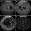
Disease-sparing of the peripapillary region in ABCA4-associated retinopathy. Sparing of the peripapillary region around the optic nerve (magenta arrowheads) from disease changes is a characteristic feature of ABCA4-associated retinopathy and becomes apparent as flecks extend centripetally across the posterior pole of the retina (A, B). Resistance of this region persists into the late atrophic stages and gradually becomes affected (C). The presence of residual circumpapillary tissue may be discernible despite the widespread atrophy (D).
Fig. 4.
Fig. 4.
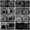
Autofluorescence subtypes of the bull’s eye maculopathy (BEM) stage of ABCA4-associated retinopathy. Confined BEM lesions are generally the earliest manifestation of macular affection in ABCA4-associated retinopathy and are highly associated with the c.5882G>A, p.(Gly1961Glu) mutation. (A–C) Small, focal lesions are typically associated with a loss of the ellipsoid zone (EZ) band and subsequent cavitation of this space in the fovea (“optical gap”). (D–F) Uniformly round BEM lesions exhibit continuous autofluorescence borders and punctate debris within the atrophy region. (G–I) Elliptical BEM lesions also exhibit smooth, continuous autofluorescent borders; however, the region inside the lesion contains less debris and are marked by a central patch of autofluorescence (“bull’s eye”) indicating prior sparing of the fovea. Much less common are centrally mottled BEM lesions (J–L) which are distinct in that they lack a hyperautofluorescent perimeter and are almost exclusive to adolescent patients.
Fig. 5.
Fig. 5.
Common ABCA4-associated retinopathy phenocopying genes and masquerading phenotypes. (A) A 35-year-old woman harboring the c.638G>C, p.(Cys213Ser) variant in PRPH2 with autofluorescent flecks across the posterior pole and peripapillary sparing phenocopying a 29-year-old ABCA4-associated retinopathy patient harboring with the C.302 + 1G> A, p.(?) variant of ABCA4. (B) A 62-year-old woman harboring a canonical splice site variant, c.582-1G>A, p.(?), in PRPH2 with a confluent distribution of autofluorescent flecks across the posterior pole and “peninsular” sparing of the fovea phenocopying a 41-year-old woman harboring the c.4457C>T, p.(Pro1486Leu) and c.4793C>A, p.(Ala1486Asp) variants of ABCA4. (C) A 60-year-old man with pattern dystrophy harboring the c.584G>A, p.(Arg195Gln) missense variant in PRPH2 with foveal sparing phenocopying a 40-year-old ABCA4 disease patient harboring the hypomorphic variant, c.5603A>T, p.(Asn1868Leu), and c.4670A>G, p.(Tyr1557Cys) variants of ABCA4. (D) A 54-year-old man with a large, circular lesion of chorioretinal atrophy and autofluorescent flecks harboring the c.571G>T, p.(Glu191*) nonsense variant in PRPH2 phenocopying a 44-year-old ABCA4 disease patient harboring the hypomorphic variant, c.5603A>T, p.(Asn1868Ile), and c.4670A>G, p.(Tyr1557Cys) variants of ABCA4. (E) A 37-year-old man with maternally inherited diabetes and deafness (MIDD) with granular autofluorescent fleck-like depositions and “bridged” sparing of the fovea phenocopying a 42-year-old ABCA4-associated retinopathy patient harboring a missense, c.2971G>C, p.(Gly991Arg), and a deep-intronic, C.570 + 1798A>G, p.(Phe191Leufs*6), variant in ABCA4. (F) A 51-year-old woman with an elliptical BEM lesion caused by the c.449C>G, p.(Ser150*) variant in CRX phenocopying a 17-year-old boy with ABCA4 disease harboring a mild missense variant, c.3113C>T, p.(Ala1038Val), and a known exon-skipping intronic variant, c.5461–10T>C, p.[Thr1821Aspfs*6,Thr1821Valfs*13] (Sangermano et al., 2016) variant in ABCA4. (G) A 57-year-old man with a uniform BEM lesion caused by the c.3423G>T, p.(Trp1141Cys) missense variant in RPGR phenocopying a 15-year-old boy with ABCA4-associated retinopathy harboring the c.5882G>A, p.(Gly1961Glu) and c.45G>A, p.(Trp15*) variant of ABCA4. (H) A 5-year-old girl with RDHl2-associated Leber congenital amaurosis (LCA) and peripapillary sparing homozygous for the missense variant, c.698T> A, p.(Val233Asp), phenocopying a 60-year-old man with end-stage ABCA4-associated retinopathy harboring the c.4139C>T, p.(Pro1380Leu) and c.4601del p.(Leu1534Trpfs*1) variants in ABCA4.
Fig. 6.
Fig. 6.
Distribution of different types of ABCA4-alleles. Unique (A) and all (B) ABCA4 variants or alleles based on data collected by Cornelis et al. (2017), supplemented with deep-intronic variant and structural variant data published since then (listed in Tables 3 and 4). The contribution of each type of variant or allele is represented. Protein truncating variants comprise nonsense, frameshift and canonical splice site variants. The complex alleles represented in these pie-charts only consist of combinations of missense variants, the most frequent of which were c.[1622T>C;3113C>T] and c.[4469G>A;5603A>T]. They do not include the complex alleles that contain noncanonical splice site variants, deep-intronic variants or protein truncating variants, when present in cis with other variants. If these had been included, ~10% of the alleles would consist of complex alleles. Most of the structural variants, deep-intronic variants and noncanonical splice site variants also result in protein truncation.
Fig. 7.
Fig. 7.
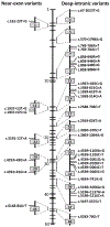
Location of deep-intronic variants in ABCA4. Left part shows variants located near exons that result in exon skipping or exon elongation. Right part shows deep-intronic variants that invariably result in the generation of pseudoexons (see Table 4).
Fig. 8.
Fig. 8.
Genotype-phenotype correlations for ABCA4-associated retinopathy. Summary of disease trajectories associated variants and genotypes of ABCA4. Overall disease severity is defined according to the spatial extent of the disease: Macular stage, disease changes are confined to the central macula; Extramacular stage, disease changes extend beyond the vascular arcades and regions nasal to the optic disc; Transitional stage, disease changes become confluent across the posterior pole initiating peripheral involvement and outer retinal atrophy; Advanced stage, multiple lesions occur and coalesce across the posterior pole. Disease trajectories are defined by the average age at which patients progress through each severity milestone. Three allele-specific trajectories are represented including patients with hypomorphic alleles, c.4253+ 43G> A, p.[ =,Ile1377Hisfs*3] and c.5603A>T, p.(Asn1868Ile) which occur only in trans with a deleterious allele, homozygous and compound heterozygous c.5882G> A, p.(Gly1961Glu) alleles, two deleterious alleles and Other two alleles which consist of all other combinations of ABCA4 alleles. The length of color-coded arrows represents the beginning and duration of defined disease severity stage. Representative autofluorescence images of patients within each trajectory group are arranged according to the age of the depicted phenotype along the time line (For interpretation of the references to color in this figure legend, the reader is referred to the Web version of this article).
Fig. 9.
Fig. 9.
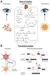
Therapeutic interventions for ABCA4-associated retinopathy. (A)Overview of all therapeutic strategies currently in clinical trials; left panel: cell replacement therapy: cells, either in a stem cell state or pre-differentiated ex vivo towards a retinal fate are directly injected into the retina; middle panel: structure formulas of compounds currently in clinical trials; right panel: gene augmentation therapy, in which wild-type ABCA4 cDNA is packaged into a lentiviral vector which is injected into the retina of subjects with ABCA4-associated retinopathy. (B) Examples of therapeutic strategies currently in preclinical development. Left panel: dual AAV-based gene augmentation, in which ABCA4 cDNA is split into two halves that are aimed to recombine once inside the target cell; right panel: AON-based modulation of pre-mRNA splicing to correct aberrant splicing processes. The figure was created with the aid of BioRender and Illustrator software. Image of the eye was adapted from pixabay.com.

References

    1. Acland GM, Aguirre GD, Ray J, Zhang Q, Aleman TS, Cideciyan AV, Pearce-Kelling SE, Anand V, Zeng Y, Maguire AM, Jacobson SG, Hauswirth WW, Bennett J, 2001. Gene therapy restores vision in a canine model of childhood blindness. Nat. Genet 28, 92–95. - PubMed
    1. Acton JH, Greenstein VC, 2013. Fundus-driven perimetry (microperimetry) compared to conventional static automated perimetry: similarities, differences, and clinical applications. Can. J. Ophthalmol 48, 358–363. - PMC - PubMed
    1. Agbaga MP, Brush RS, Mandal MN, Henry K, Elliott MH, Anderson RE, 2008. Role of Stargardt-3 macular dystrophy protein (ELOVL4) in the biosynthesis of very long chain fatty acids. Proc. Natl. Acad. Sci. U. S. A 105, 12843–12848. - PMC - PubMed
    1. Aguirre-Lamban J, Gonzalez-Aguilera JJ, Riveiro-Alvarez R, Cantalapiedra D, Avila-Fernandez A, Villaverde-Montero C, Corton M, Bianco-Kelly F, Garcia-Sandoval B, Ayuso C, 2011. Further associations between mutations and polymorphisms in the ABCA4 gene: clinical implication of allelic variants and their role as protector/risk factors. Invest. Ophthalmol. Vis. Sci 52, 6206–6212. - PubMed
    1. Ahn J, Molday RS, 2000. Purification and characterization of ABCR from bovine rod outer segments. Methods Enzymol. 315, 864–879. - PubMed

Publication types