. 2020 Nov;47(12):2765-2775.
doi: 10.1007/s00259-020-04784-0. Epub 2020 Apr 11.

Development and characterization of CD54-targeted immunoPET imaging in solid tumors

Affiliations

Development and characterization of CD54-targeted immunoPET imaging in solid tumors

Weijun Wei et al. Eur J Nucl Med Mol Imaging. 2020 Nov.

Abstract

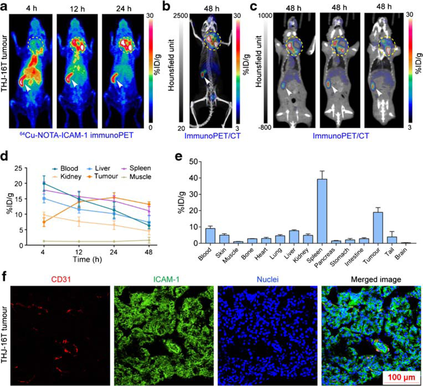
Purpose: Intercellular adhesion molecule-1 (ICAM-1, CD54) is an emerging therapeutic target for a variety of solid tumors including melanoma and anaplastic thyroid cancer (ATC). This study aims to develop an ICAM-1-targeted immuno-positron emission tomography (immunoPET) imaging strategy and assess its diagnostic value in melanoma and ATC models.

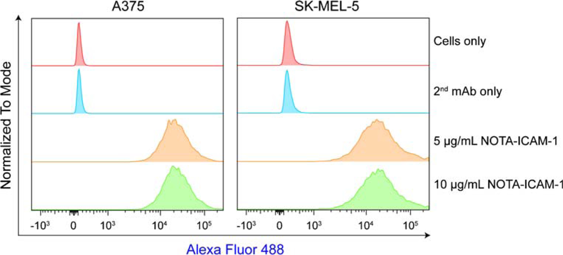
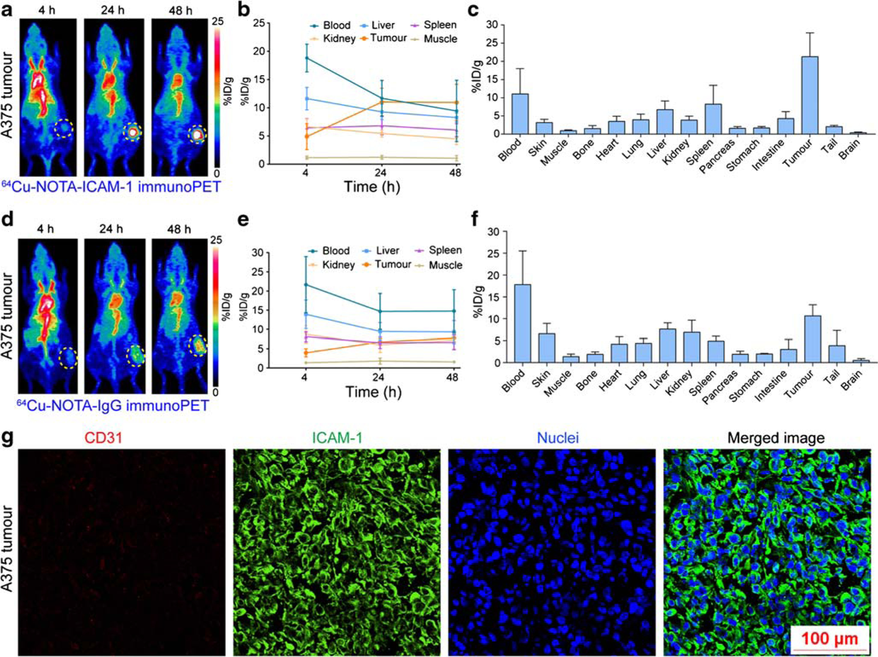
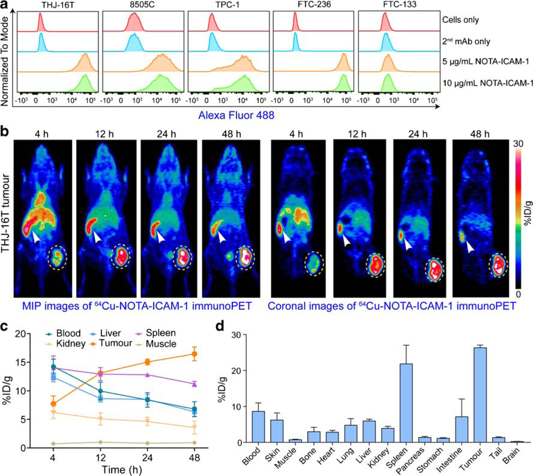
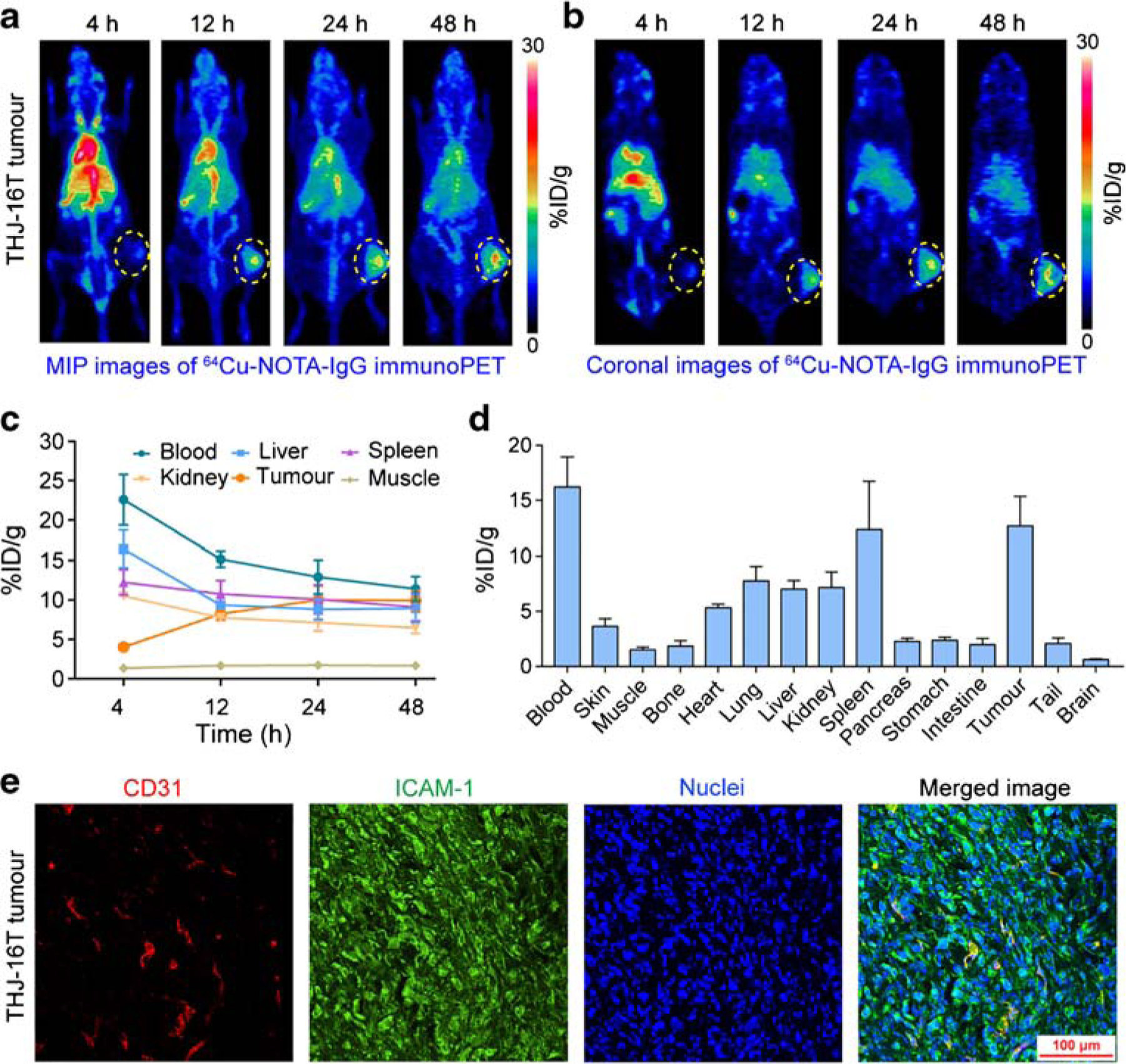
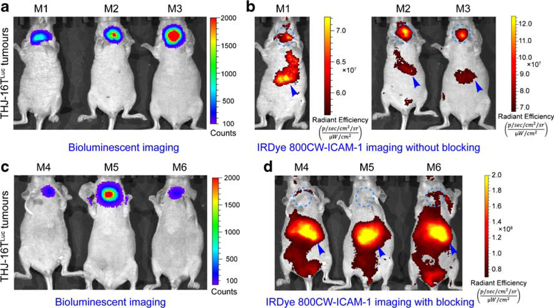
Methods: Flow cytometry was used to screen ICAM-1-positive melanoma and ATC cell lines. Melanoma and ATC models were established using A375 cell line and THJ-16T cell line, respectively. An ICAM-1-specific monoclonal antibody (R6-5-D6) and a nonspecific human IgG were radiolabeled with 64Cu and the diagnostic efficacies were interrogated in tumor-bearing mouse models. Biodistribution and fluorescent imaging studies were performed to confirm the specificity of the ICAM-1-targeted imaging probes.

Results: ICAM-1 was strongly expressed on melanoma and advanced thyroid cancer cell lines. 64Cu-NOTA-ICAM-1 immunoPET imaging efficiently delineated A375 melanomas with a peak tumor uptake of 21.28 ± 6.56 %ID/g (n = 5), significantly higher than that of 64Cu-NOTA-IgG (10.63 ± 2.58 %ID/g, n = 3). Moreover, immunoPET imaging with 64Cu-NOTA-ICAM-1 efficiently visualized subcutaneous and orthotopic ATCs with high clarity and contrast. Fluorescent imaging with IRDye 800CW-ICAM-1 also visualized orthotopic ATCs and the tumor uptake could be blocked by the ICAM-1 parental antibody R6-5-D6, indicating the high specificity of the developed probe. Finally, blocking with the human IgG prolonged the circulation of the 64Cu-NOTA-ICAM-1 in R2G2 mice without compromising the tumor uptake.

Conclusion: ICAM-1-targeted immunoPET imaging could characterize ICAM-1 expression in melanoma and ATC, which holds promise for optimizing ICAM-1-targeted therapies in the future.

Keywords: Companion diagnostics; ICAM-1; ImmunoPET; Melanoma; Thyroid cancer.


Conflict of interest statement

Conflict of interest The authors declare that they have no conflict of interest.

Figures

Fig. 1
Fig. 1
Flow cytometry assessment of ICAM-1 expression on melanoma cell lines
Fig. 2
Fig. 2
ImmunoPET imaging of ICAM-1-positive melanomas. a 64Cu-NOTA-ICAM-1 immunoPET imaging of A375-bearing nude mice. Serial maximum intensity projection images of a representative mouse at different imaging time-points were given. b Region of interest analysis of 64Cu-NOTA-ICAM-1 immunoPET imaging data. c Biodistribution study at 48 h post-injection of 64Cu-NOTA-ICAM-1. d 64Cu-NOTA-IgG immunoPET imaging of A375-bearing nude mice. Serial maximum intensity projection images of a representative mouse at different imaging time-points were given. e Region of interest analysis of 64Cu-NOTA-IgG immunoPET imaging data. f Biodistribution study at 48 h post-injection of 64Cu-NOTA-IGg. g Immunofluorescent imaging of a representative A375 tumor. The tumor section was stained for CD31 (red), ICAM-1 (green), and nuclei (blue). The tumors were indicated by yellow dotted circles
Fig. 3
Fig. 3
ImmunoPET imaging of ICAM-1 in subcutaneous anaplastic thyroid cancers. a Flow cytometry assessment of ICAM-1 expression on five thyroid cancer cell lines. b 64Cu-NOTA-ICAM-1 immunoPET imaging of subcutaneous THJ-16T-bearing R2G2 mice. Maximum intensity projection (MIP) and coronal images of a representative mouse at different imaging time-points were given. c Region of interest analysis of 64Cu-NOTA-ICAM-1 immunoPET imaging data. d Biodistribution study at 48 h post-injection of 64Cu-NOTA-ICAM-1. The tumors were indicated by yellow dotted circles and spleens were indicated by white arrowheads
Fig. 4
Fig. 4
64Cu-NOTA-IgG immunoPET imaging of ICAM-1-positive subcutaneous anaplastic thyroid cancers. a, b 64Cu-NOTA-IgG immunoPET imaging of subcutaneous THJ-16T-bearing R2G2 mice. Maximum intensity projection (MIP) and coronal images of a representative mouse at different imaging time-points were given. c Region of interest analysis of 64Cu-NOTA-IgG immunoPET imaging data. d Biodistribution study at 48 h post-injection of 64Cu-NOTA-IgG. e Immunofluorescent imaging of a subcutaneous THJ-16T tumor. The tumor section was stained for CD31 (red), ICAM-1 (green), and nuclei (blue). The tumors were indicated by yellow dotted circles
Fig. 5
Fig. 5
ImmunoPET and immunoPET/CT imaging of orthotopic anaplastic thyroid cancers. a, b 64Cu-NOTA-ICAM-1 immunoPET and immunoPET/CT imaging of a representative orthotopic THJ-16T-bearing R2G2 mouse. c ImmunoPET/CT images at different slices of the same mouse showed expansion of the tumor into the surrounding tissues and necrosis inside the tumor. d Region of interest analysis of 64Cu-NOTA-ICAM-1 immunoPET imaging data. e Biodistribution study at 48 h post-injection of 64Cu-NOTA-ICAM-1. f Immunofluorescent imaging of a representative orthotopic THJ-16T tumor. The tumor section was stained for CD31 (red), ICAM-1 (green), and nuclei (blue). The tumors were indicated by yellow dotted circles and spleens were indicated by white arrowheads
Fig. 6
Fig. 6
Bioluminescent imaging (BLI) and fluorescent imaging of orthotopic ATC models without or with R6–5-D6 blocking. a BLI of mice in the ICAM-1-targeted imaging group without R6–5-D6 blocking. The given image showed the growth of THJ-16TLuc tumors. b Fluorescent imaging of the same mice 48 h after injection of IRDye 800CW-ICAM-1. The fluorescent tracer was eliminated from the hepatobiliary system with a proportion deposited in the orthotopic THJ-16TLuc tumors. c BLI of mice in the blocking group showed comparable tumor burden. d Fluorescent imaging of the mice in the blocking group, which were injected first with a blocking dose of R6–5-D6 and then with the IRDye 800CW-ICAM-1. Fluorescent imaging acquired 48 h after injection of the tracer showed negligible signal in the thyroid areas, indicating saturation of the target by the blocking dose of R6–5-D6. The tumor areas were indicated by blue dotted circles and livers were indicated by blue arrowheads

References

    1. Reina M, Espel E. Role of LFA-1 and ICAM-1 in cancer. Cancers (Basel). 2017;9 10.3390/cancers9110153. - DOI - PMC - PubMed
    1. Yang L, Froio RM, Sciuto TE, Dvorak AM, Alon R, Luscinskas FW. ICAM-1 regulates neutrophil adhesion and transcellular migration of TNF-alpha-activated vascular endothelium under flow. Blood. 2005;106:584–92. 10.1182/blood-2004-12-4942. - DOI - PMC - PubMed
    1. Hayes SH, Seigel GM. Immunoreactivity of ICAM-1 in human tumors, metastases and normal tissues. Int J Clin Exp Pathol. 2009;2:553–60. - PMC - PubMed
    1. Hamai A, Meslin F, Benlalam H, Jalil A, Mehrpour M, Faure F, et al. ICAM-1 has a critical role in the regulation of metastatic melanoma tumor susceptibility to CTL lysis by interfering with PI3K/AKT pathway. Cancer Res. 2008;68:9854–64. 10.1158/0008-5472.CAN-08-0719. - DOI - PubMed
    1. Zhang P, Goodrich C, Fu C, Dong C. Melanoma upregulates ICAM-1 expression on endothelial cells through engagement of tumor CD44 with endothelial E-selectin and activation of a PKCalpha-p38-SP-1 pathway. FASEB J. 2014;28:4591–609. 10.1096/fj.11-202747. - DOI - PMC - PubMed

Publication types

Substances