. 2020 Apr 12;147(7):dev183954.
doi: 10.1242/dev.183954.

MMP14 is required for delamination of chick neural crest cells independently of its catalytic activity

Affiliations

MMP14 is required for delamination of chick neural crest cells independently of its catalytic activity

Cyril Andrieu et al. Development. .

Abstract

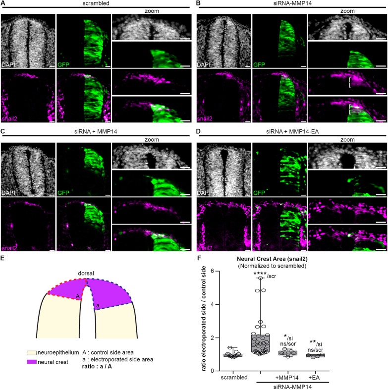
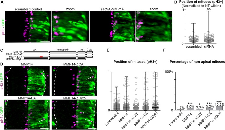
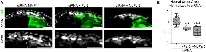
Matrix metalloproteinases have a broad spectrum of substrates ranging from extracellular matrix components and adhesion molecules to chemokines and growth factors. Despite being mostly secreted, MMPs have been detected in the cytosol, the mitochondria or the nucleus. Although most of the attention is focused on their role in matrix remodeling, the diversity of their substrates and their complex trafficking open the possibility for non-canonical functions. Yet in vivo examples and experimental demonstration of the physiological relevance of such activities are rare. Here, we have used chick neural crest (NC) cells, a highly migratory stem cell population likened to invasive cancer cells, as a model for physiological epithelial-mesenchymal transition (EMT). We demonstrate that MMP14 is required for NC delamination. Interestingly, this role is independent of its cytoplasmic tail and of its catalytic activity. Our in vivo data indicate that, in addition to being a late pro-invasive factor, MMP14 is also likely to be an early player, owing to its role in EMT.

Keywords: Delamination; EMT; MMP14; Mitosis; Neural crest; Polarity.


Conflict of interest statement

Competing interestsThe authors declare no competing or financial interests.

Figures

Fig. 1.
Fig. 1.
Expression of MMP14 during EMT of chick NC cells. (A) In situ hybridization against chick MMP14. Dorsal view of the caudal part of a stage HH13 chick embryo. Scale bar: 100 µm. (B-D) In situ hybridization for chick MMP14. Transverse sections. MMP14 expression is increased in the neural crest during EMT. Scale bars: 50 µm. s, somite; L, lateral mesoderm; np, neural plate; nt, neural tube; n, notochord. Asterisks indicate the pre-migratory NC cells.
Fig. 2.
Fig. 2.
MMP14 is required for NC delamination independently of its catalytic activity. (A-D) Immunostaining for snail 2 (magenta) after electroporation of scrambled control (A), siRNA-MMP14 (B) and co-electroporation of siRNA-MMP14 with wild-type MMP14 (C) or the non-catalytic point mutant MMP14-EA (D). Nuclei are counterstained with DAPI (grey); the electroporated side is visualized using GFP (green). Brackets indicate neural crest accumulation (B). Scale bars: 20 µm. (E) The method for measuring the area of the neural crest domain. The neural crest area on the electroporated side was divided by the neural crest area in the control side. (F) Plot of neural crest area (snail 2) with scrambled (nembryos=11, nsections=132 from three independent experiments), siRNA-MMP14 alone (nembryos=31, nsections=445 from eight independent experiments), siRNA-MMP14 with wild-type MMP14 (nembryos=7, nsections=134 from two independent experiments) and siRNA-MMP14 with MMP14-EA (nembryos=4, nsections=90 from one experiment). ANOVA with a Kruskal–Wallis Test and Dunn's multiple comparisons: ****P<0.0001 (scrambled versus siRNA), *P=0.0441 (siRNA versus siRNA+MMP14), **P=0.0030 (siRNA versus siRNA+EA) and P>0.9999 (not significant; scrambled versus siRNA+MMP14 or siRNA+EA).
Fig. 3.
Fig. 3.
MMP14 inhibition prevents the apicobasal redistribution of PCM1 during NC EMT. (A,B) immunostaining against aPKC (magenta) and snail 2 (gray) in embryos electroporated with scrambled control (A, nembryos=3) and with siRNA-MMP14 (B, nembryos=5) from one experiment. (C-D′) Immunostaining against PCM1 (magenta) and AP2 (gray) in embryos electroporated with scrambled control (C, nembryos=6) and with siRNA-MMP14 (D,D′, nembryos=5) from two independent experiments. (D′) More detail of the neural crest domain. Electroporated side is visualized using GFP (green). Bracket in D indicates the accumulation of neural crest cells. Arrowheads in C indicate the normal basolateral distribution of PCM1 on both non-electroporated and electroporated sides of embryos electroporated with the scrambled control. Arrowheads in D,D′ indicate the normal basolateral distribution of PCM1 on the control side of siRNA-MMP14 embryos. Arrows in D,D′ indicate the apical position of PCM1 on the electroporated side of siRNA-MMP14 embryos. Scale bars: 20 µm.
Fig. 4.
Fig. 4.
MMP14 is required for the occurrence of basal mitoses during NC delamination. (A,B) Immunostaining for phospho-histone 3 (pH3) (yellow) and snail 2 (magenta) in electroporated embryos with scrambled control (A) and siRNA-MMP14 (B). GFP is shown in green, nuclei are counterstained with DAPI (gray). Arrowhead in A indicates an example of basal mitosis in the electroporated side. Arrow in B indicates an example of apical mitosis in the electroporated side. Scale bars: 20 µm. (C) Diagram depicting the method of measuring the positions of mitoses along the apical-basal axis. The distance between the mitoses and the apical part of neural tube (d) was divided by the neural tube width (D). A ratio of 0 means that the mitoses were close to the apical region. A ratio of 1 means that the mitoses were close to the basal region. (D) Plot of the position of the mitoses along the apico-basal axis in the neural crest in embryos electroporated with the scrambled control (nembryos=3, nmitoses non-elec side=33, nmitoses elec side=36 from one experiment) or siRNA-MMP14 (nembryos=8, nmitoses non-elec side=51, nmitoses elec side=69, from three independent experiments). Dashed line indicates the border between the apical half and basal half. ANOVA followed by Dunn's multiple comparisons **P=0.0014 (electroporated scrambled versus electroporated siRNA), **P=0.0027 (non-electroporated siRNA versus electroporated siRNA). In the scatter dot plot, the lines indicate the median and the error bars show the extent of the whole dataset. (E) Percentage of basal mitoses in neural crest domain. Basal mitoses were defined as mitoses located within the basal half of the neural crest domain (corresponding to values between 0.5 and 1 on the graph in D). Statistics were performed according to Taillard et al. (2008), **T>6.635 (non-electroporated siRNA versus electroporated siRNA) the null hypothesis is rejected with 99% confidence. No-elec, non-electroporated side; elec, electroporated side.
Fig. 5.
Fig. 5.
Homogenous distribution of MMP14 is sufficient to promote basal mitoses independently of its catalytic activity. (A) Immunostaining against phospho-histone H3 (pH3, magenta) after electroporation with the scrambled control or siRNA-MMP14. (B) Positions of mitoses in the intermediate neural tube region with the scrambled control (nembryos=6, nmitoses=339; from two independent experiments) or siRNA-MMP14 (nembryos=11, nmitoses=362; from four independent experiments). Unpaired Mann–Whitney test, P=0.7780. (C) Diagram depicting the different forms of MMP14. (D) Immunostaining against pH3 (magenta) after overexpression of MMP14, MMP14-ΔCAT, MMP14-EA and MMP14-ΔCyto. Arrows indicate non-apical mitoses. (E) Positions of all mitoses for each condition depicted in D. Control side (nembryos=26, nmitoses=1908), MMP14 (nembryos=7, nmitoses=716), MMP14-ΔCAT (nembryos=7, nmitoses=406) and MMP14-EA (nembryos=9, nmitoses=707) from three independent experiments, and MMP14-ΔCyto (nembryos=3, nmitoses=431) from one experiment. (F) Percentages of non-apical mitoses for each condition depicted in D. Non-apical mitoses were determined as mitoses occurring more than two nuclei diameters away from the apical domain. Statistics were performed according to Taillard et al. (2008), Thresholds for T were such that, at *T>3.841, the null hypothesis is rejected with a 95% confidence, at **T>6.635, the null hypothesis is rejected with 99% confidence and at ***T>10.83, the null hypothesis is rejected with 99.9% confidence. In B,E, the lines indicate the median and the error bars show the extent of the whole dataset. Electroporated side is indicated by GFP (A,D, green). Scale bars: 35 µm.
Fig. 6.
Fig. 6.
Destabilizing apicobasal polarity is sufficient to rescue siRNA-MMP14. (A) Immunostaining for snail 2 (gray) after electroporation of siRNA-MMP14 or co-electroporation with siRNA-MMP14 and Par3. Bracket indicates accumulation of neural crest cells. (B) Plot of neural crest area (snail 2) with siRNA-MMP14 (nembryos=14, nsections=361), siRNA-MMP14 with Par3 (nembryos=6, nsections=166) and siRNA-MMP14 with Par3MO (nembryos=7, nsections=38) from two independent experiments. ANOVA and uncorrected Fisher's LSD; ***P=0.0007, ****P<0.0001. Scale bars: 25 µm. Par3MO efficiency/specificity could not be checked (see main text and Materials and Methods for details).

References

    1. Afonso C. and Henrique D. (2006). PAR3 acts as a molecular organizer to define the apical domain of chick neuroepithelial cells. J. Cell Sci. 119, 4293-4304. 10.1242/jcs.03170 - DOI - PubMed
    1. Ahlstrom J. D. and Erickson C. A. (2009). The neural crest epithelial-mesenchymal transition in 4D: a ‘tail’ of multiple non-obligatory cellular mechanisms. Development 136, 1801-1812. 10.1242/dev.034785 - DOI - PMC - PubMed
    1. Barbolina M. V. and Stack M. S. (2008). Membrane type 1-matrix metalloproteinase: substrate diversity in pericellular proteolysis. Semin. Cell Dev. Biol. 19, 24-33. 10.1016/j.semcdb.2007.06.008 - DOI - PMC - PubMed
    1. Barriga E. H., Maxwell P. H., Reyes A. E. and Mayor R. (2013). The hypoxia factor Hif-1alpha controls neural crest chemotaxis and epithelial to mesenchymal transition. J. Cell Biol. 201, 759-776. 10.1083/jcb.201212100 - DOI - PMC - PubMed
    1. Bonnans C., Chou J. and Werb Z. (2014). Remodelling the extracellular matrix in development and disease. Nat. Rev. Mol. Cell Biol. 15, 786-801. 10.1038/nrm3904 - DOI - PMC - PubMed

Publication types