Review
. 2020 Apr 7;6(4):e03719.
doi: 10.1016/j.heliyon.2020.e03719. eCollection 2020 Apr.

Commercial hydrogels for biomedical applications

Affiliations
Review

Commercial hydrogels for biomedical applications

S H Aswathy et al. Heliyon. .

Abstract

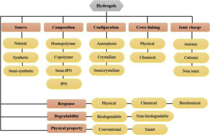
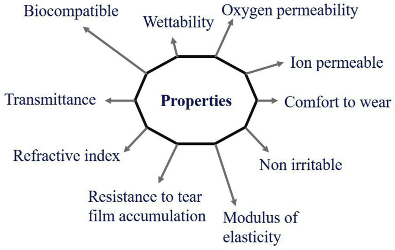
Hydrogels are polymeric networks having the ability to absorb a large volume of water. Flexibility, versatility, stimuli-responsive, soft structure are the advantages of hydrogels. It is classified based on its source, preparation, ionic charge, response, crosslinking and physical properties. Hydrogels are used in various fields like agriculture, food industry, biosensor, biomedical, etc. Even though hydrogels are used in various industries, more researches are going in the field of biomedical applications because of its resembles to living tissue, biocompatibility, and biodegradability. Here, we are mainly focused on the commercially available hydrogels used for biomedical applications like wound dressings, contact lenses, cosmetic applications, tissue engineering, and drug delivery.

Keywords: Biomedical applications; Biomedical engineering; Biotechnology; Commercial products; Hydrogels; Materials science; Nanotechnology.


Figures

Figure 1
Figure 1
Structure of hydrogel at molecular level.
Figure 2
Figure 2
Schematic representation of classification of hydrogels.
Figure 3
Figure 3
Microscopic images of outgrowth of dorsal root ganglia cells on semi synthetic hydrogel of PEG incorporated with diferent proteins. Compared to PEG alone, semi synthetic hydrogels shown better outgrowth of ganglia cells (Adapted from Berkovitch and Seliktar, 2017 with permission).
Figure 4
Figure 4
Semi-interpenetrating network made up of polyacrylamide polymer and silk fibroin chains Incorporation of fibroin improves the elasticity and softness of the hydrogel and helps in sustained release of drug (A) Schematic representation of formation of SF/PAAm semi-IPN network hydrogels (B) Typical appearance of different SF/PAAm semi-IPN hydrogels after removing from moulds (Adapted from Mandal et al., 2009 with permission).
Figure 5
Figure 5
The compression behaviour of cellulose hydrogel (yellow), Cellulose-polyacrylamide IPN hydrogel (red) and Polyacrlamide hydrogel. Only in IPN hydrogels showed good mechanical integrity after deformation (Adapted from Lin et al., 2018 with permission).
Figure 6
Figure 6
Schematic representation of in situ forming click based Hyaluronic acid hydrogel This hydrogel helps in retention of cytomodulin which induces the condrogenic differentiation (Adapted from online open access article Park et al., 2019 with permission).
Figure 7
Figure 7
Improved healing of diabetic skin wound using drug loaded thiolated PEG hydrogel compared to control (Adapted from opne access article Chen et al., 2019 with permission).
Figure 8
Figure 8
Different forms hydrogel wound dressing available in market. (a) Neoheal® hydrogel sheet used for wound dressing, (b) Amorphous gel that can be sued for necrotic wounds and burns, (c) - Hydrogel film and (d) Hydrogel impregnated gauze (Image Courtesy: (a) Kikgel, Poland, (b) Avery Dennison Medical, Longford, Ireland-(Finesse Medical Ltd) (c) Lohmann & Rauscher GmbH & Co, Germany and (d) McKesson Medical Supplies).
Figure 9
Figure 9
Properties or requirements suitable for hydrogel as contact lenses.
Figure 10
Figure 10
Commercially available hydrogel implant for drug delivery (a) Cervidil® that contains 10 mg of dinoprostone in a hydrogel and, (b) SUPPRELIN® LA, a subcutaneous implant (Image courtesy: (a) Ferring Pharmaceuticals Inc. USA, (b) Endo Pharmaceuticals, USA.
Figure 11
Figure 11
Morphological evaluation of GelrinC after implanted in patient. Faster regeneration of cartilage was achieved by using GelrinC implant Series of proton density fat suppressed images in coronal plane show development of cartilage transplant in follow-up examinations after (a) 1 week; (b) 6 months; (c) 12 months; and (d) 24 months. Arrows and circle delineate RT area (Adapted from Trattnig et al., 2015 with permission).

References

    1. Acuvue Website. https://www.acuvue.com/contact-lenses/.
    1. Aderibigbe, Blessing Atim B.B. Alginate in wound dressings. Pharmaceutics. 2018;10(2):42. - PMC - PubMed
    1. Ahmed S., Ikram S. Chitosan based scaffolds and their applications in wound healing. ALSConnect. 2016;10(1):27–37.
    1. Al-sabah A., Burnell S.E.A., Simoes I.N., Jessop Z., Blain E., Whitaker I.S. Structural and mechanical characterization of crosslinked and sterilised nanocellulose-based hydrogels for cartilage tissue engineering. Carbohydr. Polym. 2019;212(February):242–251. - PubMed
    1. Alba-Bueno F., Beltran-Masgoret À., Sanjuan C., Biarnés M., Marín J. Corneal shape changes induced by first and second generation silicone hydrogel contact lenses in daily wear. Contact Lens Anterior Eye. 2009;32(2):88–92. - PubMed

LinkOut - more resources