

Severe vision loss secondary to retinal arteriolar occlusions after multiple intravitreal brolucizumab administrations
- PMID: 32280811
- PMCID: PMC7139151
- DOI: 10.1016/j.ajoc.2020.100687
Severe vision loss secondary to retinal arteriolar occlusions after multiple intravitreal brolucizumab administrations
Abstract
Purpose: To describe a case of unilateral retinal arteriolar occlusion following multiple intravitreal brolucizumab injections for neovascular age-related macular degeneration (nAMD).
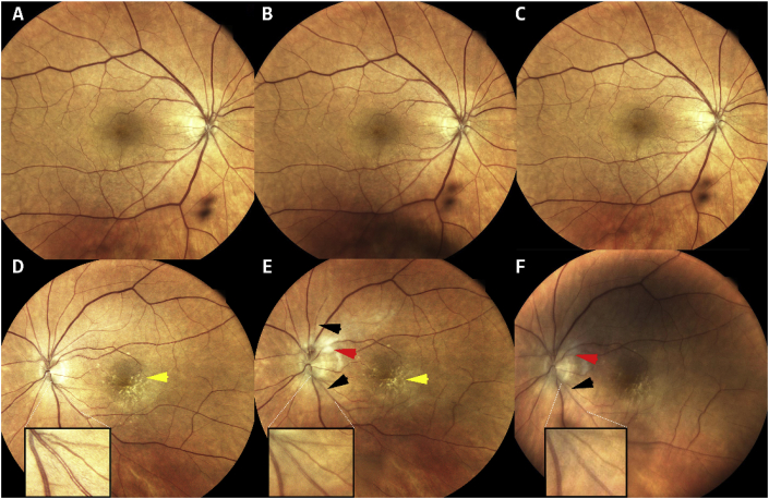
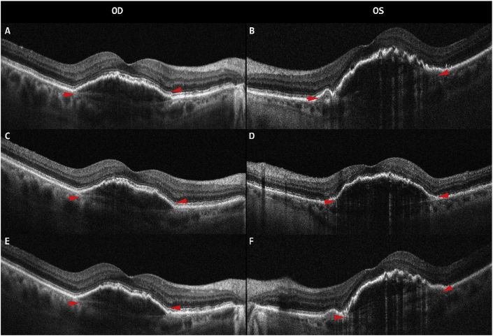
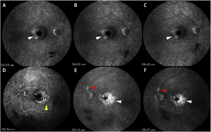
Observations: A 92-year-old Caucasian woman presented with blurry vision in her left eye (OS) after receiving the third dose of intravitreal brolucizumab. At the time of presentation, visual acuity (VA) was 20/40 in her right eye (OD) and had decreased from 20/150 to count finger (CF) at 1-foot OS. On examination, there was no evidence of active inflammation in the anterior chamber OU. Dilated fundus examination showed no vitritis in OD and 1+ vitreous cells OS, flame-shaped hemorrhage at the superior optic disc margin, and retinal whitening surrounding the proximal portion of the supero-temporal branch of the central retinal artery. There were drusen in OS and retinal pigment epithelial (RPE) changes in the maculae of OU. Intra-arteriolar greyish deposits were seen OS. Fluorescein angiography (FA) showed hyper-fluorescence in the maculae corresponding to fibrovascular pigment epithelial detachments (PED) OU. No peri-vascular leakage was noted OU. Delayed filling of multiple arterioles in early and late phases OS was observed on FA. The patient was diagnosed with retinal arteriolar occlusion associated with repeated intravitreal brolucizumab administrations.
Conclusion: Retinal arteriolar occlusion with severe vision loss, possibly secondary to inflammatory responses, can occur after subsequent intravitreal brolucizumab injections, even if no inflammation occurred after initial administrations. Vaso-occlusive disease should be considered as a potential ocular complication, with acute as well as delayed onset, following intravitreal brolucizumab therapy.
Keywords: Age-related macular degeneration; Brolucizumab; Intravitreal; Neovascular; Retinal occlusive vasculitis; Retinal vasculitis; Vaso-occlusion.
© 2020 Published by Elsevier Inc.
Conflict of interest statement
The authors declare that there are no conflicts of interest related to this manuscript.
Figures
References
-
- Hernandez-Pastor L.J., Ortega A., Garcia-Layana A., Giraldez J. Ranibizumab for neovascular age-related macular degeneration. Am J Health Syst Pharm: Offc J Am Soc Health Sys Pharama. 2008;65:1805–1814. - PubMed
-
- Wong W.L., Su X., Li X. Global prevalence of age-related macular degeneration and disease burden projection for 2020 and 2040: a systematic review and meta-analysis. Lancet Glob health. 2014;2:e106–e116. - PubMed
-
- Dugel P.U., Jaffe G.J., Sallstig P. Brolucizumab versus aflibercept in participants with neovascular age-related macular degeneration: a randomized trial. Ophthalmology. 2017;124:1296–1304. - PubMed
-
- Dugel P.U., Koh A., Ogura Y. HAWK and HARRIER: phase 3, multicenter, randomized, double-masked trials of brolucizumab for neovascular age-related macular degeneration. Ophthalmology. 2020;127:72–84. - PubMed
Publication types
LinkOut - more resources
Full Text Sources
Medical
