. 2020 Oct 21;30(11):872-880.
doi: 10.1093/glycob/cwaa033.

The O-GlcNAc modification promotes terminal differentiation of human corneal epithelial cells

Affiliations

The O-GlcNAc modification promotes terminal differentiation of human corneal epithelial cells

Nicole M McColgan et al. Glycobiology. .

Abstract

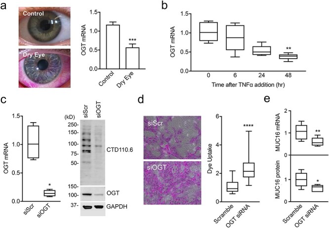
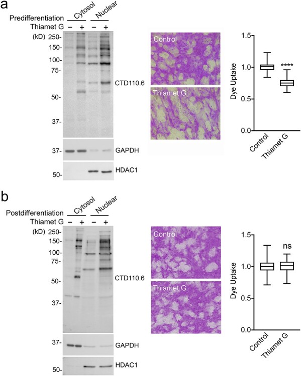
Dynamic modification of nuclear and cytoplasmic proteins with O-linked β-N-acetylglucosamine (O-GlcNAc) plays an important role in orchestrating the transcriptional activity of eukaryotic cells. Here, we report that the O-GlcNAc modification contributes to maintaining ocular surface epithelial homeostasis by promoting mucin biosynthesis and barrier function. We found that induction of human corneal epithelial cell differentiation stimulated the global transfer of O-GlcNAc to both nuclear and cytosolic proteins. Inflammatory conditions, on the other hand, were associated with a reduction in the expression of O-GlcNAc transferase at the ocular surface epithelia. Loss- and gain-of-function studies using small interfering RNA targeting O-GlcNAc transferase, or Thiamet G, a selective inhibitor of O-GlcNAc hydrolase, respectively, revealed that the presence of O-GlcNAc was necessary to promote glycocalyx barrier function. Moreover, we found that Thiamet G triggered a correlative increase in both surface expression of MUC16 and apical epithelial cell area while reducing paracellular permeability. Collectively, these results identify intracellular protein O-glycosylation as a novel pathway responsible for promoting the terminal differentiation of human corneal epithelial cells.

Keywords: O-GlcNAc modification; cell differentiation; cornea; epithelium; transmembrane mucin.


Figures

Fig. 1
Fig. 1
Induction of corneal epithelial differentiation promotes O-GlcNAcylation. (A) Phase contrast and immunofluorescence micrographs showing MUC16 distribution (green) in human corneal epithelial cells before and after induction of differentiation for 7 days. Nuclei were stained by 4′,6-diamidino-2-phenylindole (DAPI) (blue). Representative images are shown (bars, 100 μm). (B) Cell extracts were subjected to SDS-PAGE and immunoblot analysis using antibodies to MUC16 and GAPDH. (C) Protein isolated from cytoplasmic and nuclear fractions before and after induction of differentiation was subjected to immunoblot analysis using CTD110.6, GAPDH (cytoplasmic control) and HDAC1 (nuclear control) antibodies.
Fig. 2
Fig. 2
Inflammatory stress reduces OGT expression at the ocular surface. (A) Rose bengal staining of epithelial cells is observed at the ocular surface of a patient with dry eye. OGT expression in human conjunctival epithelium as determined using data from a public microarray depository. (B) RNA was isolated from differentiated cultures of human corneal epithelial cells treated with TNFα, and the OGT mRNA abundance was determined by quantitative polymerase chain reaction (qPCR). (C) Cells were transfected before differentiation with OGT-specific siRNA (siOGT) or scramble siRNA (siScr). After the treatments, OGT mRNA abundance was determined by qPCR and aliquots of cell lysates were resolved by SDS-PAGE and analyzed by immunoblot using CTD110.6, OGT and GAPDH antibodies. (D) Glycocalyx barrier function after transfection with siOGT or siScr was determined by the rose bengal penetration assay. (E) Expression and biosynthesis of MUC16 was determined by qPCR and immunoblot, respectively. All experiments were performed at least in triplicate. The data in (A) represent the mean ± SD. The box and whisker plots in B, C, D and E show the 25th and 75th percentiles (box), the median (horizontal line in box), and the minimum and maximum data values (whiskers). Significance was determined using unpaired t test (A and E), Kruskal–Wallis with Dunn’s post hoc test (B) or Mann Whitney test (c and d). *P < 0.05; **P < 0.01; ***P < 0.001; ****P < 0.0001.
Fig. 3
Fig. 3
The O-GlcNAc modification functions during early differentiation. (A) Thiamet G was administered to human corneal epithelial cells before differentiation for 7 days. Protein isolated from cytoplasmic and nuclear fractions was subjected to immunoblot analysis using CTD110.6, GAPDH and HDAC1 antibodies. Glycocalyx barrier function was determined by the rose bengal penetration assay. (B) Same as (A) but Thiamet G was administered for 48 h after the induction of differentiation. All experiments were performed at least in triplicate. The box and whisker plots show the 25th and 75th percentiles (box), the median (horizontal line in box) and the minimum and maximum data values (whiskers). Significance was determined using unpaired t test. ****P < 0.0001; ns, nonsignificant.
Fig. 4
Fig. 4
Thiamet G stimulates mucin biosynthesis and paracellular barrier function. (A) Thiamet G was administered to human corneal epithelial cells before differentiation for 7 days. MUC16 (green) was localized by immunofluorescence microscopy and the fluorescence intensity per cell quantified using ImageJ. Representative en face images of nonpermeabilized epithelial cells are shown to the right. Nuclei were stained by 4′,6-diamidino-2-phenylindole (DAPI) (blue). Representative images are shown (bars, 200 μm). (B) Morphometric analyses of cell area were performed using ImageJ. A positive linear correlation was observed between MUC16 intensity and cell area following incubation with Thiamet G. (C) Thiamet G was added basolaterally to cells grown in Transwell cell culture inserts. Following a 7-day treatment, TEER values were significantly increased compared to control. Macromolecular permeability was estimated by measuring FD-4 in the basolateral compartment at the end of the experiments. For each condition in (A) and (B), eight images from two independent tissue chamber slides were analyzed. Experiments in (C) were performed at least in triplicate. The box and whisker plots show the 25th and 75th percentiles (box), the median (horizontal line in box) and the minimum and maximum data values (whiskers). Significance was determined using Mann Whitney test (A and B) or unpaired t test (C). **P < 0.01; ***P < 0.001; ****P < 0.0001.

References

    1. Akimoto Y, Kawakami H, Yamamoto K, Munetomo E, Hida T, Hirano H. 2003. Elevated expression of O-GlcNAc-modified proteins and O-GlcNAc transferase in corneas of diabetic Goto-Kakizaki rats. Invest Ophthalmol Vis Sci. 44:3802–3809. - PubMed
    1. Andres LM, Blong IW, Evans AC, Rumachik NG, Yamaguchi T, Pham ND, Thompson P, Kohler JJ, Bertozzi CR. 2017. Chemical modulation of protein O-GlcNAcylation via OGT inhibition promotes human neural cell differentiation. ACS Chem Biol. 12:2030–2039. - PMC - PubMed
    1. Argueso P, Gipson IK. 2012. Assessing mucin expression and function in human ocular surface epithelia in vivo and in vitro. Methods Mol Biol. 842:313–325. - PMC - PubMed
    1. Argueso P, Guzman-Aranguez A, Mantelli F, Cao Z, Ricciuto J, Panjwani N. 2009. Association of cell surface mucins with galectin-3 contributes to the ocular surface epithelial barrier. J Biol Chem. 284:23037–23045. - PMC - PubMed
    1. Argueso P, Spurr-Michaud S, Russo CL, Tisdale A, Gipson IK. 2003. MUC16 mucin is expressed by the human ocular surface epithelia and carries the H185 carbohydrate epitope. Invest Ophthalmol Vis Sci. 44:2487–2495. - PubMed

Publication types

Substances