Review
. 2020 Oct-Dec;26(5-6):438-454.
doi: 10.1177/1073858420912404. Epub 2020 Apr 13.

If Human Brain Organoids Are the Answer to Understanding Dementia, What Are the Questions?

Affiliations
Review

If Human Brain Organoids Are the Answer to Understanding Dementia, What Are the Questions?

Lezanne Ooi et al. Neuroscientist. 2020 Oct-Dec.

Abstract

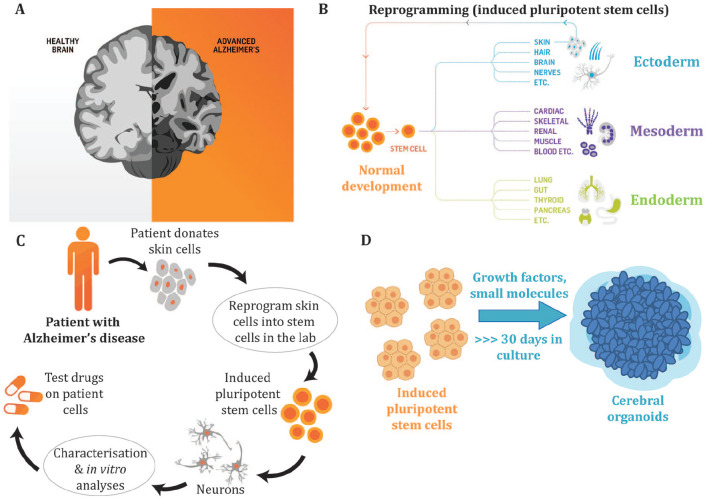
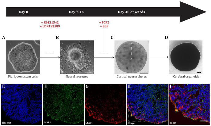
Because our beliefs regarding our individuality, autonomy, and personhood are intimately bound up with our brains, there is a public fascination with cerebral organoids, the "mini-brain," the "brain in a dish". At the same time, the ethical issues around organoids are only now being explored. What are the prospects of using human cerebral organoids to better understand, treat, or prevent dementia? Will human organoids represent an improvement on the current, less-than-satisfactory, animal models? When considering these questions, two major issues arise. One is the general challenge associated with using any stem cell-generated preparation for in vitro modelling (challenges amplified when using organoids compared with simpler cell culture systems). The other relates to complexities associated with defining and understanding what we mean by the term "dementia." We discuss 10 puzzles, issues, and stumbling blocks to watch for in the quest to model "dementia in a dish."

Keywords: Alzheimer’s disease; cerebral; cortical; dementia; disease model; induced pluripotent stem cells; neurodegeneration; organoids.


Conflict of interest statement

Declaration of Conflicting Interests: The author(s) declared the following potential conflicts of interest with respect to the research, authorship, and/or publication of this article: MV is the scientific founder of Skin2NeuronPty Ltd and has a financial interest in the company.

Figures

Figure 1.
Figure 1.
The experimental paradigm using cerebral organoids to investigate Alzheimer’s disease. (A) Alzheimer’s disease (AD) is the most prevalent form of dementia and is characterized by neuronal cell death to the cortex and hippocampus of the brain resulting in impaired memory, cognition, and behavior. The underlying molecular mechanisms that result in neurodegeneration remain to be defined, with patient-derived induced pluripotent stem cells (iPSCs) offering a biologically relevant model to better understand the biological basis of AD. The AD brain shows a reduced volume due to the loss of synapses and neurons. (B) During normal development pluripotent stem cells differentiate into multiple cell types of the body, including cells from the three germ layers, the ectoderm, mesoderm, and endoderm. Terminally differentiated cells, such as skin cells, can be reprogrammed to generate iPSCs that have the capacity to differentiate into multiple cell types of the body. (C) To model AD in a dish, patients donate cells (such as skin cells) that are reprogrammed into iPSCs. The iPSCs can then be differentiated into neurons or other cell types. The use of AD patient–derived iPSCs allows researchers to generate cells of a specific lineage, including neurons and glial cells, to characterize disease phenotypes that may be a result of the patient’s genetic makeup and to develop new therapeutics via drug screening. (D) Cerebral organoids are three-dimensional stem cell cultures that have the advantage over traditional two-dimensional culture approaches as they allow the stem cells to self-organize into structures, form signaling networks and develop cell-cell interactions that better mimic in vivo neurodevelopment. These three-dimensional culture systems allow us to better model and further interrogate the roles that different neural and glial cell types play in neurodegeneration. The cerebral organoids contain a mixture of neural cell types that represent the anatomical structure of the brain.
Figure 2.
Figure 2.
Generalized schematic of cerebral organoid differentiation based on Lancaster and Knoblich (2014). (A) Small molecule inhibitors are added to human pluripotent stem cells to drive differentiation toward a cortical phenotype. Scale bar = 200 µm. (B) Following neural induction, neural rossettes are harvested for three-dimensional culture as neurospheres. Scale bar = 200 µm. (C) Cortical neurospheres are cultured in suspension to allow for maturation and expansion. Following 2 weeks, neurospheres are maintained for longer times to allow for further self-organization and differentiation. Cortical neurospheres self-organize into distinct structures. Neural rosettes observed in vitro during cerebral organoid culture (highlighted in the rectangle) represent the neural tube formation in vivo during human neurodevelopment. Scale bar = 200 µm. (D) Cerebral organoids are maintained for long-term culture allowing for neural differentiation that recapitulates human brain development, and continue to grow in size. Scale bar = 200 µm. Cerebral organoids can be cryosectioned and characterized by immunocytochemistry, using antibodies directed at specific neuronal and glial markers to show the heterogeneous populations of cells contained within an individual organoid. A representative example of a cerebral organoid derived from a healthy individual shows the nuclear stain, Hoechst, blue (E); the mature neuronal marker microtubule associated protein 2 (MAP2), green (F); the astrocyte marker glial fibrillary acidic protein (GFAP), red (G); the merged overlay (H; scale bar = 25 µm); and a magnified image to show the mix of neurons and astrocytes in the organoid (I; scale bar = 50 µm).
Figure 3.
Figure 3.
Genetic risk for Alzheimer’s disease. Mutations in APP, PSEN1, and PSEN2 that cause younger-onset AD (YOAD; also known as “familial AD”) are rare with the vast majority of genetic risk for late onset (LOAD; also known as “sporadic AD”) arising through common variants in multiple genes that each increase risk but which individually are not causative. Figure modified from Karch and Goate (2015).
Figure 4.
Figure 4.
Summary of the amyloid, tau, and inflammation hypotheses for Alzheimer’s disease. Amyloid precursor protein (APP) is a membrane protein that is proteolyticaly cleaved by multiple enzymes. The non-amyloidgenic pathway proceeds when APP is cleaved by the activity of α-secretase (α-sec) to soluble APP α (sAPPα) and APP carboxy terminal fragment α (APP-CTFα), these products are cleaved by γ-secretase (γ-sec) to produce truncated Aβ (p3) and the cytoplasmic polypeptide named AICD. The amyloidgenic pathway occurs when APP undergoes cleavage by β-secretase (β-sec) to form sAPPβ and APP-CTFβ. These proteins are cleaved by γ-sec to form AICD and amyloid-β (Aβ). In YOAD, mutations in APP, PSEN1, and PSEN2 there is an increase in the 42-residue Aβ42, relative to Aβ40, which leads to an increase in Aβ oligomers and amyloid plaque formation (Thinakaran and Koo 2008). Tau binds to microtubules and is important in cytoskeletal function. In AD, tau is hyperphosphorylated resulting in cytoskeletal dysfunction. Hyperphosphorylated tau detaches from microtubules and forms paired helical filaments that aggregate and form neurofibrillary tangles (NFTs). Inflammation is hypothesized to contribute to cognitive loss in AD. Astrocytes and microglia are activated in the AD brain, releasing pro-inflammatory cytokines (e.g., tumor necrosis factor-α [TNFα], interleukin [IL]-1β, IL-6, interferon-γ [IFNγ]) and chemokines (e.g., MIP-1α and MIP-1β) resulting in neuronal death, either by directly damaging neurons or by failing in their normal function to clear aggregates from the brain (Azizi and others 2015). There are multiple underlying molecular pathways leading to AD; intersecting pathways between amyloid, tau, inflammation, and other processes contribute to a complex mechanism that drives neurodegeneration.
Figure 5.
Figure 5.
Assessing Aβ and tau pathology in human brain organoids. (A) The localization of Aβ and phosphorylated tau (p-tau) has been visualized in organoids by immunocytochemistry (Choi and others 2014; Gonzalez and others 2018; Lee and others 2016; Lin and others 2018; Park and others 2018; Pavoni and others 2018; Raja and others 2016). (B) β-sheet aggregates have been stained with the fluorescent Thioflavin-S (Thio S) dye. Thio S staining was proposed to identify tau pathology in AD organoids (Raja and others 2016), though the precise molecular identity of the aggregates needs to be confirmed. (C) Enzyme-linked immunosorbent assays (ELISAs) have been used to quantify secreted Aβ in the organoid medium (Choi and others 2014; Gonzalez and others 2018; Lee and others 2016; Lin and others 2018; Park and others 2018; Pavoni and others 2018; Raja and others 2016). (D) Protein levels of Aβ and p-tau have been compared in AD and control organoids by western blotting semiquantitative analysis (Choi and others 2014; Gonzalez and others 2018; Lin and others 2018; Park and others 2018; Raja and others 2016). See also Table 1.
Figure 6.
Figure 6.
Functional assessment of cerebral organoids by calcium imaging. (A) A representative image of a culture derived from a YOAD cerebral organoid loaded with the ratiometric calcium indicator Fura2-AM. A 9-month-old organoid was seeded onto a glass coverslip for Ca2+ imaging. The image shows the overlay of 340 nm and 380 nm channels; each colored circle corresponds to a region of interest (ROI) represented in (B). When Ca2+ binds to the indicator, fluorescence at 340 nm increases, while 380 nm fluorescence decreases. The 340/380 ratio is thus used as a measurement of Ca2+ responses to drugs or agonists or to assess spontaneous activity. (B) Relative change in fluorescence intensity over time of specific ROIs from (A) using Fura2-AM. Neurons can be stimulated with chemicals that are perfused into the bath chamber and the Ca2+ responses recorded. The culture was exposed to the excitatory neurotransmitter glutamate (20 µM; perfusion indicated by the horizontal black bar) to elicit a Ca2+ response, followed by high K+ (60 mM; perfusion indicated by the horizontal black bar) to mimic membrane depolarization. Responses may be fast or slow transient increases or prolonged increases that do no return to baseline within the timeframe of the experiment.
Figure 7.
Figure 7.
Electrophysiological assessment of cerebral organoids by microelectrode arrays. (A) Organoids can be seeded in well chambers (B) on top of regularly spaced arrays of electrodes. (C) Organoids are electrically active; shown are recordings from two different organoids at 25 kHz, whereby spikes identify electrophysiological activity. The recordings show a difference in spontaneous activity from the two organoids, which can be quantified via various parameters, such as spike amplitude and firing rate. Experiments can be designed to compare organoid responses to chemical or electrical stimulation and can provide information on neuronal network dynamics and the formation of functional circuits.
Figure 8.
Figure 8.
Schematic showing opportunities for the use of brain organoids in the drug development pipeline. In particular, stem cell–derived culture models have the potential to improve lead optimization for “formal preclinical” and clinical development.

References

    1. Abner EL, Nelson PT, Kryscio RJ, Schmitt FA, Fardo DW, Woltjer RL, and others. 2016. Diabetes is associated with cerebrovascular but not Alzheimer’s disease neuropathology. Alzheimers Dement 12(8):882–9. - PMC - PubMed
    1. Allum N, Allansdottir A, Gaskell G, Hampel J, Jackson J, Moldovan A, and others. 2017. Religion and the public ethics of stem-cell research: attitudes in Europe, Canada and the United States. PLoS One 12(4):e0176274. - PMC - PubMed
    1. Arber C, Lovejoy C, Wray S. 2017. Stem cell models of Alzheimer’s disease: progress and challenges. Alzheimers Res Ther 9(1):42. - PMC - PubMed
    1. Azizi G, Navabi SS, Al-Shukaili A, Seyedzadeh MH, Yazdani R, Mirshafiey A. 2015. The role of inflammatory mediators in the pathogenesis of Alzheimer’s disease. Sultan Qaboos Univ Med J 15(3):e305–e316. - PMC - PubMed
    1. Bayne T, Seth AK, Massimini M. 2020. Are there islands of awareness? Trends Neurosci 43(1):6–16. - PubMed

Publication types