. 2020 Mar 25:(157):10.3791/60653.
doi: 10.3791/60653.

Assessment of Acute Wound Healing using the Dorsal Subcutaneous Polyvinyl Alcohol Sponge Implantation and Excisional Tail Skin Wound Models

Affiliations

Assessment of Acute Wound Healing using the Dorsal Subcutaneous Polyvinyl Alcohol Sponge Implantation and Excisional Tail Skin Wound Models

Meredith J Crane et al. J Vis Exp. .

Abstract

Wound healing is a complex process that requires the orderly progression of inflammation, granulation tissue formation, fibrosis, and resolution. Murine models provide valuable mechanistic insight into these processes; however, no single model fully addresses all aspects of the wound healing response. Instead, it is ideal to use multiple models to address the different aspects of wound healing. Here, two different methods that address diverse aspects of the wound healing response are described. In the first model, polyvinyl alcohol sponges are subcutaneously implanted along the mouse dorsum. Following sponge retrieval, cells can be isolated by mechanical disruption, and fluids can be extracted by centrifugation, thus allowing for a detailed characterization of cellular and cytokine responses in the acute wound environment. A limitation of this model is the inability to assess the rate of wound closure. For this, a tail skin excision model is utilized. In this model, a 10 mm x 3 mm rectangular piece of tail skin is excised along the dorsal surface, near the base of the tail. This model can be easily photographed for planimetric analysis to determine healing rates and can be excised for histological analysis. Both described methods can be utilized in genetically altered mouse strains, or in conjunction with models of comorbid conditions, such as diabetes, aging, or secondary infection, in order to elucidate wound healing mechanisms.


Figures

Figure 1:
Figure 1:. Schematic of murine wound healing models.
(A) Side (left) and top (right) view of dehydrated PVA sponges measuring 8 mm x 8 mm x 0.4 mm. (B) Illustration of a mouse demonstrating the placement of the dorsal midline incision (central red line) and six 1 cm x 1 cm x 0.5 cm PVA sponges in subcutaneous pockets. (C) Schematic demonstrating the size and placement of an excisional skin wound (10 mm x 3 mm red rectangle) on the dorsal surface of the tail. (D) Wound fluid isolated from three sponges that were retrieved from the subcutaneous space 7 days after implantation. (E) The appearance of sponges retrieved from the wound 7 days after implantation.
Figure 2:
Figure 2:. The systemic and cellular response to PVA sponge implantation.
(A) A time course of IL-6 levels in the plasma demonstrates that PVA sponge implantation induced a systemic inflammatory response. The concentration of IL-6 was measured using a multiplex bead-based immunoassay. (B) The number of cells isolated from PVA sponges wound increased over time. (C) A time course demonstrates the accumulation of neutrophils, monocytes, and monocyte-derived macrophages in PVA sponge wounds over time. (D) A representative gating strategy of cells isolated from PVA sponge wounds demonstrates how to identify leukocyte subsets by flow cytometric analysis. Gates are defined as follows: (i and ii) doublet exclusion, (iii) dead cell exclusion using an amine-reactive fixable viability dye, (iv) debris exclusion by FSC-A and SSC-A, (v) CD45+ hematopoietic cells, (vi) Ly6G+ neutrophils, (vii) Siglec-F+ eosinophils, (viii) Ly6GSiglec-F F4/80+ monocytes/macrophages, (ix) F4/80+Ly6Chi monocytes, and (x) F4/80+Ly6Clow macrophages. Gates were placed according to fluorescence-minus one (FMO) controls. The data shown in A–C are the mean ± SEM, n = 6–10 mice per group in (A), n = 8–9 mice in (B), and n = 6–8 mice in (C). All data are combined from 2–3 independent experiments.
Figure 3:
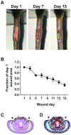
Figure 3:. Assessment of excisional tail skin wound healing.
(A) Representative photographs of excisional tail skin wounds taken 1, 7, and 15 days post-wounding. (B) The rate of closure of excisional tail skin wounds. Wounds were photographed every other day. Image processing software was used to trace the wound bed margins and calculate the wound area at indicated time points. The wound area is presented as a fraction of the wound area measured on day 1. Representative images of tail cross sections that were paraffin-embedded, sectioned, and stained with (C) H&E or (D) Masson’s Trichrome. The wound was located on the dorsal surface of the tail cross section; lateral wound margins are indicated by arrowheads. The data shown in (B) are the mean ± SEM, n = 8 mice per group. Data are combined from two independent experiments.

References

    1. Gottrup F, Agren MS, Karlsmark T Models for use in wound healing research: a survey focusing on in vitro and in vivo adult soft tissue. Wound Repair and Regeneration: Official Publication of the Wound Healing Society [and] the European Tissue Repair Society. 8 (2), 83–96 (2000). - PubMed
    1. Elliot S, Wikramanayake TC, Jozic I, Tomic-Canic M A Modeling Conundrum: Murine Models for Cutaneous Wound Healing. Journal of Investigative Dermatology. 138 (4), 736–740 (2018). - PubMed
    1. Eming SA, Martin P, Tomic-Canic M Wound repair and regeneration: Mechanisms, signaling, and translation. Science Translational Medicine. 6 (265), 265sr6 (2014). - PMC - PubMed
    1. Huber-Lang M, Lambris JD, Ward PA Innate immune responses to trauma. Nature Immunology. 19 (4), 327–341 (2018). - PMC - PubMed
    1. Novak ML, Koh TJ Phenotypic Transitions of Macrophages Orchestrate Tissue Repair. The American Journal of Pathology. 183 (5), 1352–1363 (2013). - PMC - PubMed

Publication types

Substances

LinkOut - more resources