

Coronavirus Disease 2019 (COVID-19) CT Findings: A Systematic Review and Meta-analysis
- PMID: 32283052
- PMCID: PMC7151282
- DOI: 10.1016/j.jacr.2020.03.006
Coronavirus Disease 2019 (COVID-19) CT Findings: A Systematic Review and Meta-analysis
Abstract
Purpose: To date, considerable knowledge gaps remain regarding the chest CT imaging features of coronavirus disease 2019 (COVID-19). We performed a systematic review and meta-analysis of results from published studies to date to provide a summary of evidence on detection of COVID-19 by chest CT and the expected CT imaging manifestations.
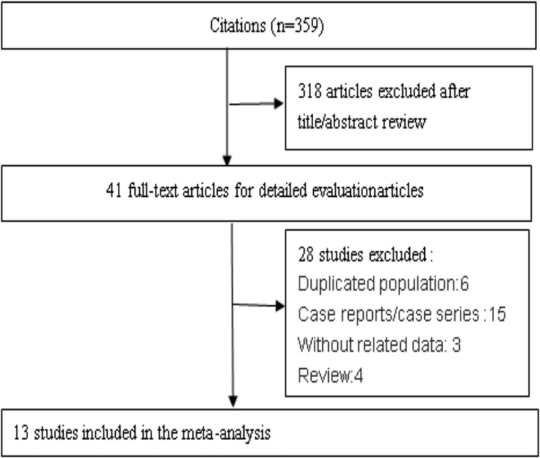
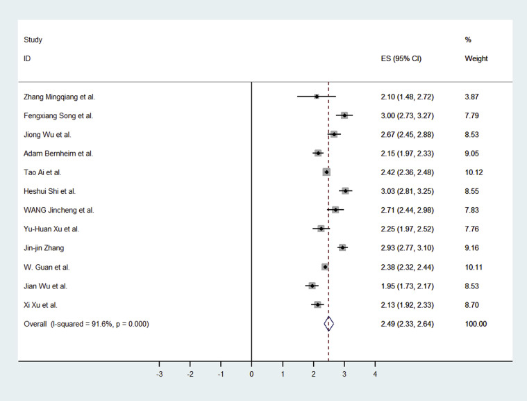
Methods: Studies were identified by searching PubMed database for articles published between December 2019 and February 2020. Pooled CT positive rate of COVID-19 and pooled incidence of CT imaging findings were estimated using a random-effect model.
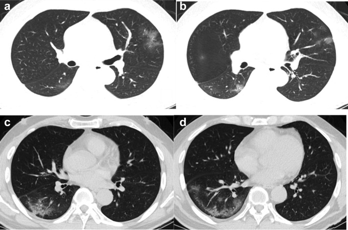
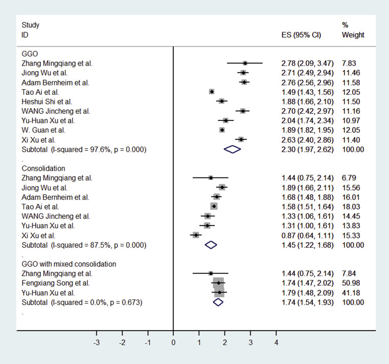
Results: A total of 13 studies met inclusion criteria. The pooled positive rate of the CT imaging was 89.76% and 90.35% when only including thin-section chest CT. Typical CT signs were ground glass opacities (83.31%), ground glass opacities with mixed consolidation (58.42%), adjacent pleura thickening (52.46%), interlobular septal thickening (48.46%), and air bronchograms (46.46%). Other CT signs included crazy paving pattern (14.81%), pleural effusion (5.88%), bronchiectasis (5.42%), pericardial effusion (4.55%), and lymphadenopathy (3.38%). The most anatomic distributions were bilateral lung infection (78.2%) and peripheral distribution (76.95%). The incidences were highest in the right lower lobe (87.21%), left lower lobe (81.41%), and bilateral lower lobes (65.22%). The right upper lobe (65.22%), right middle lobe (54.95%), and left upper lobe (69.43%) were also commonly involved. The incidence of bilateral upper lobes was 60.87%. A considerable proportion of patients had three or more lobes involved (70.81%).
Conclusions: The detection of COVID-19 chest CT imaging is very high among symptomatic individuals at high risk, especially using thin-section chest CT. The most common CT features in patients affected by COVID-19 included ground glass opacities and consolidation involving the bilateral lungs in a peripheral distribution.
Keywords: COVID-19; CT imaging findings; ground glass opacities; meta-analysis; thin-section chest CT.
Copyright © 2020 American College of Radiology. Published by Elsevier Inc. All rights reserved.
Figures
Comment in
-
Tomographic Findings and Thrombogenic Effects of COVID-19.J Am Coll Radiol. 2020 Dec;17(12):1545. doi: 10.1016/j.jacr.2020.06.034. Epub 2020 Jul 13. J Am Coll Radiol. 2020. PMID: 32735918 Free PMC article. No abstract available.
-
[COVID-19 and pleural effusions].Rev Mal Respir. 2021 Feb;38(2):219-221. doi: 10.1016/j.rmr.2021.01.007. Epub 2021 Jan 19. Rev Mal Respir. 2021. PMID: 33516596 Free PMC article. French. No abstract available.
References
-
- Higgins J.P., Thompson S.G. Quantifying heterogeneity in a meta-analysis. Stat Med. 2002;21:1539–1558. - PubMed
Publication types
MeSH terms
LinkOut - more resources
Full Text Sources
Medical
Miscellaneous
