. 2020 Jun:178:104793.
doi: 10.1016/j.antiviral.2020.104793. Epub 2020 Apr 10.

Remdesivir and SARS-CoV-2: Structural requirements at both nsp12 RdRp and nsp14 Exonuclease active-sites

Affiliations

Remdesivir and SARS-CoV-2: Structural requirements at both nsp12 RdRp and nsp14 Exonuclease active-sites

Ashleigh Shannon et al. Antiviral Res. 2020 Jun.

Abstract

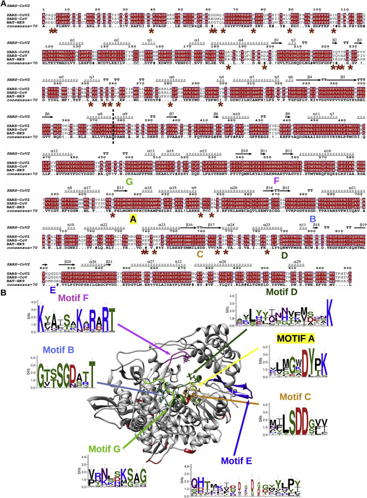
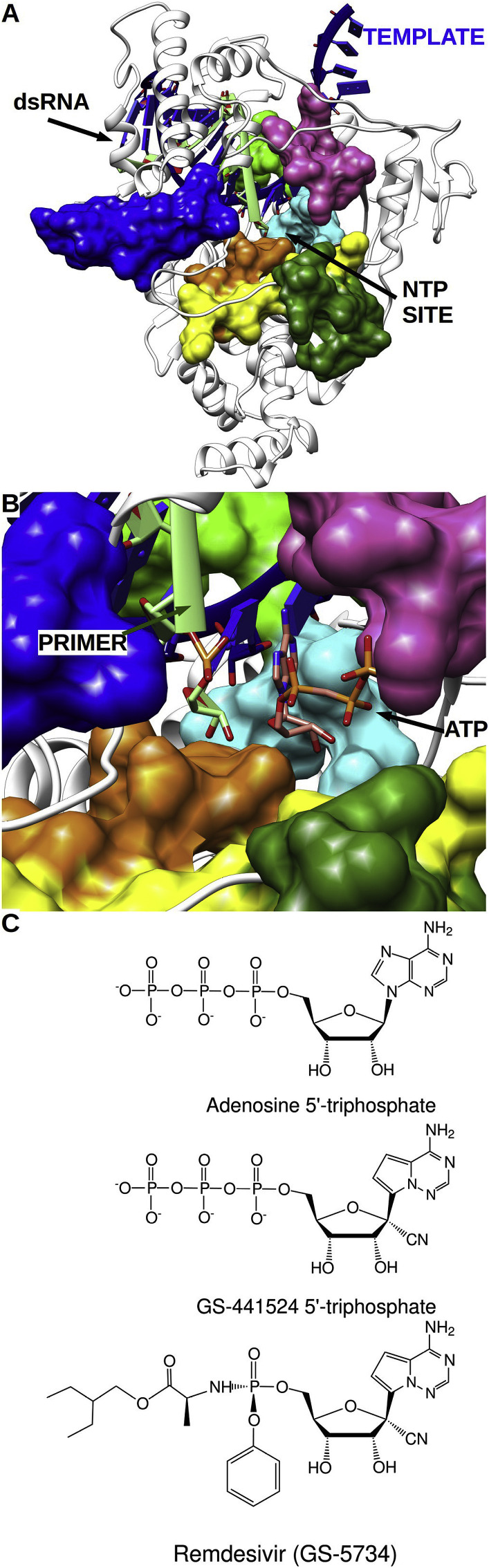
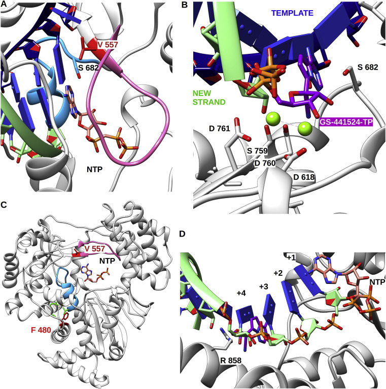
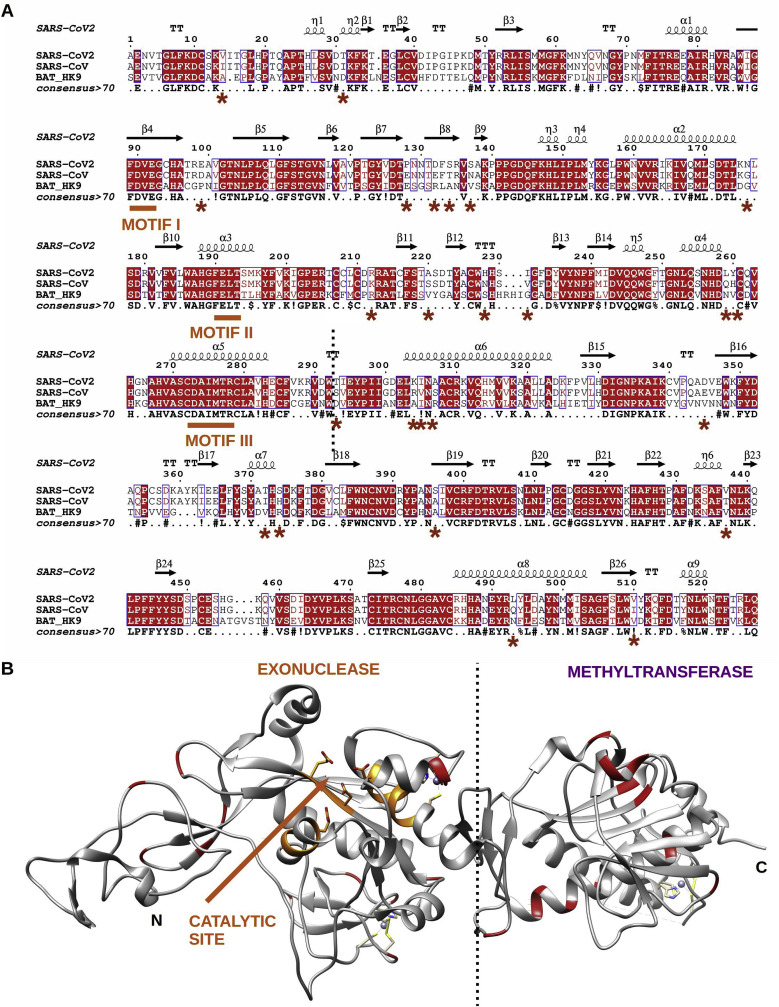
The rapid global emergence of SARS-CoV-2 has been the cause of significant health concern, highlighting the immediate need for antivirals. Viral RNA-dependent RNA polymerases (RdRp) play essential roles in viral RNA synthesis, and thus remains the target of choice for the prophylactic or curative treatment of several viral diseases, due to high sequence and structural conservation. To date, the most promising broad-spectrum class of viral RdRp inhibitors are nucleoside analogues (NAs), with over 25 approved for the treatment of several medically important viral diseases. However, Coronaviruses stand out as a particularly challenging case for NA drug design due to the presence of an exonuclease (ExoN) domain capable of excising incorporated NAs and thus providing resistance to many of these available antivirals. Here we use the available structures of the SARS-CoV RdRp and ExoN proteins, as well as Lassa virus N exonuclease to derive models of catalytically competent SARS-CoV-2 enzymes. We then map a promising NA candidate, GS-441524 (the active metabolite of Remdesivir) to the nucleoside active site of both proteins, identifying the residues important for nucleotide recognition, discrimination, and excision. Interestingly, GS-441524 addresses both enzyme active sites in a manner consistent with significant incorporation, delayed chain termination, and altered excision due to the ribose 1'-CN group, which may account for the increased antiviral effect compared to other available analogues. Additionally, we propose structural and function implications of two previously identified RdRp resistance mutations in relation to resistance against Remdesivir. This study highlights the importance of considering the balance between incorporation and excision properties of NAs between the RdRp and ExoN.

Keywords: COVID-19; Coronavirus; Exonuclease; Mutation; Nucleotide analogue; RNA-Dependent RNA polymerase; Remdesivir; Resistance.


Figures

Fig. 1
Fig. 1
(A) Amino acid sequence alignment of selected coronavirus nsp12 protein, including: SARS-CoV2 (YP_009724389), SARS-CoV (NC_004718); BatHK9 (NC_009021). SARS-CoV2 nsp12 secondary structure (derived from PDB: 6NUR) is indicated on top. Fully conserved residues are in red font and boxed, whereas partially conserved residues are displayed in red font (above 70% conservation). A consensus sequence is generated using same criteria uppercase is identity, “.” is no consensus, ! is any of I,V; $ is any of L,M, % is any of F,Y; # is any of N,D,Q,E,B,Z. Red star marked the mutation between SARS-CoV2 and SARS-CoV. The NiRAN and the polymerase domains are separated by a dashed line. The polymerase conserved motifs (A–G) are shown under the consensus sequence. Alignments were made using Seaview (Gouy et al., 2010) and Espript (Gouet et al., 2003) (B) Cartoon representation of nsp12 SARS-CoV (PDB: 6NUR). The motifs are colored as follows: A, yellow; B, cyan; C, orange; D, forest green; E, blue; F, pink; G, light green. Around the structure are WebLogo (Crooks et al., 2004) highlighting the A-G motifs. All structural models were made using Chimera (Pettersen et al., 2004).
Fig. 2
Fig. 2
(A) Cartoon representation of nsp12 SARS-CoV-2 with spatial arrangement of RdRp motifs complexed with a modelled RNA template (blue) and neo synthesised (light green). Models are derived from either PDB model 6NUR or 6M71; the latter has a 2.9 Å resolution (biorxiv.org/content/10.1101/2020.03.16.993386v1.full.pdf) and they exhibit a RMSD of 0.612 Å over 790 amino acids, which means that the model is valid in both. Color code is the same as in Fig. 1. (B) Close up of the NTP entry and catalytic sites with NTP in position to be incorporated. Position of RNA and NTP was obtained by superimposition of references crystal structures of poliovirus and T7 RNA polymerases elongation complexes (PDB 4K4S and 1S76). (C) Chemical structures of nucleotides discussed throughout the manuscript including: (a) ATP (b) GS-441524-TP (c) Remdesivir.
Fig. 3
Fig. 3
Cartoon representation of nsp12 SARS-CoV polymerase domain (PDB: 6NUR) pre-incorporation of GS-441524-TP and mutation mapping. (A) Top view of V 557 with RNA strands and NTP in pre-incorporation position. V 557 forms a hydrophobic wall upon which the template base is stacked. (B) Close up of RNA strands with GS-441524-TP in a pre-incorporation position at the catalytic site. Template strand is in blue, newly synthesised strand is in light green, and GS-441524-TP in purple. Key residues of active site (from motif A, B and C) are shown in stick and labelled. (C) Remdesivir resistance mutations highlighted in red on the cartoon representation of nsp12 SARS-CoV polymerase domain. F 480 faces a patch of hydrophobic residues shown in green that indirectly impact motif B (in cyan). V 557 is located on the side of the template groove and is at the end of motif F (in pink). NTP in pre-incorporation position is shown in stick. (D) View of the newly synthesised RNA in mix representation stick from position 1 to 4 and ribbon for position five and above, for clarity reason template RNA is not shown. Shown is a NTP in a pre-incorporation position and newly nascent RNA. In position +4 the conserved R 858 is poised to make a steric clash with the 1′-CN group of GS-441524 once it has been incorporated and translocated.
Fig. 4
Fig. 4
(A) Amino acid sequence alignment of selected coronavirus nsp14 proteins: SARS-CoV-2 (YP_009724389), SARS-CoV (NC_004718); BatHK9 (NC_009021). SARS-CoV nsp14 secondary structure (derived from PDB: 5NFY) is indicated on top. Fully conserved residues are in red and boxed, whereas partially conserved residues are displayed in red only (above 70% conservation). A consensus sequence is generated using same criteria as in Fig. 1. Residues marked with a red star show polymorphisms between SARS-CoV-2 and SARS-CoV. The N-terminal exonuclease (ExoN) and C-terminal methyltransferase domains are separated by a dashed vertical line. The ExoN catalytic residues (motifs I, II, and III) are underlined in orange. (B) Cartoon representation of the crystal structure of the nsp14 SARS-CoV (PDB: 5NFY). The nsp14 ExoN domain, catalytic site is highlighted in orange; and star residues are shown in red. None of the mutations are in the vicinity of the catalytic site.
Fig. 5
Fig. 5
The excision step catalyzed by nsp14. Close-up of nsp14 ExoN active site (PDB: 5NFY). Modelling of active site ions and RNA was done using a superimposition of the related Lassa virus ExoN structure complexed with RNA and ions (PDB: 4FVU and 4GV9). (A) Reference position of RNA and ions in the excision of an RNA 3′-end nucleotide. (B) Position of RNA and ions in the case of GS-441524-MP excision. GS-441524-MP is structurally superimposed to the ribose of nucleotide in position of A. The distorted base of GS-441524-MP would force the ribose to move in place of the ions preventing the proper distances for an efficient two metal ion catalysis to happen. (C) Same view as B detailed with catalytic (DEED) and binding residues (FHN) in stick and transparent surface.

References

    1. Agostini M.L. Coronavirus susceptibility to the antiviral remdesivir (GS-5734) is mediated by the viral polymerase and the proofreading exoribonuclease. mBio. 2018;9 - PMC - PubMed
    1. Agostini M.L. Small-Molecule antiviral β-d-N4-hydroxycytidine inhibits a proofreading-intact coronavirus with a high genetic barrier to resistance. J. Virol. 2019;93 - PMC - PubMed
    1. Booth C.M. Clinical features and short-term outcomes of 144 patients with SARS in the greater Toronto area. J. Am. Med. Assoc. 2003;289:2801–2809. - PubMed
    1. Bouvet M. RNA 3’-end mismatch excision by the severe acute respiratory syndrome coronavirus nonstructural protein nsp10/nsp14 exoribonuclease complex. Proc. Natl. Acad. Sci. U. S. A. 2012;109:9372–9377. - PMC - PubMed
    1. Campagnola G., McDonald S., Beaucourt S., Vignuzzi M., Peersen O.B. Structure-function relationships underlying the replication fidelity of viral RNA-dependent RNA polymerases. J. Virol. 2015;89:275–286. - PMC - PubMed

MeSH terms