Review
. 2020 Jul 15:747:144655.
doi: 10.1016/j.gene.2020.144655. Epub 2020 Apr 10.

The MID1 gene product in physiology and disease

Affiliations
Review

The MID1 gene product in physiology and disease

Rossella Baldini et al. Gene. .

Abstract

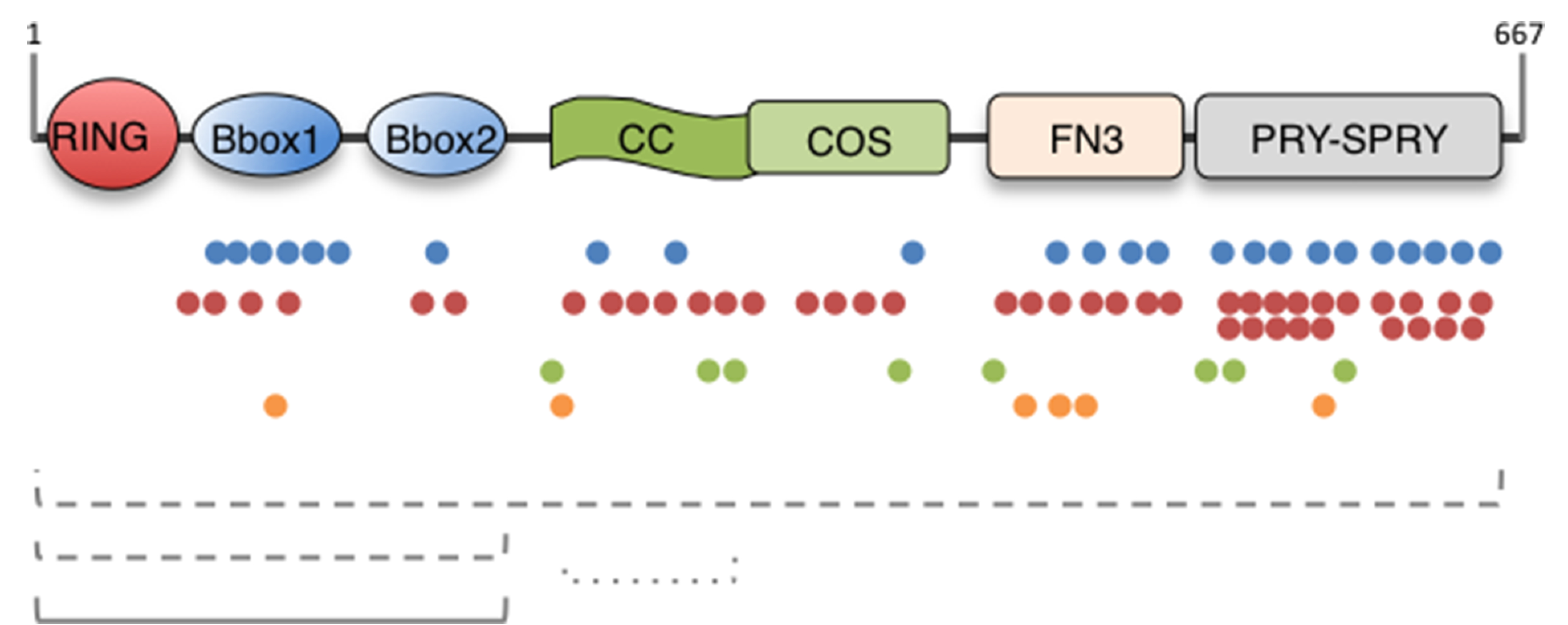
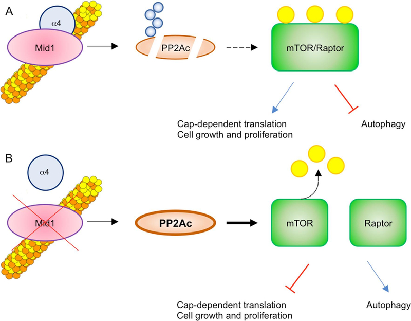
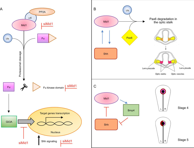
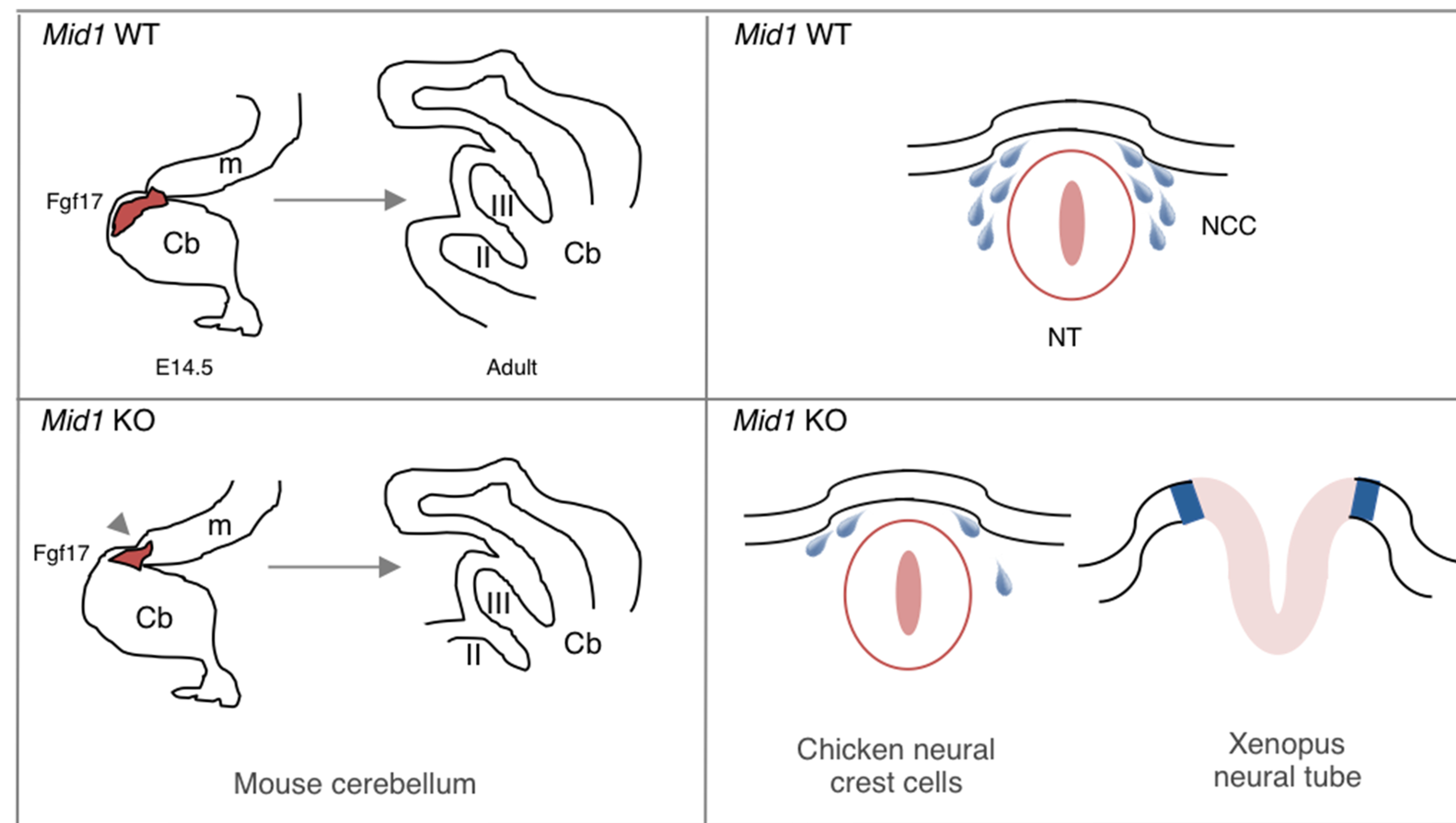
MID1 is an E3 ubiquitin ligase of the Tripartite Motif (TRIM) subfamily of RING-containing proteins, hence also known as TRIM18. MID1 is a microtubule-binding protein found in complex with the catalytic subunit of PP2A (PP2Ac) and its regulatory subunit alpha 4 (α4). To date, several substrates and interactors of MID1 have been described, providing evidence for the involvement of MID1 in a plethora of essential biological processes, especially during embryonic development. Mutations in the MID1 gene are responsible of the X-linked form of Opitz syndrome (XLOS), a multiple congenital disease characterised by defects in the development of midline structures during embryogenesis. Here, we review MID1-related physiological mechanisms as well as the pathological implication of the MID1 gene in XLOS and in other clinical conditions.


Conflict of interest statement

Declaration of Competing Interest The authors declare that they have no known competing financial interests or personal relationships that could have appeared to influence the work reported in this paper.

Figures

Fig. 1.
Fig. 1.. MID1 protein domain structure and OS-associated mutations.
The domain composition of the MID1/TRIM18 protein is depicted. The MID1 protein is 667 residue-long and the limits of the single domains are following in brackets: RING (10–59), Really Interesting New Gene domain; B-Box (B1, 114–164; B2, 170–212), B-Box domain; CC (219–319), Coiled-coil; COS (320–380), C-terminal subgroup one signature; FN3 (382–472), Fibronectin type III repeat; PRY (483–528), domain associated with SPRY domains; SPRY (538–657), SPla and the RYanodine Receptor. Below the scheme, colour dots represent the different mutations reported so far in OS patients: blue dots, missense mutations; red dots, nonsense and truncating mutations; green dots, splice site mutations; orange dots, inframe indels. The dashed lines represent deletions and rearrangements; the continuous line represents duplications. (For interpretation of the references to colour in this figure legend, the reader is referred to the web version of this article.)
Fig. 2.
Fig. 2.. MID1 regulates mTORC1 signalling through PP2A.
A) The microtubular pool of PP2Ac is the target of MID1 activity, leading to its proteasomal-dependent degradation upon poly-ubiquitination (Blue circles). Since mTOR/Raptor association is dependent on PP2A, the degradation of the latter leads to an increased formation of the mTOR/Raptor complex, activating its signalling pathway (phosphorylated form, yellow circles). B) MID1 loss-of-function reduces PP2Ac degradation, leading to de-phosphorylation and decreased association of mTOR and Raptor, causing a drop of active mTORC1 complex formation and signalling. (For interpretation of the references to colour in this figure legend, the reader is referred to the web version of this article.)
Fig. 3.
Fig. 3.. Model of Shh pathway regulated by MID1.
A) MID1 is involved in the proteasome-dependent cleavage of the Fu kinase domain, leading to GLI3A (activator) translocation into the nucleus. This process activates Shh target genes expression increasing Shh signalling. Silencing of MID1 (red writings) impairs Fu cleavage thus reducing Shh signalling. B) In Xenopus, an overlapping expression of mid1 (in pink) and pax6 (in yellow) is observed in the optic stalk, in early stages during the development of the visual system. mid1 is expressed within the forming optic stalk under the control of Shh. Here, mid1 regulates the ubiquitination and proteasomal degradation of Pax6 protein that is cleared from the optic stalk region, setting the border between the optic stalk and the retina via Mid1. C) In chicken development, both Shh (dark red) and cMid1 (pink) are initially expressed bilaterally in the Hensen’s node (black) until stage 5. cMid1 then induces the expression of Bmp4 (green) on the right side of the node. Bmp4 represses right-sided Shh expression, thus restricting Shh to the left side of the node. Left-sided Shh represses cMid1 expression on the left, restricting it to the right side of the node, together with Bmp4. (For interpretation of the references to colour in this figure legend, the reader is referred to the web version of this article.)
Fig. 4.
Fig. 4.. Distribution of MID1 during embryonic development.
In the upper part, expression of MID1 in specific developmental stages is summarised. The coloured lines indicate the models used in the studies as indicated on the right-hand side. In the bottom part a schematic representation of Mid1 distribution at different stages of embryonic development is shown (pink shading); Carnegie stages (CS) are indicated. A) Mid1 distribution is restricted to the right side of the Hensen’s node; B) the cranial region of the neural plate (np) displays the strongest expression of Mid1 at CS9; C) Mid1 is mainly transcribed in the proliferating compartments of telencephalic vesicle (te), dorsal midbrain (mb) and hindbrain (hb); D) at late embryonic stages, high levels of Mid1 transcript are particularly described in the developing hindbrain and midbrain. Mid1 mRNA is also present in the heart (he) and in several organs of the urogenital system (us). (For interpretation of the references to colour in this figure legend, the reader is referred to the web version of this article.)
Fig. 5.
Fig. 5.. Mutations in Mid1 determine neuroanatomical defects during embryonic development.
Left-hand side, mouse Mid1 knock-out causes a defective morphogenesis of the cerebellum (cb) and anterior midbrain (m) during development, including the rostralisation of the midbrain/hindbrain boundary (arrowhead) during midgestation (embryonic day 14.5, E14.5) and the consequent malformation of adult cerebellar most anterior lobes (lobes II and III). Right-hand side, Mid1 is implicated in proper neurulation event during embryonic development: defects in cranial neural crest cells (NCC) migration observed in chicken and in neural tube (NT) closure in Xenopus are associated with silencing of Mid1.

References

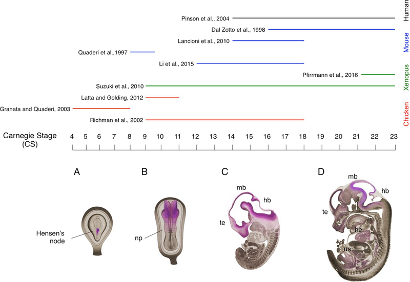
    1. Abramyan J, Richman JM, 2018. Craniofacial development: discoveries made in the chicken embryo. Int. J. Dev. Biol 62, 97–107. - PubMed
    1. Aranda-Orgilles B, Trockenbacher A, Winter J, Aigner J, Kohler A, Jastrzebska E, Stahl J, Muller EC, Otto A, Wanker EE, et al., 2008b. The Opitz syndrome gene product MID1 assembles a microtubule-associated ribonucleoprotein complex. Hum. Genet 123, 163–176. - PMC - PubMed
    1. Aranda-Orgilles B, Aigner J, Kunath M, Lurz R, Schneider R, Schweiger S, 2008a. Active transport of the ubiquitin ligase MID1 along the microtubules is regulated by protein phosphatase 2A. PLoS ONE 3, e3507. - PMC - PubMed
    1. Aranda-Orgilles B, Rutschow D, Zeller R, Karagiannidis AI, Kohler A, Chen C, Wilson T, Krause S, Roepcke S, Lilley D, et al., 2011. Protein phosphatase 2A (PP2A)-specific ubiquitin ligase MID1 is a sequence-dependent regulator of translation efficiency controlling 3-phosphoinositide-dependent protein kinase-1 (PDPK-1). J. Biol. Chem 286, 39945–39957. - PMC - PubMed
    1. Arigoni M, Barutello G, Riccardo F, Ercole E, Cantarella D, Orso F, Conti L, Lanzardo S, Taverna D, Merighi I, et al., 2013. miR-135b coordinates progression of ErbB2-driven mammary carcinomas through suppression of MID1 and MTCH2. Am. J. Pathol 182, 2058–2070. - PubMed

Substances