

Identification of Cardiac MRI and Bio-Marker Thresholds for One-Year Survival in Pre-Capillary Pulmonary Hypertension: Prospective Study
- PMID: 32283599
- PMCID: PMC7230686
- DOI: 10.3390/medicina56040167
Identification of Cardiac MRI and Bio-Marker Thresholds for One-Year Survival in Pre-Capillary Pulmonary Hypertension: Prospective Study
Abstract
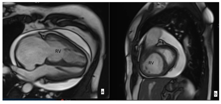
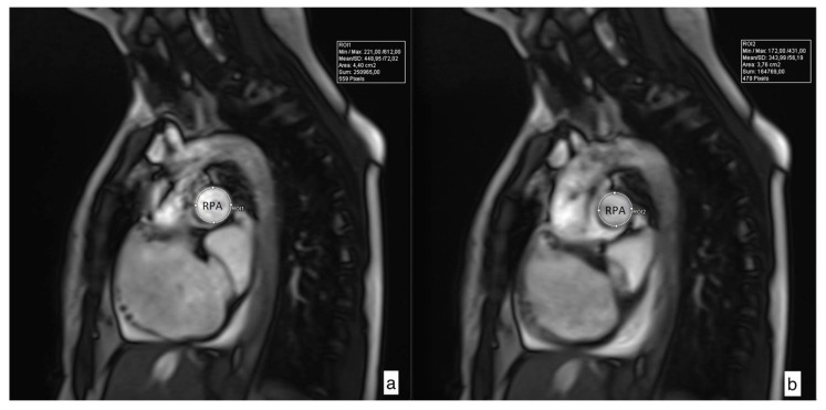
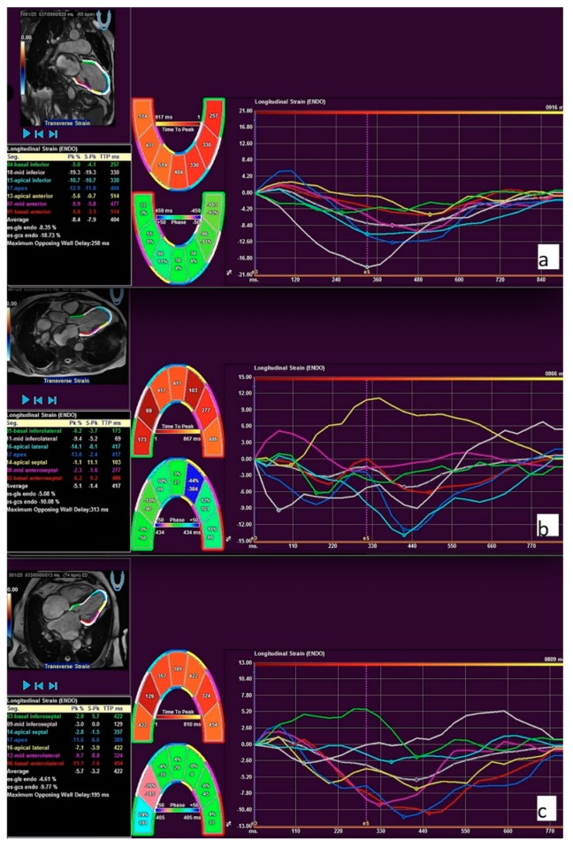
Background and objectives: Non-invasive imaging of the heart has an important place in the diagnosis and management of pulmonary arterial hypertension (PAH). The aim of this study was to establish the thresholds of cardiac magnetic resonance imaging (CMRI)-derived biventricular deformation, function parameters, and levels of N-terminal pro brain natriuretic peptide (NT-proBNP) for the prediction of survival of pre-capillary pulmonary hypertension (PHprecap) patients. Materials and Methods: In total, 64 incident PHprecap cases, who underwent CMRI, were consecutively enrolled in a prospective cohort study. Patients underwent a systemic evaluation, including measurement of NT-proBNP, two-dimensional (2D) echocardiography, six-minute walk test (6MWT), CMRI with feature tracking (FT), and right-heart catheterization (RHC). Patients were divided into two groups according to one-year survival (survival and non-survival groups). Survival analysis was performed. Results: One-year survival was 79.6%. The distribution between age, sex, mean pulmonary artery pressure (mPAP), New York Heart Association (NYHA) functional class, and 6MWT did not differ between the groups. Survival was significantly lower in the PAH group associated with connective tissue disease (CTD-PAH), where 44% (n = 4) of patients died during the first year. Univariate analysis revealed that severely reduced right-ventricle (RV) ejection fraction (EF) <25.5%, left-ventricle global longitudinal strain (LV GLS) >-14.18%, and right pulmonary artery (RPA) relative area change (RAC) <19%, and severely increased NT-proBNP level >1738 (ng/L) indicate an increased risk of death in PHprecap patients. Conclusions: Impaired RV systolic function and LV global longitudinal strain, decrease of pulmonary artery distensibility, and CTD-PAH etiology, together with high NT-proBNP level, impair prognosis in pre-capillary PH patients. These findings are important for the risk stratification and management of pre-capillary pulmonary hypertension patients.
Keywords: cardiac magnetic resonance; feature tracking; pulmonary hypertension; right ventricle.
Conflict of interest statement
All authors declare that they have no competing interests.
Figures
References
MeSH terms
Substances
LinkOut - more resources
Full Text Sources
Medical
Research Materials
Miscellaneous
