. 2020 Jul;26(7):803-813.
doi: 10.1261/rna.074385.119. Epub 2020 Apr 13.

An ancient evolutionary connection between Ribonuclease A and EndoU families

Affiliations

An ancient evolutionary connection between Ribonuclease A and EndoU families

Arcady Mushegian et al. RNA. 2020 Jul.

Abstract

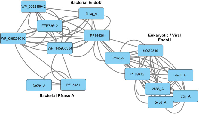
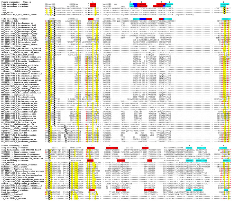
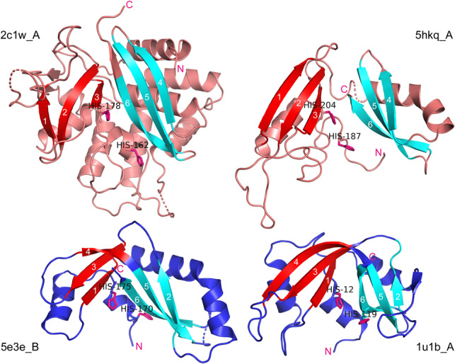
The ribonuclease A family of proteins is well studied from the biochemical and biophysical points of view, but its evolutionary origins are obscure, as no sequences homologous to this family have been reported outside of vertebrates. Recently, the spatial structure of the ribonuclease domain from a bacterial polymorphic toxin was shown to be closely similar to the structure of vertebrate ribonuclease A. The absence of sequence similarity between the two structures prompted a speculation of convergent evolution of bacterial and vertebrate ribonuclease A-like enzymes. We show that bacterial and homologous archaeal polymorphic toxin ribonucleases with a known or predicted ribonuclease A-like fold are distant homologs of the ribonucleases from the EndoU family, found in all domains of cellular life and in viruses. We also detected a homolog of vertebrate ribonucleases A in the transcriptome assembly of the sea urchin Mesocentrotus franciscanus These observations argue for the common ancestry of prokaryotic ribonuclease A-like and ubiquitous EndoU-like ribonucleases, and suggest a better-grounded scenario for the origin of animal ribonucleases A, which could have emerged in the deuterostome lineage, either by an extensive modification of a copy of an EndoU gene, or, more likely, by a horizontal acquisition of a prokaryotic immunity-mediating ribonuclease gene.

Keywords: protein folding; ribonuclease A; ribonuclease EndoU.


Figures

FIGURE 1.
FIGURE 1.
Network of prokRNase A-related and EndoU-related conserved PFAM domains and sequence models based on select queries from the NR and PDB databases. The arc between domains/models indicates a statistically supported match obtained by the HHPred or HHBoost searches.
FIGURE 2.
FIGURE 2.
Multiple alignment of RNase A-like and EndoU-like protein sequences. Unique identifiers in the NR or PDB databases are shown before each sequence. In the secondary structure lines, h stands for a helical structure and s stands for an elongated structure (a strand). Vertebrate and sea urchin RNase A sequences are superimposed on the alignment to maximize the overlap of the structurally equivalent secondary structure elements. Conserved catalytic histidines are shown in white-on-black type, conserved hydrophobic residues (I, F, L, M, V, W, Y) are indicated by yellow shading, conserved residues with the propensity to make turns or kinks in the main chain (A, G, S, P) are indicated by bold red type, and catalytic lysine in animal RNases A is shown by asterisks in the secondary structure line. The strands that belong to the same β-sheet in EndoU family, or to the same wing of a sheet in RNases A, are shaded with the same color, and dark blue shading indicates the crease connecting the two wings in RNases A. The numbering of the β-strands is modified to consider only structurally equivalent strands, accounting for the contributions of the long strand to both wings in RNases A (see text for details).
FIGURE 3.
FIGURE 3.
Spatial structure and topology of EndoU, prokRNase A, and animal RNase A proteins. (Upper left) X. laevis EndoU (2c1w_A); (upper right) E.coli EndoU-like toxin (5hkq_A); (lower left) Y. kristensenii RNase A-like toxin (5e3e_B); (lower right) bovine pancreatic ribonuclease (1u1b_A). Known or putative catalytic histidines are shown in all structures. The β-sheets or “wings” of a creased sheet are colored in cyan and red, to match the shading of the strands in Figure 2. The amino-termini in all chains are closely followed by two conserved histidines (or by single His-12 in 1u1b_A), and the carboxy-termini in all chains are located immediately downstream from the conserved Strand 6.

References

    1. Altschul SF, Madden TI, Schaffer AA, Zhang J, Zhang Z, Miller W, Lipman DJ. 1997. Gapped BLAST and PSI-BLAST: a new generation of protein database search programs. Nucleic Acids Res 25: 3389–3402. 10.1093/nar/25.17.3389 - DOI - PMC - PubMed
    1. Anfinsen CB. 1973. Principles that govern the folding of protein chains. Science 181: 223–230. 10.1126/science.181.4096.223 - DOI - PubMed
    1. Ardelt W, Shogen K, Darzynkiewicz Z. 2008. Onconase and amphinase, the antitumor ribonucleases from Rana pipiens oocytes. Curr Pharm Biotechnol 9: 215–225. 10.2174/138920108784567245 - DOI - PMC - PubMed
    1. Avey HP, Boles MO, Carlisle CH, Evans SA, Morris SJ, Palmer RA, Woolhouse BA, Shall S. 1967. Structure of ribonuclease. Nature 213: 557–562. 10.1038/213557a0 - DOI - PubMed
    1. Batot G, Michalska K, Ekberg G, Irimpan EM, Joachimiak G, Jedrzejczak R, Babnigg G, Hayes CS, Joachimiak A, Goulding CW. 2017. The CDI toxin of Yersinia kristensenii is a novel bacterial member of the RNase A superfamily. Nucleic Acids Res 45: 5013–5025. 10.1093/nar/gkx230 - DOI - PMC - PubMed

Publication types

LinkOut - more resources