. 2020 May 21;64(6):e02135-19.
doi: 10.1128/AAC.02135-19. Print 2020 May 21.

Detection of Protein Aggregation in Live Plasmodium Parasites

Affiliations

Detection of Protein Aggregation in Live Plasmodium Parasites

Arnau Biosca et al. Antimicrob Agents Chemother. .

Abstract

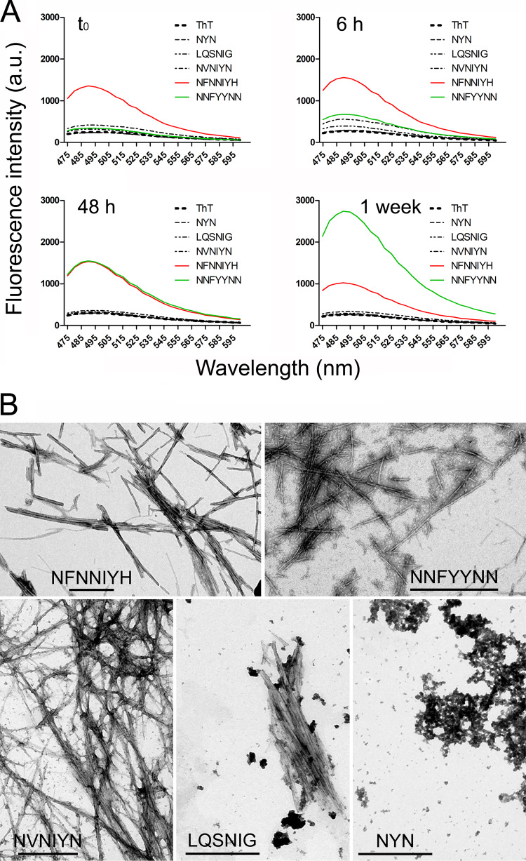
The rapid evolution of resistance in the malaria parasite to every single drug developed against it calls for the urgent identification of new molecular targets. Using a stain specific for the detection of intracellular amyloid deposits in live cells, we have detected the presence of abundant protein aggregates in Plasmodium falciparum blood stages and female gametes cultured in vitro, in the blood stages of mice infected by Plasmodium yoelii, and in the mosquito stages of the murine malaria species Plasmodium berghei Aggregated proteins could not be detected in early rings, the parasite form that starts the intraerythrocytic cycle. A proteomics approach was used to pinpoint actual aggregating polypeptides in functional P. falciparum blood stages, which resulted in the identification of 369 proteins, with roles particularly enriched in nuclear import-related processes. Five aggregation-prone short peptides selected from this protein pool exhibited different aggregation propensity according to Thioflavin-T fluorescence measurements, and were observed to form amorphous aggregates and amyloid fibrils in transmission electron microscope images. The results presented suggest that generalized protein aggregation might have a functional role in malaria parasites. Future antimalarial strategies based on the upsetting of the pathogen's proteostasis and therefore affecting multiple gene products could represent the entry to new therapeutic approaches.

Keywords: malaria; protein aggregation.


Figures

FIG 1
FIG 1
ProteoStat staining for the detection of intracellular protein aggregates in live in vitro cultures of P. falciparum blood stages and female gametes. (A) RBC-invading merozoite. (B) Egressed merozoites. (C and D) Intraerythrocytic blood stages: schizont (asterisk), trophozoites (arrows), and rings (arrowheads). The black arrows in phase contrast images indicate the boundary of two RBCs infected by trophozoite stages, to highlight the lack of fluorescence in the host RBC cytosol. (E to G) Gametocytes. (H to J) Egressed female gametes (arrowheads), which lack the RBC membrane otherwise stained by Oregon Green 488.
FIG 2
FIG 2
ProteoStat staining for the detection of intracellular protein aggregates in live P. berghei stages in Anopheles gambiae mosquitoes. (A) Male gametes. (B and C) Ookinetes. (D) Oocysts and sporozoites. The arrowhead indicates a stream of sporozoites leaving the oocyst; a blow-up of this region is shown in the inset of the protein aggregates panel (inset scale bar, 5 μm).
FIG 3
FIG 3
Flow cytometry analysis of ProteoStat-stained desynchronized P. falciparum cultures. The fraction of ProteoStat-positive RBCs and pRBCs is indicated (%), the latter consisting of late ring/early trophozoites and schizonts, the three stages represented in the cartoons.
FIG 4
FIG 4
Flow cytometry sorting of ProteoStat-stained proteins in live P. falciparum blood stages. (A) Scheme of the process. (B) Histograms showing the intensity of ProteoStat signal versus the number of events, for the sample before sorting (left panel) and the resulting ProteoStat+ and ProteoStat fractions. (C) Dot plot showing the intensity of the ProteoStat signal versus the size of each event, for the sample before sorting (left panel) and the resulting ProteoStat+ and ProteoStat fractions. (D) To monitor ProteoStat fluorescence, pictures at ×600 magnification were taken in the bright field and fluorescence channel BP596-660 upon excitation with a 488-nm laser. (E) Silver-stained SDS-PAGE fractionation of the ProteoStat+ sample. (F) Schematic graph representing the LC-MS/MS analysis of Coomassie blue-stained bands excised from a gel run in parallel to that of panel E. The results obtained are reported in Table S1 in the supplemental material.
FIG 5
FIG 5
Isolation of P. falciparum aggregative proteins insoluble in 0.1% SDS. (A) Scheme of the process. (B) Silver-stained SDS-PAGE fractionation of the 0.1% SDS-resistant sample. (C) Schematic graph representing the LC-MS/MS analysis of Coomassie blue-stained material not entering the stacking gel, excised from a gel run in parallel to that of panel B. The results obtained are reported in Table S3.
FIG 6
FIG 6
Analysis of P. falciparum aggregative proteins insoluble in 0.1% SDS. (A) Venn diagram (http://bioinformatics.psb.ugent.be/webtools/Venn/) showing the intersection between the proteins from the P. falciparum proteome containing a prion-forming domain identified by the PLAAC algorithm and the proteins identified in 0.1% SDS-resistant aggregates and in ProteoStat-stained aggregates sorted by flow cytometry. (B to D) Gene ontology analysis of the P. falciparum proteins identified in 0.1% SDS-resistant aggregates classified according to: biological process (B), molecular function (C), and cellular component (D).
FIG 7
FIG 7
Peptide characterization. (A) ThT fluorescence analysis of the peptides after different incubation times. (B) Transmission electron microscopy images of the peptides. Scale bars, 500 nm.

References

    1. World Health Organization. 2019. World malaria report, 2019. World Health Organization, Geneva, Switzerland.
    1. Mbengue A, Bhattacharjee S, Pandharkar T, Liu H, Estiu G, Stahelin RV, Rizk SS, Njimoh DL, Ryan Y, Chotivanich K, Nguon C, Ghorbal M, Lopez-Rubio JJ, Pfrender M, Emrich S, Mohandas N, Dondorp AM, Wiest O, Haldar K. 2015. A molecular mechanism of artemisinin resistance in Plasmodium falciparum malaria. Nature 520:683–687. doi:10.1038/nature14412. - DOI - PMC - PubMed
    1. Alonso PL, Tanner M. 2013. Public health challenges and prospects for malaria control and elimination. Nat Med 19:150–155. doi:10.1038/nm.3077. - DOI - PubMed
    1. Prudêncio M, Rodriguez A, Mota MM. 2006. The silent path to thousands of merozoites: the Plasmodium liver stage. Nat Rev Microbiol 4:849–856. doi:10.1038/nrmicro1529. - DOI - PubMed
    1. Cowman AF, Crabb BS. 2006. Invasion of red blood cells by malaria parasites. Cell 124:755–766. doi:10.1016/j.cell.2006.02.006. - DOI - PubMed

Publication types