. 2020 Apr 21;117(16):9022-9031.
doi: 10.1073/pnas.1914353117. Epub 2020 Apr 13.

The T1D-associated lncRNA Lnc13 modulates human pancreatic β cell inflammation by allele-specific stabilization of STAT1 mRNA

Affiliations

The T1D-associated lncRNA Lnc13 modulates human pancreatic β cell inflammation by allele-specific stabilization of STAT1 mRNA

Itziar Gonzalez-Moro et al. Proc Natl Acad Sci U S A. .

Abstract

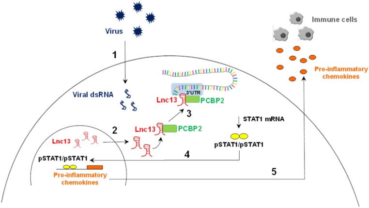
The vast majority of type 1 diabetes (T1D) genetic association signals lie in noncoding regions of the human genome. Many have been predicted to affect the expression and secondary structure of long noncoding RNAs (lncRNAs), but the contribution of these lncRNAs to the pathogenesis of T1D remains to be clarified. Here, we performed a complete functional characterization of a lncRNA that harbors a single nucleotide polymorphism (SNP) associated with T1D, namely, Lnc13 Human pancreatic islets harboring the T1D-associated SNP risk genotype in Lnc13 (rs917997*CC) showed higher STAT1 expression than islets harboring the heterozygous genotype (rs917997*CT). Up-regulation of Lnc13 in pancreatic β-cells increased activation of the proinflammatory STAT1 pathway, which correlated with increased production of chemokines in an allele-specific manner. In a mirror image, Lnc13 gene disruption in β-cells partially counteracts polyinosinic-polycytidylic acid (PIC)-induced STAT1 and proinflammatory chemokine expression. Furthermore, we observed that PIC, a viral mimetic, induces Lnc13 translocation from the nucleus to the cytoplasm promoting the interaction of STAT1 mRNA with (poly[rC] binding protein 2) (PCBP2). Interestingly, Lnc13-PCBP2 interaction regulates the stability of the STAT1 mRNA, sustaining inflammation in β-cells in an allele-specific manner. Our results show that the T1D-associated Lnc13 may contribute to the pathogenesis of T1D by increasing pancreatic β-cell inflammation. These findings provide information on the molecular mechanisms by which disease-associated SNPs in lncRNAs influence disease pathogenesis and open the door to the development of diagnostic and therapeutic approaches based on lncRNA targeting.

Keywords: inflammation; lncRNA; pancreatic β-cell; polymorphism; type 1 diabetes.


Conflict of interest statement

The authors declare no competing interest.

Figures

Fig. 1.
Fig. 1.
Lnc13 is expressed in pancreatic β-cells, up-regulated by viral infections, and correlated with STAT1 expression in human pancreatic islets. (A) Lnc13 expression was analyzed in the human β-cell line EndoC-βH1, in human pancreatic islets, and in a set of human tissues (heart, liver, muscle, brain, spleen, kidney, intestine, colon, thymus, lung, and stomach). Lnc13 expression was determined by Q-PCR and normalized by the housekeeping gene β-actin. Results are means ± SEM of three experimental replicates. (B) Human EndoC-βH1 cells were left untransfected (—white bars) or transfected with PIC (1 µg/mL; gray bars) for 24 h. Expression of Lnc13, STAT2, and IRF7 (positive controls), INS and PAX6 (negative controls) was assayed by Q-PCR and normalized by the housekeeping gene β-actin. Results are means ± SEM of four independent experiments; ***P < 0.001 vs. untreated cells; Student’s t test. (C) Human EndoC-βH1 cells were left uninfected (—) or infected with the CVB5 (CVB5; MOI 5) for 24 h. Lnc13 expression was determined by Q-PCR and normalized by the housekeeping gene β-actin. Results are means ± SEM of five independent experiments; **P < 0.01; Student’s t test. (D) EndoC-βH1s were kept untreated (i.e., 0 h) or treated with PIC for 8 or 24 h. Relative Lnc13 and STAT1 expressions were determined by Q-PCR and normalized by the housekeeping gene β-actin. Results are means ± SEM of six independent experiments; ***P < 0.001 vs. time 0 h; Student’s t test. (E) Human EndoC-βH1 cells were left uninfected (—) or infected with the CVB5 (CVB5) for 24 h. STAT1 expression was determined by Q-PCR and normalized by the housekeeping gene β-actin. Results are means ± SEM of five independent experiments. **P < 0.01; Student’s t test. (F) Lnc13 and STAT1 expressions were determined in 43 human pancreatic islets of nondiabetic individuals. Expression values were normalized by the housekeeping gene β-actin. Spearman’s correlation analysis was performed to check for correlation between Lnc13 and STAT1 expression values; Spearman’s R = 0.51 (0.24–0.0.71); P < 0.001.
Fig. 2.
Fig. 2.
The T1D-associated SNP genotype in the Lnc13 gene correlates with STAT1 expression in human pancreatic islets, and overexpression of Lnc13 in β-cells leads to an up-regulation of STAT1 mRNA expression in an allele-specific manner. (A) Expression of STAT1 in human pancreatic islets stratified by the genotype of the T1D-associated SNP rs917997 in the Lnc13 gene. Results are means ± SEM of 15 samples with the homozygous risk genotype (CC), 26 samples with the heterozygous genotype (CT), and two samples with the homozygous protective genotype (TT) genotype. **P < 0.01; Student’s t test. (B and C) EndoC-βH1 cells were left untransfected (NT) or transfected with pCMV6, pLnc13-C, or pLnc13-T. After 48 h, expression levels of Lnc13 (B) and STAT1 (C) were determined by Q-PCR and normalized by the housekeeping gene β-actin. Results are means ± SEM of four independent experiments; ***P < 0.001 and *P < 0.05 vs. pCMV6-transfected cells; †††P < 0.001 vs. pLnc13-C-transfected cells; Student’s t test. (D) STAT1 mRNA expression in pLnc13-C- and pLnc13-T-transfected β-cells corrected by Lnc13 expression values to control for differences in Lnc13 allele stability. Results are means ± SEM of four independent experiments; **P < 0.01; Student’s t test.
Fig. 3.
Fig. 3.
Overexpression of Lnc13 up-regulates proinflammatory chemokine production in an allele-specific manner in pancreatic β-cells through STAT1 signaling activation. (AD) EndoC-βH1 cells were transfected with pLnc13-C or pLnc13-T, and mRNA levels of CXCL10, CXCL9, CCL5, and CXCL1 were determined by Q-PCR and normalized by the housekeeping gene β-actin and corrected by Lnc13 expression values to control for differences in Lnc13 allele stability. Results are means ± SEM of four independent experiments; **P < 0.01 and *P < 0.05 vs. pLnc13-C-transfected cells; Student’s t test. (E and F) EndoC-βH1 cells were transfected with pLnc13-C or pLnc13-T, and CXCL10 (E), and CCL5 (F) protein release to the medium was determined by ELISA in cell supernatants. CXCL10 and CCL5 values (pg/mL) were corrected by Lnc13 expression values to control for differences in Lnc13 allele stability. Results are means ± SEM of six and three independent experiments, respectively. *P < 0.05 vs. pLnc13-C-transfected cells; Student’s t test. (G) Human EndoC-βH1 cells were transfected with pCMV6 or with pLnc13-C and, subsequently, left untreated (NT), treated with intracellular PIC (1 µg/mL) for 24 h (PIC), or treated with PIC and ruxolitinib for 24 h (PIC + Inhib). CXCL10 mRNA expression was measured by Q-PCR and normalized by the housekeeping gene β-actin. The results are means ± SEM of three independent experiments; **P < 0.01 and *P < 0.05 vs. NT and transfected with the same plasmid; †††P < 0.001, ††P < 0.01, and P < 0.05 as indicated; ANOVA followed by Student’s t test followed with Bonferroni correction. (H) EndoC-βH1s were transfected with pCMV6 or pLnc13-C, chromatin was fragmented and precipitated with anti-STAT1 or anti-IgG (as the negative control), and CXCL10 promoter or a control region (Oct4 gene body) was amplified by Q-PCR. Results are means ± SEM of four independent experiments; *P < 0.05 as indicated; ANOVA followed by Student’s t test followed with Bonferroni correction. (I) Supernatants of pCMV6- or pLnc13-C-transfected EndoC-βH1 cells were used to determine chemotactic migration of Jurkat cells using a transwell system and a fluorescence-based assay. Supernatant of PIC-transfected cells was used as the positive control. The results are means ± SEM of three independent experiments. *P < 0.05 vs. pCMV6-transfected cells; Student’s t test.
Fig. 4.
Fig. 4.
Lnc13 gene disruption in pancreatic β-cells partially counteracts the effect of PIC in STAT1 and proinflammatory chemokine expression up-regulation. (A) Lnc13 disruption was performed by generating a deletion of 1,698 bp using the CRISPR-Cas9 technique and single guide RNAs (sgRNAs) targeting the Lnc13 gene. The presence of the deletion was confirmed by PCR using a pair of primers located inside the deleted region (for detection of unedited cells; wild type forward (WTF) and wild type reverse (WTR)) and a pair of primers located outside the deleted region (for detection of edited cells; mutF and mutR). (B) EndoC-βH1 cells were transfected with an empty px459 vector (CRISPR-Ctrl) or with a vector harboring the Lnc13 targeting sgRNAs (CRISPR-Lnc13). Lnc13 expression was determined by Q-PCR and normalized by the housekeeping gene β-actin. The results are means ± SEM of three independent cell populations. **P < 0.01; Student’s t test. (CE) Control and Lnc13-disrupted mixed cell populations were exposed to intracellular PIC for 24 h, STAT1 (C), CXCL10 (D), and CCL5 (E) expressions were determined by Q-PCR and normalized by the housekeeping gene β-actin. The results are represented as fold induction and are means ± SEM of three independent cell populations. ***P < 0.001, **P < 0.01 and *P < 0.05 vs. nontreated cells; †††P < 0.0001, and P < 0.05 as indicated; Student’s t test.
Fig. 5.
Fig. 5.
Upon exposure to PIC, Lnc13 translocates from the nucleus and facilitates the interaction between the PCBP2 and the 3′-UTR of STAT1 mRNA. (A) EndoC-βH1 cells were exposed to PIC (1 µg/mL) for 24 h, and relative Lnc13 expression was determined in nucleus and whole extracts. Expression of MALAT1 and RPLPO was used as a control for nucleus and whole cell fractions, respectively. Amounts of specific nuclear RNA were measured by Q-PCR and compared to the total amount of RNA in the whole cell. Results are represented as a logarithm (nucleus/whole) and are means ± SEM of three independent experiments; *P < 0.05; Student’s t test. (B) EndoC-βH1s were exposed to PIC (1 µg/mL) for 24 h and cytoplasmic (Cyt), nuclear (Nuc), and chromatin (Chro) fractions were purified. PCBP2 protein expression was determined in all cellular compartments by Western blot and protein expression of Hsp90, HDAC1, and H3 was used as a control for cytoplasmic, nuclear, and chromatin fractions, respectively. The results are representative of three independent experiments. (C) EndoC-βH1 cells were left nontransfected (NT) or transfected with PIC for 24 h. RNA immunoprecipitation was performed using a specific antibody for PCBP2 and Lnc13, and STAT1 expression was determined in PCBP2-bound RNA by Q-PCR. Results are means ± SEM of three independent experiments, and the amounts of Lnc13 and STAT1 are expressed as relative to the input. IgG was used as the negative control; ***P < 0.001 and **P < 0.01 as indicated; ANOVA followed by Student’s t test with Bonferroni correction. (D) In vitro transcribed biotinylated 3′-UTR region of STAT1 was incubated with cellular extracts overexpressing Lnc13-C+PCBP2, Lnc13-T+PCBP2, or PCBP2. Afterward, 3′-UTR-STAT1-bound proteins were purified using streptavidin beads, and PCBP2 and Hsp90 (as the negative control) were detected by Western blot. Incubation with streptavidin beads alone was used as the negative control. The results are representative of three independent experiments. (E) Densitometry results for purified PCBP2 amounts in RNA pull down experiments are represented as means ± SEM of three independent experiments. ANOVA followed by Student’s t test with Bonferroni correction; ***P < 0.001 and *P < 0.01 vs. negative control; †††P < 0.001 and P < 0.05 as indicated. (F) Lnc13 antisense purification was performed in nontreated (NT) and PIC-treated nuclear and cytoplasmic fractions of EndoC-βH1 cells. Lnc13-bound 3′-UTR-STAT1 amounts were determined by Q-PCR using specific primers. A nonrelated similar lncRNA was used as the negative control (Ctrl). Results are represented as fold enrichment and are means ± SEM of four independent experiments. *P < 0.05 as indicated; ANOVA followed by Student’s t test with Bonferroni correction. (G) RNA-protein interaction assay. Cells were transfected with pPCBP2 alone, pPCBP2+pLnc13-C, pPCBP2+pLnc13-T, or pPCBP2+pLnc13-delSNP. Cell lysates were incubated with in vitro transcribed 3′-UTR-STAT1 molecules, and a native agarose gel electrophoretic mobility shift assay was performed (Lower). After electrophoresis, proteins were transferred to a nitrocellulose membrane for PCBP2 visualization (Upper). The 3′-UTR-STAT1 RNA molecule alone was loaded as the Ctrl. The results are representative of three independent experiments. (H) EndoC-βH1 cells were transfected with a plasmid encoding Lnc13-C or a mutant Lnc13 in which the region containing the T1D-associated SNP was deleted (pLnc13-delSNP). STAT1, CXCL10, and CCL5 expressions were determined by Q-PCR and normalized by the housekeeping gene β-actin and corrected by Lnc13 expression values. Results are means ± SEM of four independent experiments. P < 0.05 as indicated; Student’s t test.
Fig. 6.
Fig. 6.
Lnc13 participates in virus-induced pancreatic β-cell inflammation by stabilizing STAT1 mRNA through interaction with the PCBP2 protein. Viral infections in pancreatic β-cells lead to the generation of viral dsRNA that is recognized by viral dsRNA cytoplasmic receptors (1). Viral dsRNA induces a decrease in DCP2 expression that leads to an increase in Lnc13 RNA levels. In addition, viral dsRNA provokes the translocation of Lnc13 from the nucleus to the cytoplasm (2). Once in the cytoplasm, Lnc13 interacts with PCBP2 to bind the 3′UTR of STAT1 mRNA and induce its stabilization (3). Increased STAT1 mRNA levels lead to increased protein and provide the substrate for generation of activated STAT1 (phosphorylated STAT1; pSTAT1). pSTAT1 forms homodimers and translocates to the nucleus, promoting the expression of several proinflammatory chemokines (4). Chemokines are then released by pancreatic β-cells, attracting immune cells into the islets (5). In genetically susceptible individuals harboring the T1D risk genotype in Lnc13 (rs917997*CC), the function of Lnc13 is affected, leading to an excessive antiviral inflammatory response that contributes to β-cell destruction in T1D.

Comment in

  • Role of long non-coding RNA in T1DM.
    Greenhill C. Greenhill C. Nat Rev Endocrinol. 2020 Jul;16(7):344-345. doi: 10.1038/s41574-020-0368-2. Nat Rev Endocrinol. 2020. PMID: 32358540 No abstract available.

References

    1. Eizirik D. L., Colli M. L., Ortis F., The role of inflammation in insulitis and beta-cell loss in type 1 diabetes. Nat. Rev. Endocrinol. 5, 219–226 (2009). - PubMed
    1. Santin I., Eizirik D. L., Candidate genes for type 1 diabetes modulate pancreatic islet inflammation and β-cell apoptosis. Diabetes Obes. Metab. 15 (suppl. 3), 71–81 (2013). - PubMed
    1. Dotta F., et al. , Coxsackie B4 virus infection of beta cells and natural killer cell insulitis in recent-onset type 1 diabetic patients. Proc. Natl. Acad. Sci. U.S.A. 104, 5115–5120 (2007). - PMC - PubMed
    1. Op de Beeck A., Eizirik D. L., Viral infections in type 1 diabetes mellitus–Why the β cells? Nat. Rev. Endocrinol. 12, 263–273 (2016). - PMC - PubMed
    1. Rodriguez-Calvo T., Enterovirus infection and type 1 diabetes: Unraveling the crime scene. Clin. Exp. Immunol. 195, 15–24 (2019). - PMC - PubMed

Publication types

MeSH terms