. 2020 Apr 28;117(17):9621-9629.
doi: 10.1073/pnas.1907799117. Epub 2020 Apr 13.

Chitin perception in plasmodesmata characterizes submembrane immune-signaling specificity in plants

Affiliations

Chitin perception in plasmodesmata characterizes submembrane immune-signaling specificity in plants

Cécilia Cheval et al. Proc Natl Acad Sci U S A. .

Abstract

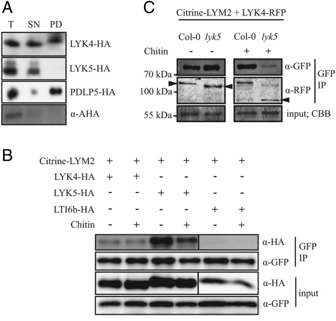
The plasma membrane (PM) is composed of heterogeneous subdomains, characterized by differences in protein and lipid composition. PM receptors can be dynamically sorted into membrane domains to underpin signaling in response to extracellular stimuli. In plants, the plasmodesmal PM is a discrete microdomain that hosts specific receptors and responses. We exploited the independence of this PM domain to investigate how membrane domains can independently integrate a signal that triggers responses across the cell. Focusing on chitin signaling, we found that responses in the plasmodesmal PM require the LysM receptor kinases LYK4 and LYK5 in addition to LYM2. Chitin induces dynamic changes in the localization, association, or mobility of these receptors, but only LYM2 and LYK4 are detected in the plasmodesmal PM. We further uncovered that chitin-induced production of reactive oxygen species and callose depends on specific signaling events that lead to plasmodesmata closure. Our results demonstrate that distinct membrane domains can integrate a common signal with specific machinery that initiates discrete signaling cascades to produce a localized response.

Keywords: immunity; membrane domain; plasmodesmata; receptor signaling.


Conflict of interest statement

The authors declare no competing interest.

Figures

Fig. 1.
Fig. 1.
LYK4 and LYK5 regulate plasmodesmal permeability in response to chitin. (A) Microprojectile bombardment into leaf tissue of 5- to 6‐wk‐old Arabidopsis shows that Col-0 and lyk3 but not lym2-1, lyk4, and lyk5-2 exhibit reduced movement of GFP to neighboring cells in response to chitin. Data were collected from six biological replicates, and the number of cells showing GFP has been normalized to the mean of the mock-treated data within genotypes. These data are summarized in box plots in which the line within the box marks the median, the box signifies the upper and lower quartiles, and the whiskers represent the minimum and maximum within 1.5 × interquartile range. Notches represent approximate 95% confidence intervals. The number of bombardment sites (n) counted is ≥84. Asterisks indicate statistical significance compared with control conditions. ***P < 0.001. (B) Confocal images of aniline blue-stained plasmodesmal callose in leaves of 5- to 6‐week‐old Col-0 plants, as well as lym2-1, lyk4, lyk5-2, and cerk1-2 mutants. Images were acquired 30 min postinfiltration with water or chitin. (Scale bars, 15 µm.) (C) Quantification of plasmodesmata-associated fluorescence of aniline blue stained callose using automated image analysis. Col-0 and the cerk1-2 mutant show an increase in aniline blue stained plasmodesmal callose 30 min post chitin treatment. In lym2-1, lyk4, and lyk5-2, this response is not detected. This correlates with the movement phenotype and identifies that chitin-triggered plasmdesmata closure is caused by callose deposition at plasmodesmata. The fluorescence intensity is summarized in box plots in which the line within the box marks the median, the box signifies the upper and lower quartiles, the minimum and maximum within 1.5 × interquartile range. Number of images (n) is ≥31, and *** indicates P < 0.001 when chitin-treated samples were compared with mock treatments within genotypes.
Fig. 2.
Fig. 2.
LYK4 and LYK5 can associate with LYM2 but only LYK4 is detected in plasmodesmata. (A) Western blot analysis of purified plasmodesmata fractions from N. benthamiana tissue expressing LYK4-HA, LYK5-HA, or PDLP5-HA. Total (T) (extracts from ground tissue), supernatant (SN) (all cellular material excluding cell walls), and plasmodesmatal (PD) (membranes released from purified cell walls) extracts were separated by SDS/PAGE and probed with anti-HA to determine the presence of LYK4-HA, LYK5-HA, and PDLP5-HA in each fraction or with anti–H+-ATPase (AHA) to detect PD-excluded H+-ATPases. (B) Western blot analysis of immunoprecipitated proteins from N. benthamiana tissue expressing Citrine-LYM2 and LYK4-HA, LYK5-HA, or LTI6b-HA. LYK4-HA and LYK5-HA are detected in detergent-extracted fractions by IP of Citrine-LYM2. Input and immunoprecipitated (IP) samples were probed α-GFP and α-HA antibodies as indicated. CBB, Coomassie brilliant blue. (C) Western blot analysis of immunoprecipitated protein extracts from Arabidopsis protoplasts expressing Citrine-LYM2 and LYK4-RFP. LYK4-RFP is detected in samples from both Col-0 and lyk5-2 protoplasts. Input and immunoprecipitated (IP) samples were probed with anti-GFP and anti-RFP antibodies as indicated. LYK4-RFP bands of different sizes are indicated by arrowheads; size markers are indicated to the left. (AC) Experiments were repeated three times with similar results.
Fig. 3.
Fig. 3.
LYK5 dynamically associates with LYK4 in the PM. (A) Western blot analysis of immunoprecipitated (IP) protein extracts from N. benthamiana tissue expressing LYK4-GFP and LYK5-HA or LTI6b-HA. LYK4-GFP was immunoprecipitated from detergent-extracted fractions and probed with anti-GFP and anti-HA to detect LYK4-GFP and LYK5-HA or LTI6b-HA, respectively. Experiments were repeated three times with similar results. (B) FRET-FLIM analysis of LYK4-GFP in the presence of acceptors LYK5-RFP or BRI1-RFP and the presence and absence of chitin. Fluorescence lifetime was measured in N. benthamiana tissue transiently coexpressing the indicated constructs as donors or acceptors. Box plots represent GFP fluorescence-weighted average lifetime (τAv, ns): the line within the box marks the median, the box signifies the upper and lower quartiles, the whiskers represent the minimum and maximum within 1.5 × interquartile range. Data were analyzed by ANOVA with a post hoc Tukey multiple comparison of means (P value < 0.01). Samples with the same letter code are not significantly different. Number of images (n) analyzed is ≥19. (C) FRET-FLIM analysis of LYK4-GFP at the plasmodesmal PM and the PM in the presence and absence of LYK5-RFP in N. benthamiana tissue. Plasmodesmata were marked by coexpression of Citrine-LYM2 and ROI were defined around plasmodesmata (PD) and in the PM for analysis. Box-plots represent GFP fluorescence-weighted average lifetime (τAv, ns): the line within the box marks the median, the box signifies the upper and lower quartiles, and the whiskers represent the minimum and maximum within 1.5 × interquartile range. Asterisks indicate statistical significance compared with control conditions (***P < 0.001). Number of ROIs (n) analyzed is ≥27. (D) % mobile fraction of LYM2, LYK4 and LYK5 as measured by FRAP assays. For Citrine-LYM2 FRAP measurements were taken for the plasmodesmata-located (PD) and PM-located pools of receptor. For LYK4-RFP and LYK5-RFP, FRAP measurements were taken in the PM. Error bars are SE. *P < 0.05; **P < 0.001. Number of FRAP experiments analyzed is ≥43.
Fig. 4.
Fig. 4.
LYM2 accumulates at PD in response to chitin. (A) Single-plane confocal images of N. benthamiana tissue expressing Citrine-LYM2 before and after chitin treatment. Left shows Citrine-LYM2 in water-treated tissue, and Right shows Citrine-LYM2 30 min post chitin treatment. Arrowheads indicate example plasmodesmata. (Scale bars, 10 μm.) (B) Quantification of the PD index of Citrine-LYM2 in N. benthamiana after mock and chitin treatments. Number of images analyzed is ≥18. (C) Fluorescence anisotropy (r) of cytosolic GFP, PM located Citrine-LYM2, and plasmodesmata-located (PD) Citrine-LYM2. Number of images analyzed is ≥11. (B and C) Box plots: the line within the box marks the median, the box signifies the upper and lower quartiles, and the whiskers represent the minimum and maximum within 1.5 × interquartile range. Samples with the same letter code (a, b, or c) are not significantly different (P < 0.001).
Fig. 5.
Fig. 5.
CPK-dependent phosphorylation of RBOHD is required for plasmodesmata closure in response to chitin. (A) Microprojectile bombardment into leaf tissue shows that rbohd mutants do not show a reduction in GFP movement to neighboring cells in response to chitin. RBOHD mutant variants RBOHDS39A/S339A/S343A (S39A/S339A/S343A) and RBOHDS163A (S163A) exhibit a reduction in movement of GFP to neighboring cells in response to chitin while the RBOHD phosphosite mutant variants RBOHDS343A/S347A (S343A/S347A) and RBOHDS133A (S133A) do not. (B) ROS produced by seedlings treated with chitin. Col-0, RBOHDS133A, RBOHDS163A, cpk6-1 and cpk11-2 seedlings all produce significantly more ROS than the rbohd mutant. Different letters (a, b, and c) indicate statistically significant groups when the chitin-triggered ROS are compared between genotypes (P < 0.05, analysis excluding cerk1-2); *** indicates significantly more ROS produced (P < 0.001) when chitin treatment is compared with water controls within genoptypes (all genotypes). Number of seedlings measured is ≥19. (C) Microprojectile bombardment into leaf tissue shows that cpk6-1 and cpk11-2 mutants do not show a reduction in GFP movement to neighboring cells in response to chitin. cpk5 mutants show constitutively reduced movement that increases upon chitin treatment and bik1 mutants behave like Col-0. (A and C) The number of cells showing GFP has been normalized to the mean of the mock data within genotypes. Box plots: the line within the box marks the median, the box signifies the upper and lower quartiles, and the minimum and maximum within 1.5 × interquartile range. Notches represent approximate 95% CIs. ***P < 0.001 (number of bombardment sites counted ≥ 89).
Fig. 6.
Fig. 6.
Possible mechanisms for LYM2-mediated chitin signaling in the plasmodesmal PM. This cartoon illustrates two possibilities for some key elements of LysM protein chitin signaling in the PM and plasmodesmal PM. Top represents the relevant associations and localizations we have identified under mock conditions (absence of fungus). Here, LYK5 (green) interacts with LYK4 (light blue), and LYM2 (orange) in the PM and LYK5 mediates modification of a pool of LYK4. This could occur via a population of bipartite complexes (A) or a tripartite LYM2-LYK4-LYK5 complex (B) in the PM. CPK5 negatively regulates callose synthesis in the plasmodesmal PM via a specific phosphorylation pattern (P) (white) of RBOHD. In response to chitin (Lower, presence of fungus), a pool of LYK4 and LYM2 dissociate from LYK5. LYK5 associates with CERK1 (dark blue) (A) or both CERK1 and LYK4 (B) to mediate signaling at the PM, and LYM2 accumulates at plasmodesmata, where it forms a higher-order complex or a signaling platform. This complex recruits LYK4 and CPK6 and -11 (brown) to phosphorylate (P) (white) RBOHD (yellow) at Ser133 and Ser347 and induces callose (blue) synthesis via a glucan synthase-like enzyme (GSL) (purple) to close PD. The PM LYK5-containing complex signals, in part, via RLCKs that phosphorylate (P) (white) RBOHD (yellow) at Ser39, Ser339, and Ser343 (P) (white). While not represented here, RLCKs might constitutively associate with LysM receptor complexes in the PM as for LRR-RKs (47).

Similar articles

Cited by

References

    1. Ott T., Membrane nanodomains and microdomains in plant-microbe interactions. Curr. Opin. Plant Biol. 40, 82–88 (2017). - PubMed
    1. Gronnier J., Gerbeau-Pissot P., Germain V., Mongrand S., Simon-Plas F., Divide and rule: Plant plasma membrane organization. Trends Plant Sci. 23, 899–917 (2018). - PubMed
    1. Lingwood D., Simons K., Lipid rafts as a membrane-organizing principle. Science 327, 46–50 (2010). - PubMed
    1. Płóciennikowska A., Hromada-Judycka A., Borzęcka K., Kwiatkowska K., Co-operation of TLR4 and raft proteins in LPS-induced pro-inflammatory signaling. Cell. Mol. Life Sci. 72, 557–581 (2015). - PMC - PubMed
    1. Jacobs S. R., Damania B., NLRs, inflammasomes, and viral infection. J. Leukoc. Biol. 92, 469–477 (2012). - PMC - PubMed

Publication types

MeSH terms

LinkOut - more resources