. 2020 Jan 10;21(2):442.
doi: 10.3390/ijms21020442.

Cell-Penetrating Peptides Enhance the Activity of Human Fibroblast Growth Factor 2 by Prolonging the Retention Time: A New Vision for Drug-Delivery Systems

Affiliations

Cell-Penetrating Peptides Enhance the Activity of Human Fibroblast Growth Factor 2 by Prolonging the Retention Time: A New Vision for Drug-Delivery Systems

Jaehoon Lee et al. Int J Mol Sci. .

Abstract

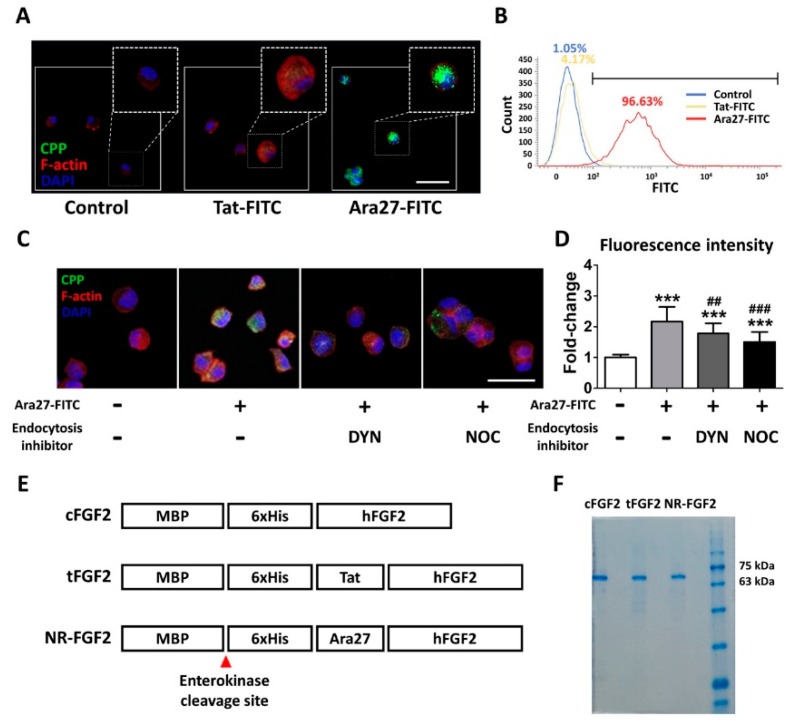
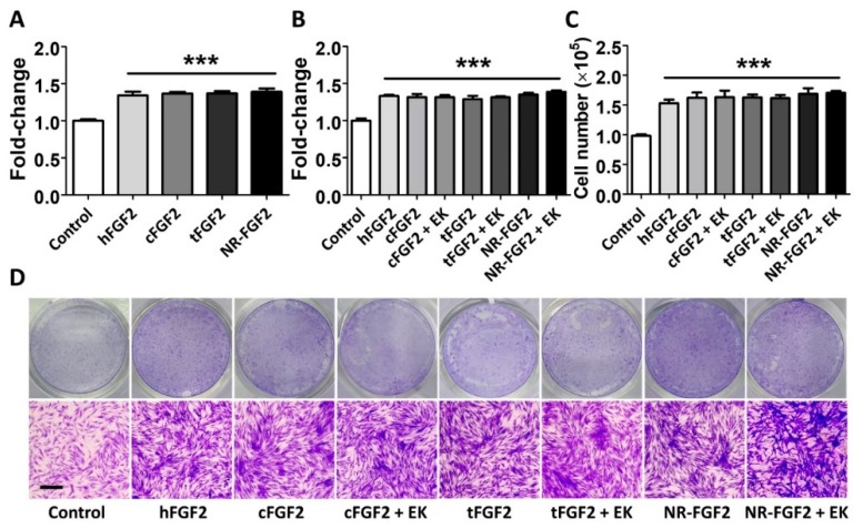
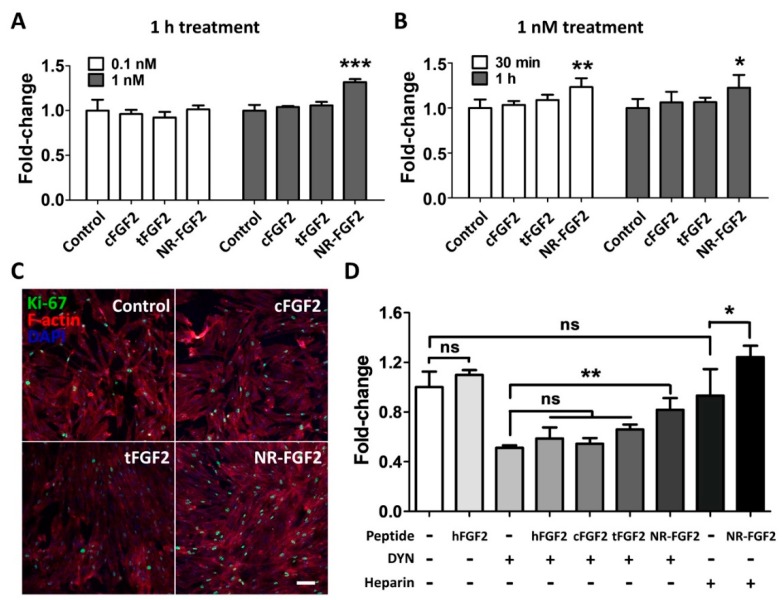
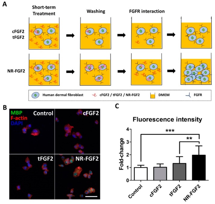
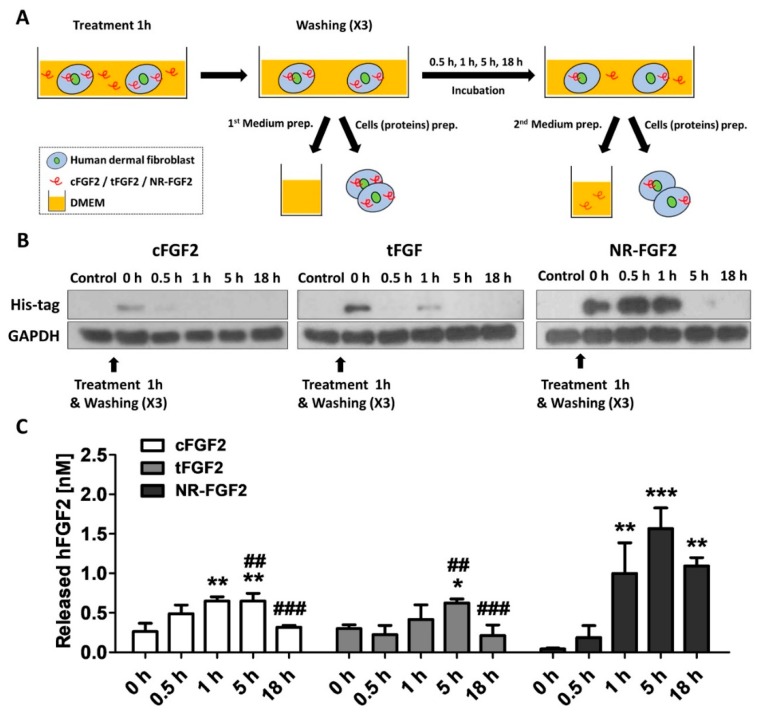
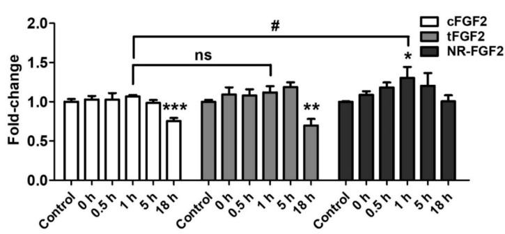
Cell-penetrating peptides (CPPs) are defined by their ability to deliver cargo into cells and have been studied and developed as a promising drug-delivery system (DDS). However, the issue of whether the CPPs that have already entered the cells can be re-released or reused has not been studied. The purpose of this research was to construct CPP-conjugated human fibroblast growth factor 2 (hFGF2) and investigate whether they can be re-released from the cell membrane for reuse. This study combined hFGF2 with Tat or Ara27, a newly developed CPP derived from the zinc knuckle (CCHC-type) family protein of Arabidopsis. Human dermal fibroblast (HDF) was treated with Tat-conjugated hFGF2 (tFGF2) and Ara27-conjugated hFGF2 (NR-FGF2) for both long and short durations, and the effects on cell growth were compared. Furthermore, tFGF2 and NR-FGF2 re-released from the cells were quantified and the effects were evaluated by culturing HDF in a conditioned medium. Interestingly, the proliferation of HDF increased only when NR-FGF2 was treated for 1 h in endocytosis-independent manner. After 1 h, NR-FGF2 was significantly re-released, reaching a maximum concentration at 5 h. Furthermore, increased proliferation of HDF cultured in the conditioned medium containing re-released NR-FGF2 was discovered. While previous studies have focused on the delivery of cargo and its associated applications, this study has revealed that combinations of superior CPPs and therapeutics can be expected to prolong both the retention time and the cell-penetrating capacity, even in the presence of external factors. Therefore, CPPs can be applied in the context of topical drugs and cosmetics as a new DDS approach.

Keywords: Ara27; cell-penetrating peptides; drug-delivery system; fibroblast growth factor 2; re-release.


Conflict of interest statement

The authors declare no conflicts of interest. The founding sponsors had no role in the design of the study; in the collection, analysis, or interpretation of the data; in the writing of the manuscript; or in the decision to publish the results. NeoRegen Biotech Co., Ltd. supported the salary for J.S., S.P., and J.L.

Figures

Figure 1
Figure 1
A newly developed cell-penetrating peptide (CPP), Ara27, and the purification of CPP-conjugated human fibroblast growth factor 2 (hFGF2). (A) Human dermal fibroblast (HDF) was treated with Tat-fluorescein isothiocyanate (FITC) and Ara27-FITC at 1 μM for 1 h; consequently, more Ara27-FITC than Tat-FITC enters the cells (scale bar = 50 μm, white). (B) The cell-penetrating ability of Ara27-FITC was also validated by fluorescence activated cell sorter. (C,D) The mechanism of the cell-penetrating ability of Ara27 was confirmed by dynasore and nocodazole (n > 80) (E) Control hFGF2 (cFGF2), Tat-conjugated hFGF2 (tFGF2), and Ara27-conjugated hFGF2 (NR-FGF2) were composed of maltose binding protein (MBP), his-tag for purification, CPPs, and functional hFGF2. (F) Fusion proteins were purified via Ni-NTA affinity chromatography and were analyzed by sodium dodecyl sulfate-polyacrylamide gel electrophoresis (SDS-PAGE). The results are the means of at least three independent experiments (mean + SD). *** p<0.001 versus the control group and ## p<0.01 and ### p<0.001 versus the Ara27-FITC-treated group.
Figure 2
Figure 2
Effects of CPPs or MBP of fusion proteins on hFGF2 activity during long-term culture. HDF was cultured with normal hFGF2 and CPP-conjugated hFGF2 for five days and analyzed using a WST-1 cell viability assay and Crystal Violet staining. (A) tFGF2 and NR-FGF2 did not show significant differences to hFGF2. (B) The presence of MBP did not affect the hFGF2 activity. (C,D) This effect was also confirmed by counting the cell number and Crystal Violet staining (scale bar = 100 μm, black). The results are the means of at least three independent experiments (mean + SD). *** p<0.001 versus the control group.
Figure 3
Figure 3
Effects of short-term treatment of NR-FGF2 on HDF. HDF was treated with cFGF2, tFGF2, and NR-FGF2 at 0.1 nM or 1 nM for 1 h and maintained for five days. (A) The proliferation of HDF significantly increased only when NR-FGF2 was treated at a concentration of 1 nM. (B) When CPP-conjugated hFGF2 was treated for 30 min, cell proliferation significantly increased only in HDF pretreated with NR-FGF2. (C) The expression of Ki-67 in the nucleus increased when NR-FGF2 was treated for a short time (scale bar = 100 μm, white). (D) Despite inhibiting the endocytosis pathway, the short-term treatment ability of NR-FGF2 was maintained. The results are the means of at least three independent experiments (mean + SD). * p<0.05, ** p<0.01, and *** p<0.001 versus the control group. ns, not significant.
Figure 4
Figure 4
Cell-penetrating ability of NR-FGF2. (A) The enhanced cell-penetrating ability of Ara27 increased the residence time of hFGF2 and, second, that this could continue to affect cells (B,C) As a result of the short-term treatment of CPP-conjugated hFGF2, NR-FGF2 sufficiently penetrated the cells within 1 h (n > 80; scale bar = 500 μm, white). The results are the means of at least three independent experiments (mean + SD). ** p<0.01 and *** p<0.001 versus the control group.
Figure 5
Figure 5
Re-release of NR-FGF2 that had penetrated HDF. (A) Experimental schematic diagram: (i) CPP-conjugated hFGF2 was treated in HDF for 1 h; (ii) after washing, the medium and cell lysate were harvested for presenting the initial (0 h) amount of hFGF2; (iii) the medium and cell lysate were obtained by culture time (0.5, 1, 5, and 18 h); and (iv) hFGF2 in the medium or cell lysate was analyzed by ELISA or western blot method, respectively. (B) NR-FGF2 remained in the cells for up to 5 h, whereas cFGF2 and tFGF2 were absent after 1 h. (C) At 1 h, NR-FGF2 was significantly re-released as compared with cFGF2 and tFGF2, and the concentration was maximized at 5 h. The results are the means of at least three independent experiments (mean + SD). * p<0.05, ** p<0.01, and *** p<0.001 versus the control group and ## p<0.01 and ### p<0.001 versus the NR-FGF2-treated group.
Figure 6
Figure 6
Mechanism of short-term treatment effects of NR-FGF2. (A) CPP-conjugated hFGF2 existed for at least 72 h under experimental conditions. (B) It was confirmed that the short-term treatment ability of NR-FGF2 was not due to its binding to the cell membrane by heparin washing. (C) The inhibition of endocytosis pathways did not affect cell penetration of NR-FGF2. (D) Re-released NR-FGF2 was increased over time and the maximum concentration was achieved at 5 h. The results are the means of at least three independent experiments (mean + SD). *** p<0.001 versus the control group.
Figure 7
Figure 7
Effects of re-released NR-FGF2. CPP-conjugated hFGF2 was pretreated at 1 nM for 1 h, and HDF was cultured in the conditioned medium obtained at a specific time for five days. The results showed that only the cell growth of HDF cultured in the conditioned medium obtained 1 h after the pretreatment of NR-FGF2 was significantly increased. The results are the means of at least three independent experiments (mean + SD). * p<0.05, ** p<0.01, and *** p<0.001 versus the control group and # p<0.05 versus the NR-FGF2-treated group. ns, not significant.

Similar articles

Cited by

References

    1. Komin A., Russell L., Hristova K.A., Searson P. Peptide-based strategies for enhanced cell uptake, transcellular transport, and circulation: Mechanisms and challenges. Adv. Drug Deliv. Rev. 2017;110:52–64. doi: 10.1016/j.addr.2016.06.002. - DOI - PubMed
    1. Su Y., Xie Z., Kim G.B., Dong C., Yang J. Design strategies and applications of circulating cell-mediated drug delivery systems. ACS Biomater. Sci. Eng. 2015;1:201–217. doi: 10.1021/ab500179h. - DOI - PMC - PubMed
    1. Yoo J.H., Shanmugam S., Thapa P., Lee E.-S., Balakrishnan P., Baskaran R., Yoon S.-K., Choi H.-G., Yong C.S., Yoo B.K. Novel self-nanoemulsifying drug delivery system for enhanced solubility and dissolution of lutein. Arch. Pharm. Res. 2010;33:417–426. doi: 10.1007/s12272-010-0311-5. - DOI - PubMed
    1. Veronese F.M., Pasut G. PEGylation, successful approach to drug delivery. Drug Discov. Today. 2005;10:1451–1458. doi: 10.1016/S1359-6446(05)03575-0. - DOI - PubMed
    1. Hara T., Iriyama S., Makino K., Terada H., Ohya M. Mathematical description of drug movement into tumor with EPR effect and estimation of its configuration for DDS. Colloids Surf. B Biointerfaces. 2010;75:42–46. doi: 10.1016/j.colsurfb.2009.08.013. - DOI - PubMed

MeSH terms

Substances

LinkOut - more resources