. 2020 May;6(5):522-532.
doi: 10.1038/s41477-020-0633-3. Epub 2020 Apr 13.

An RNA thermoswitch regulates daytime growth in Arabidopsis

Affiliations

An RNA thermoswitch regulates daytime growth in Arabidopsis

Betty Y W Chung et al. Nat Plants. 2020 May.

Abstract

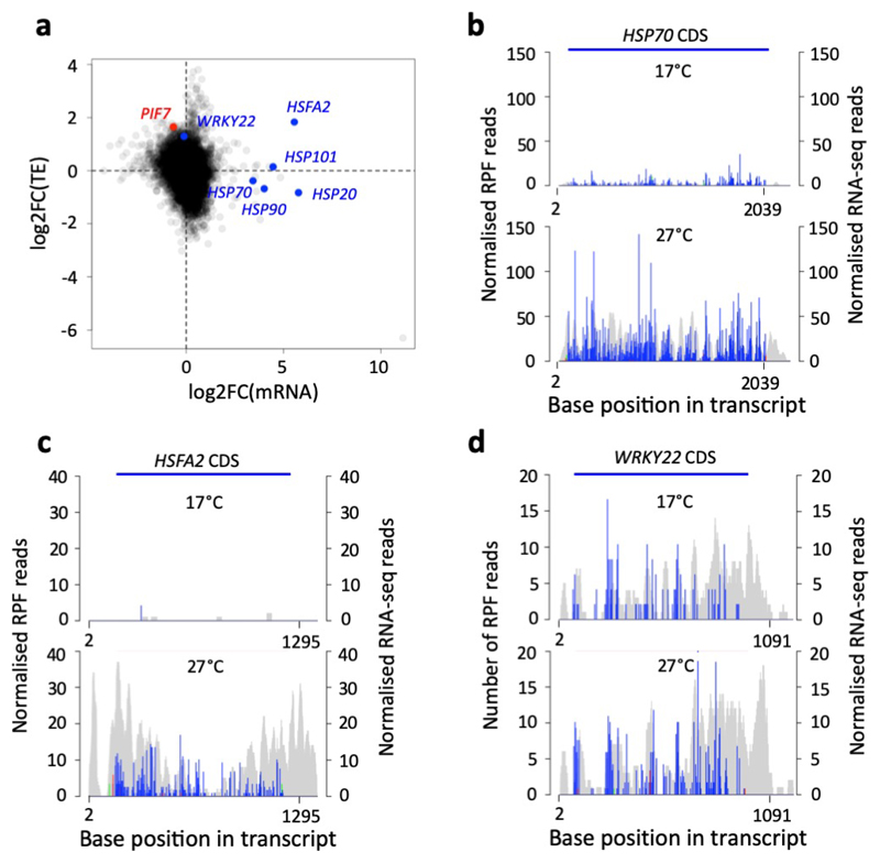
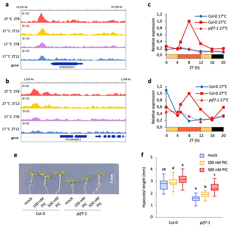
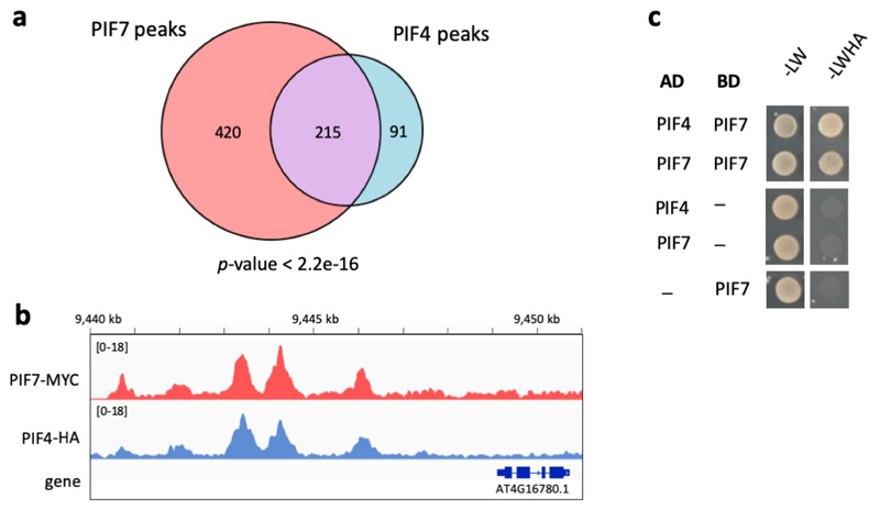
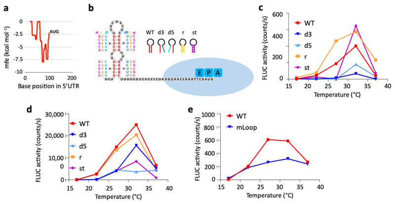
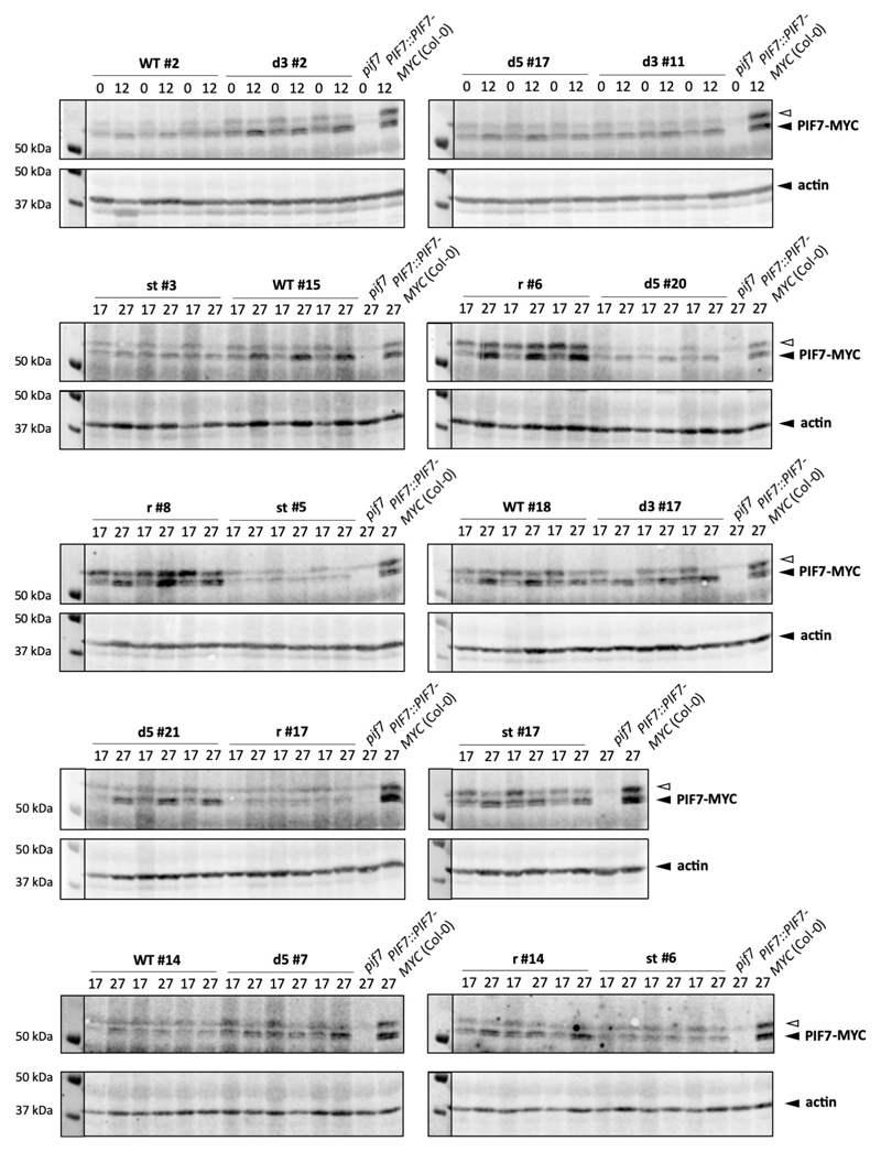
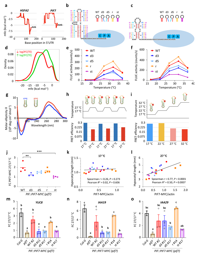
Temperature is a major environmental cue affecting plant growth and development. Plants often experience higher temperatures in the context of a 24 h day-night cycle, with temperatures peaking in the middle of the day. Here, we find that the transcript encoding the bHLH transcription factor PIF7 undergoes a direct increase in translation in response to warmer temperature. Diurnal expression of PIF7 transcript gates this response, allowing PIF7 protein to quickly accumulate in response to warm daytime temperature. Enhanced PIF7 protein levels directly activate the thermomorphogenesis pathway by inducing the transcription of key genes such as the auxin biosynthetic gene YUCCA8, and are necessary for thermomorphogenesis to occur under warm cycling daytime temperatures. The temperature-dependent translational enhancement of PIF7 messenger RNA is mediated by the formation of an RNA hairpin within its 5' untranslated region, which adopts an alternative conformation at higher temperature, leading to increased protein synthesis. We identified similar hairpin sequences that control translation in additional transcripts including WRKY22 and the key heat shock regulator HSFA2, suggesting that this is a conserved mechanism enabling plants to respond and adapt rapidly to high temperatures.


Conflict of interest statement

Conflict of interest:

The authors declared that they have no conflict of interest.

Figures

Extended Data Fig. 1
Extended Data Fig. 1. PIF7, HSFA2 and WRKY22 show enhanced translation at warm temperature.
a Scatter plot of log fold changes in TE and mRNA abundance observed in Ribo-seq with parallel RNA-seq. b-d Histograms of 5′ end positions of normalized 28-nucleotide RPF reads (blue, green and red for frames 0, 1 and 2, left axis) and RNA-Seq reads (grey, right axis) mapped to the HSP70 (b), HSFA2 (c) and WRKY22 (d) transcript.
Extended Data Fig. 2
Extended Data Fig. 2. PIF7-MYC protein accumulation in response to warm temperature
a-d Western blots of PIF7-MYC protein used for the quantification shown in Fig. 1m. The PIF7::PIF7-MYC line is in the Col-0 background. Actin levels are shown as loading control. e, f Western blot (e) and quantification (f) of PIF7-MYC protein of an independent transgenic PIF7:: PIF7-MYC line in Col-0 background at ZT0 (dawn) and ZT12; seedlings were grown in LD at constant 17°C or with a 27 °C midday for 7 d. Protein levels were normalised to actin. Bars represent the mean, error bars indicate the SEM (n = 3). The experiment was repeated once with similar results. g-i Western blots (g) and quantification (h) of PIF7-MYC protein as well as PIF7-MYC transcript levels (i) of PIF7::PIF7-MYC (Col-0) seedlings grown at constant 17 °C for 7 d and then either shifted to 27 °C at ZT4 (= 0 h) or kept at 17 °C for the indicated amount of time. Actin levels are shown as loading control. Protein levels were normalised to actin and expressed relative to levels at 0 h, transcript levels were normalised to PP2A and expressed relative to levels at 0 h. Data points represent the mean, error bars indicate the SEM (n = 3). The experiment was repeated once with similar results. j, k Western blot of PIF7-MYC protein at ZT8 and ZT12 of PIF7::PIF7-MYC (Col-0) seedlings grown in LD at constant 17°C (j) or with a 27°C midday (k). Seedlings were treated with 100 μM cyclo-heximide (CHX), 50 μM MG132, a combination of the two or mock-treated at ZT4 on the day of sampling. Actin levels are shown as loading control. Two biological replicates are shown. The experiment was repeated once with similar results. The open arrow indicates an unspecific signal. Asterisks indicate significant differences to 17 °C control treatment (Two-sided Student’s t-test, * p < 0.05, ** p < 0.01, *** p < 0.001).
Extended Data Fig. 3
Extended Data Fig. 3. Additional thermomorphogenesis phenotypes in pif mutants.
a-d Hypocotyl length of 7-d-old Col-0 and pif mutant seedlings grown in LD at constant 17 °C, 22 °C and 27 °C (a) (n = 15), in SD at constant 17 °C, 22 °C and 27 °C (b) (n = 23, except for pif4 17 °C and 22 °C with n = 21 and pif7 27 °C with n = 22), in SD at constant 17 °C or with a daytime temperature at 27 °C (c) (n = 24 except for Col-0 27 °C with n= 19 and pif7 27 °C with n = 22) and in LD at constant 17 °C or with a warm midday of 27 °C (d) (n = 20 except for Col-0 17 °C and pif7 27 °C with n = 22), respectively. Seedlings were grown at 40 μmol m-2 s-1 in LD and 80 μmol m-2 s-1 in SD. e, f Flowering time of Col-0 and pif mutant plants grown in LD at constant 17 °C or with a warm 37 °C midday (n = 12 except for pif4 pif7 with n = 11). Flowering time was scored as leaves at bolting (e) and days to bolting (f). g-i Hypocotyl length (g, h; n = 20-25) and stomatal index (SI) (i; n = 12) of 7-d-old and 14-d-old seedlings of two independent PIF7::PIF7-MYC complementation lines in the pif7-1 background, respectively. Seedlings were grown in LD at 17 °C with a warm midday of 27 °C. Box plots display the 25th and 75th percentile with the median as centre value and whiskers representing 1.5 times the IQR. Letters indicate significance groups; samples with the same letters are not significantly different (2-way ANOVA followed by two-sided Tukey test, p < 0.05). Asterisks indicate samples that are significantly different to Col-0 wild type (One-way ANOVA followed by two-sided Dunnett’s test, * p < 0.05, ** p < 0.01, *** p < 0.001). All experiments were repeated once with similar results.
Extended Data Fig. 4
Extended Data Fig. 4. The pif7 mutant lacks induction of a subset of temperature-responsive genes
a, b Average log fold change between expression at 27 °C and 17 °C for genes differentially expressed in pif7-1 (n = 1007) (a) and genes of cluster 7 identified in Fig. 3b (n = 293) (b). Box plots display the 25th and 75th percentile with the median as centre value and whiskers representing 1.5 times the IQR. Asterisks indicate significant differences (Two-sided Student’s t-test, * p < 0.05, ** p < 0.01, *** p < 0.001).
Extended Data Fig. 5
Extended Data Fig. 5. PIF7 affects auxin biosynthesis.
a, b IGV browser view of PIF7-MYC binding at the YUC8 (a) and YUC9 (b) promoters. c, d Relative expression of YUC8 (c) and YUC9 (d) observed in the RNA-seq experiment displayed in Fig. 3. Data are expressed relative to Col-0 27 °C at ZT8. e, f Hypocotyl length of 7-d-old Col-0 or pif7-1 seedlings treated with 100 or 500 nM picloram (PIC) or mock-treated (n = 24). Box plots display the 25th and 75th percentile with the median as centre value and whiskers representing 1.5 times the IQR. Letters indicate significance groups; samples with the same letters are not significantly different (2-way ANOVA followed by two-sided Tukey test, p < 0.05). The experiment was repeated once with similar results.
Extended Data Fig. 6
Extended Data Fig. 6. PIF7 and PIF4 are likely to interact.
a Venn diagram showing the overlap between PIF7-MYC and PIF4-HA ChIP-seq peaks. p-value was obtained by Fisher’s exact test for the independence of the two gene sets in comparison with the genomic background (n = 33554). b IGV browser view of PIF7-MYC and PIF4-HA binding in the ATHB2 promoter. c Yeast-2-hybrid assay testing interaction of PIF4 and PIF7 proteins expressed as fusions to a GAL4 binding domain (BD) or activation domain (AD). Empty vectors expressing BD and AD served as negative controls. The experiment was repeated once with similar results.
Extended Data Fig. 7
Extended Data Fig. 7. Hairpin structures in the 5’ UTR confer responsiveness to warm temperature.
a mfe plot of the WRKY22 5’UTR using a 40 nt sliding window. b Predicted hairpin structure in the WRKY22 5’ UTR; mutated sequences used in in vitro studies are indicated in boxes. c-e In vitro translation of 5’UTR hairpin::FLUC RNA fusions at different temperatures, using FLUC activity as read-out. Translation assays with WRKY22 (c) and 5’-capped PIF7 (d) wild-type (WT), 3’ and 5’ disrupted (d3, d5), reconstituted (r) and stabilised (st) hairpin constructs as well as with PIF7 WT and mutated hairpin loop (mLoop) constructs (e) were performed. Data points represent the mean of two technical replicates. The experiments were repeated twice with similar results.
Extended Data Fig. 8
Extended Data Fig. 8. Mutations in the 5’ UTR hairpin affect PIF7-MYC protein accumulation
Western blots of PIF7-MYC protein of independent PIF7::PIF7-MYC transgenic lines harbouring wild-type (WT), 3’ and 5’ disrupted (d3, d5), reconstituted (r) and stabilised (st) hairpin sequences. Seedlings were grown in LD at constant 17°C or with a 27°C midday. Actin levels are shown as loading control. Blots were used for quantifications shown in Fig. 4j-l and Supplementary Figure 9b. The experiment was repeated once with similar results.
Extended Data Fig. 9
Extended Data Fig. 9. PIF7-MYC protein accumulation and hypocotyl elongation of transgenic PIF7::PIF7-MYC lines harbouring mutant hairpin sequences.
a Quantification of PIF7-MYC protein at ZT12 in independent PIF7::PIF7-MYC transgenic lines harbouring wild-type (WT), 3’ and 5’ disrupted (d3, d5), reconstituted (r) and stabilised (st) hairpin sequences. Seedlings were grown in LD at constant 17°C (left) or with a 27°C midday (right). Protein levels were normalised to actin and levels were expressed relative to the levels of the PIF7::PIF7-MYC (Col-0) line used in previous experiments to allow for comparisons across blots. Data points represent the mean, error bars indicate the SEM (n = 3). b Hypocotyl length of the transgenic lines analysed in (a) (n = 25). Seedlings were grown in LD at constant 17°C (left) or with a 27°C midday (right) for 7 d. Box plots display the 25th and 75th percentile with the median as centre value and whiskers representing 1.5 times the IQR. The experiment was repeated once with similar results.
Figure 1
Figure 1. Arabidopsis responds rapidly to daytime warm temperature cycles, and this is mirrored by changes in translational efficiency of genes such as PIF7 within 15 minutes.
a Hypocotyl growth rates of 7-d-old Col-0 wild-type seedlings in long days (LD) at constant 17 °C or with a 27 °C midday (n = 8). Black and yellow bars indicate subjective day and night, respectively, red hatching indicates warm temperatures. The experiment was repeated twice with similar results. b, c Hypocotyl length of 7-d-old Col-0 seedlings grown as in (a) (n = 22 for 17 °C, n = 24 for 27 °C). The experiment was repeated twice with similar results. d-f Petiole length of the 4th true leaf (d, e) and total leaf numbers at flowering (f) of 30-d-old adult Col-0 plants grown at constant 17 °C or with a 27 °C and 37 °C midday, respectively (n = 15; n = 13 for 17°C). The experiments were repeated once with similar results. g Schematic representation of ribosome profiling. Plants grown in liquid media either remained in 17 °C or were shifted to 27 °C for 15 minutes followed by snap-freezing in liquid nitrogen for ribosome profiling. h Meta-translatome generated by riboSeqR where 5’-end position of all ribosome-protected fragments (RPFs) relative to start and stop codons were mapped to the transcriptome. Red, green and blue bars indicate the proportion of 28-nucleotide RPF reads mapped to frames 0, 1 and 2, respectively. Most RPFs mapped to the 0 position (blue colour). i Volcano plot of the fold change (FC) in translational efficiency (TE) between 27 °C and 17 °C for transcripts detected in (g) with a cut-off at P < 0.01. Statistical analysis was performed using xtail as described . j Bubble plot of gene ontology (GO) terms significantly enriched among genes with significantly altered TE identified in (i). GO term enrichment calculated from a single replicate using STRING . A Fisher’s exact test with multiple testing correction was employed for statistical analysis. k Histograms of 5′ end positions of normalized 28-nucleotide RPF reads (blue, green and red, left axis) and RNA-Seq reads (grey, right axis) mapped to the PIF7 transcript. Filtered and normalised RPF reads were 66 and 127 at 17 °C and 27 °C, respectively. l-n PIF7-MYC protein (l, m) and transcript (n) levels in 7-d-old transgenic PIF7::PIF7-MYC (Col-0) seedlings grown as in (a). Actin levels are shown as loading control. Protein levels were quantified from western blots and normalised to actin (n = 4), transcript levels were measured by qPCR and normalised to PP2A (n = 3). The open arrow indicates an unspecific signal. Black and yellow bars indicate subjective day and night, respectively, red hatching indicates warm temperatures. Shading (a) and error bars (c, e, f, m, n) indicate the standard error of the mean (SEM) around the mean value. Box plots display the 25th and 75th percentile with the median as centre value and whiskers representing 1.5 times the inter-quartile range (IQR). Asterisks indicate significant differences to 17 °C control treatment (Two-sided Students t-test * p < 0.05, ** p < 0.01, *** p < 0.001).
Figure 2
Figure 2. PIF7 is necessary for thermomorphogenesis in response to warm daytime temperature cycles.
a, b Hypocotyl length of 7-d-old Col-0 and pif mutant seedlings grown in LD at constant 17 °C or with a 22 °C and 27 °C midday, respectively (n = 24 except for pif7 17 °C with n = 23, pif4 pif7 17 °C with n = 20 and pif4 27 °C with n = 23). The experiment was repeated twice with similar results. c Hypocotyl growth rates of Col-0 and pif7 mutant seedlings grown in LD with a warm 27 °C midday. Lines represent the mean, shading indicates the SEM (n = 8). Black and yellow bars indicate subjective day and night, respectively, red hatching indicates warm temperatures. The experiment was repeated twice with similar results. d Stomatal index of the abaxial cotyledon epidermis of 14-d-old Col-0 and pif mutant seedlings grown in LD at constant 17°C or with a 27 °C midday (n = 10). The experiment was repeated once with similar results. e, f Petiole length of the 4th true leaf of 30-d-old adult Col-0 and pif mutant plants grown as in (d) (n = 15). The experiment was repeated once with similar results. Box plots display the 25th and 75th percentile with the median as centre value and whiskers representing 1.5 times the IQR. Letters indicate significance groups; samples with the same letters are not significantly different (Two-way ANOVA followed by two-sided Tukey test, p < 0.05).
Figure 3
Figure 3. PIF7 directly activates the warm temperature transcriptome in response to daytime thermal cycles.
a Clustering of RNA-seq data of Col-0 and pif7-1 seedlings grown in LD with a 27 °C midday (ZT4-ZT12) compared to Col-0 seedlings grown at 17 °C. Expression profiles of differentially expressed genes (DEGs) in Col-0 at 27 °C compared to 17 °C are shown, with profiles of the same genes in pif7-1 compared to Col-0 at 27 °C slotted in below. Blue bars indicate genes bound by PIF7-MYC in ChIP-seq experiments. b Zoom-in of cluster 7 shown in (a). c, d Venn diagrams showing the overlap between genes regulated by temperature and misregulated in pif7-1 (c) and the overlap between genes bound by PIF7-MYC and misregulated in pif7-1 (d). p-values were obtained by Fisher’s exact test for the independence of the two gene sets in comparison with the genomic background (n = 33554). e De-novo motif under PIF7-MYC ChIP-seq peaks identified using MEME. E-value was calculated as the product of p-values using the MEME package . f IGV browser view of PIF7-MYC binding in the ATHB2 promoter. g Binding intensity profiles for PIF7-MYC ChIP-seq peaks at ZT8 and ZT12, 17 °C and 27 °C, respectively.
Figure 4
Figure 4. Thermosensitive hairpin structures in the HSFA2 and PIF7 5’UTRs enhance translation in response to warm temperature.
a Minimal free energy (mfe) plot of the HSFA2 and PIF7 5’UTRs using a 40 nt sliding window. b, c Predicted hairpin structures in the 5’ UTR of HSFA2 (b) and PIF7 (c); mutated sequences used in in vitro studies (see below for details) are indicated in boxes. d Density plot of lowest mfe in the 5’ UTRs of genes with reduced and enhanced TE. e, f In vitro translation of HSFA2 (e) and PIF7 (f) 5’UTR hairpin::firefly luciferase (FLUC) RNA fusions at different temperatures, using FLUC activity as read-out. Wild-type (WT), 3’ and 5’ disrupted (d3, d5), reconstituted (r) and stabilised (st) hairpin sequences were tested. Data points represent the mean of two technical replicates. The experiments were repeated three times with similar results. g Circular dichroism spectrum of an RNA molecule containing the putative PIF7 5’ UTR hairpin sequence shown in (c) at 17 °C and 27 °C. The experiment was repeated once with similar results. h, i FRET efficiencies of an RNA molecule containing the putative PIF7 5’UTR hairpin sequence shown in (c) tagged with 6-carboxyfluorescein and 6-carboxytetramethylrhodamine fluorophores; FRET was measured during multiple shifts between 17 °C and 27 °C (h) and over a temperature gradient from 17°C to 32°C (i). j-l Fold change (FC) in PIF7-MYC protein levels between 27 °C and 17 °C (j) as well as correlations between PIF7-MYC levels and hypocotyl length at 17 °C (k) and 27 °C (l) at ZT12 in independent PIF7::PIF7-MYC (pif7-1) transgenic lines harbouring the different hairpin mutants depicted in (c). Asterisks indicate significant differences to WT (One-way ANOVA followed by two-sided Dunnett’s test, * p < 0.05, ** p < 0.01, *** p < 0.001). Lines in j represent the mean in FC obtained from multiple independent transgenic lines (n = 4 except for d3 with n = 3). Data points in k and l represent mean hypocotyl length (n = 20) plotted against mean protein level (n = 3) for each transgenic line. The experiment was repeated once with similar results. m-o Fold change in YUC8 (m), IAA19 (n) and IAA29 (o) transcript levels between 27 °C and 17 °C at ZT12 in the indicated genotypes. Bars represent the mean, error bars represent the SEM (n = 3). Letters indicate significance groups; samples with the same letters are not significantly different (One-way ANOVA followed by two-sided Tukey test, p < 0.05). The experiment was repeated once with similar results.

Comment in

  • Warm days, relaxed RNA.
    van Gelderen K, Pierik R. van Gelderen K, et al. Nat Plants. 2020 May;6(5):438-439. doi: 10.1038/s41477-020-0643-1. Nat Plants. 2020. PMID: 32284550 No abstract available.

References

    1. Quint M, et al. Molecular and genetic control of plant thermomorphogenesis. Nature Plants. 2016;2:15190. - PubMed
    1. Scheffers BR, et al. The broad footprint of climate change from genes to biomes to people. Science. 2016;354:aaf7671. - PubMed
    1. Nusinow DA, et al. The ELF4-ELF3-LUX complex links the circadian clock to diurnal control of hypocotyl growth. Nature. 2011;475:398–402. - PMC - PubMed
    1. Mizuno T, et al. Ambient Temperature Signal Feeds into the Circadian Clock Transcriptional Circuitry Through the EC Night-Time Repressor in Arabidopsis thaliana. Plant & cell physiology. 2014;0:1–19. - PubMed
    1. Box MS, et al. ELF3 Controls Thermoresponsive Growth in Arabidopsis. Current Biology. 2014 - PubMed

MeSH terms