. 2020 Apr;6(4):416-426.
doi: 10.1038/s41477-020-0634-2. Epub 2020 Apr 13.

A mobile ELF4 delivers circadian temperature information from shoots to roots

Affiliations

A mobile ELF4 delivers circadian temperature information from shoots to roots

Wei Wei Chen et al. Nat Plants. 2020 Apr.

Abstract

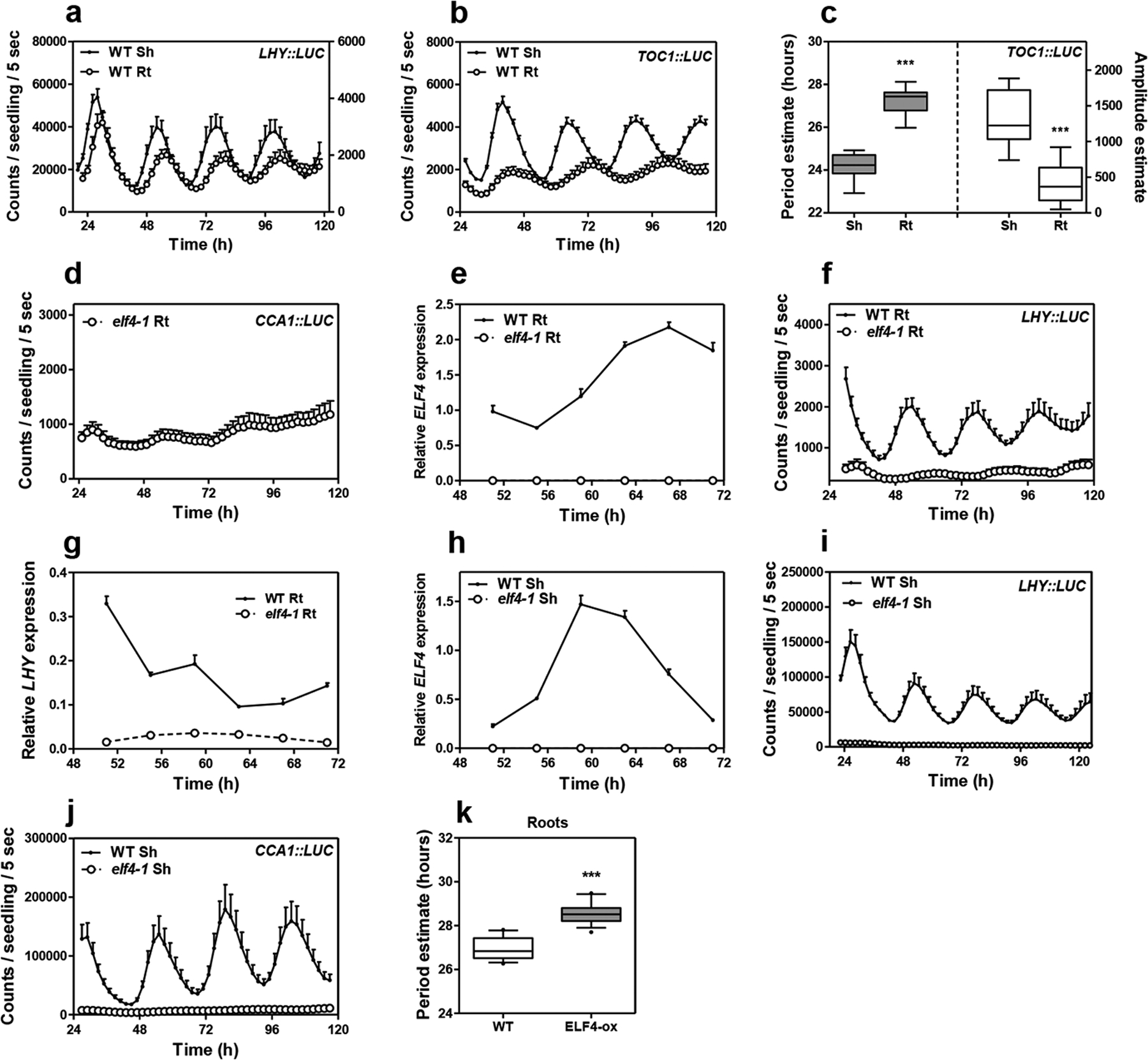
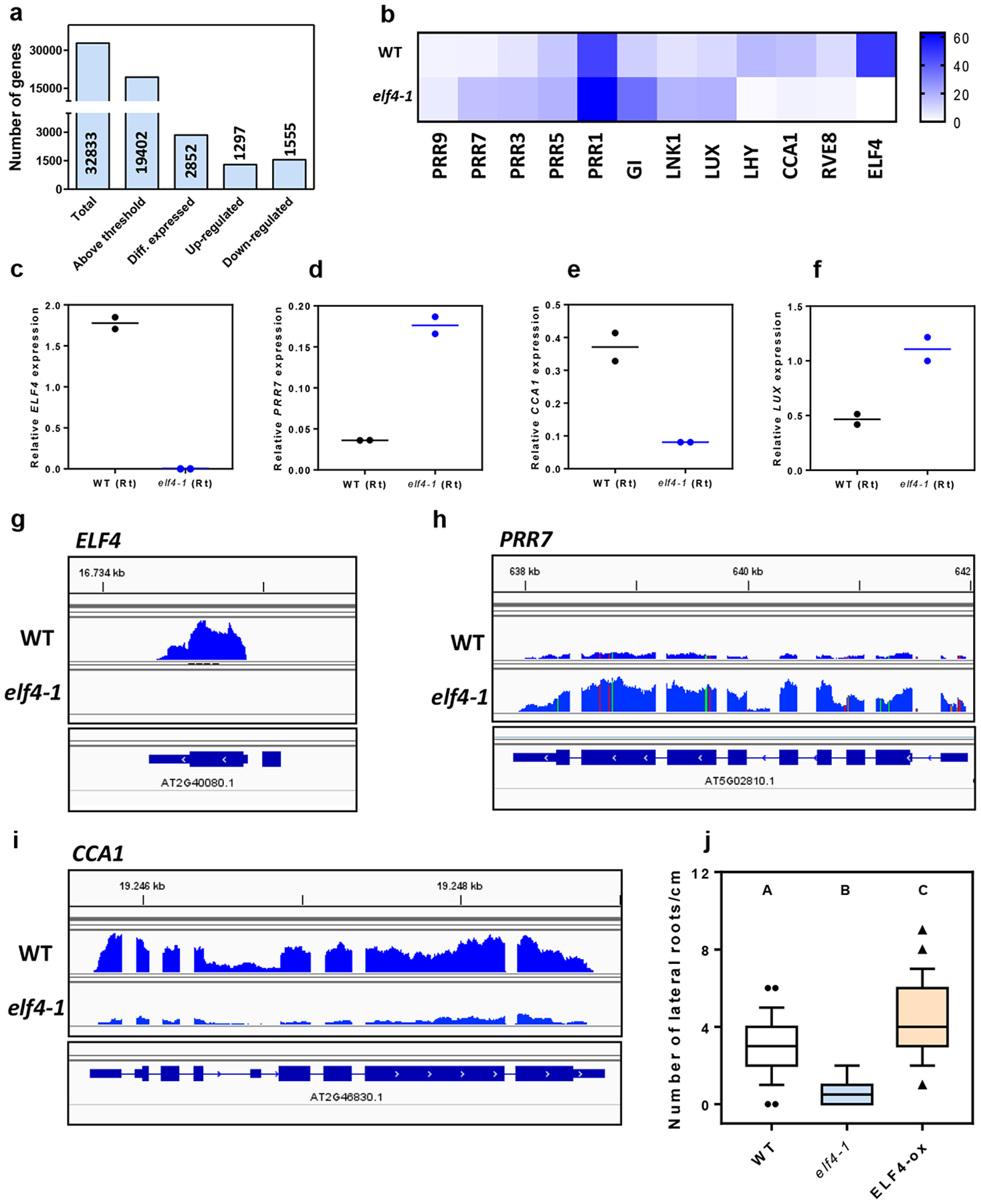
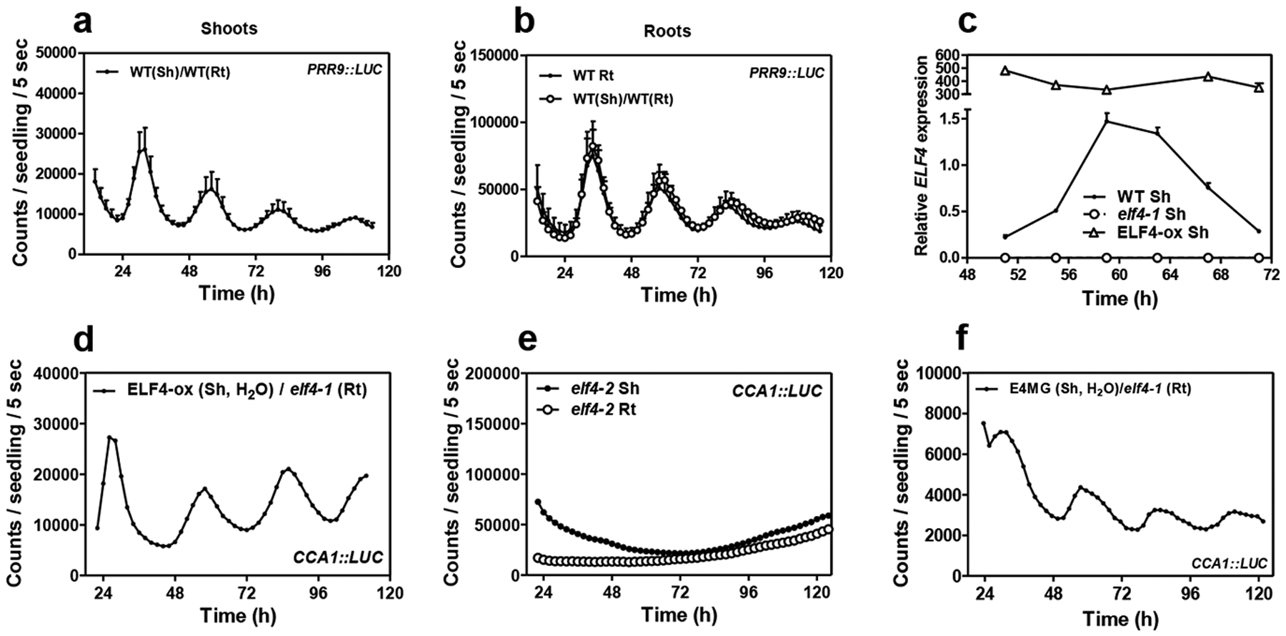
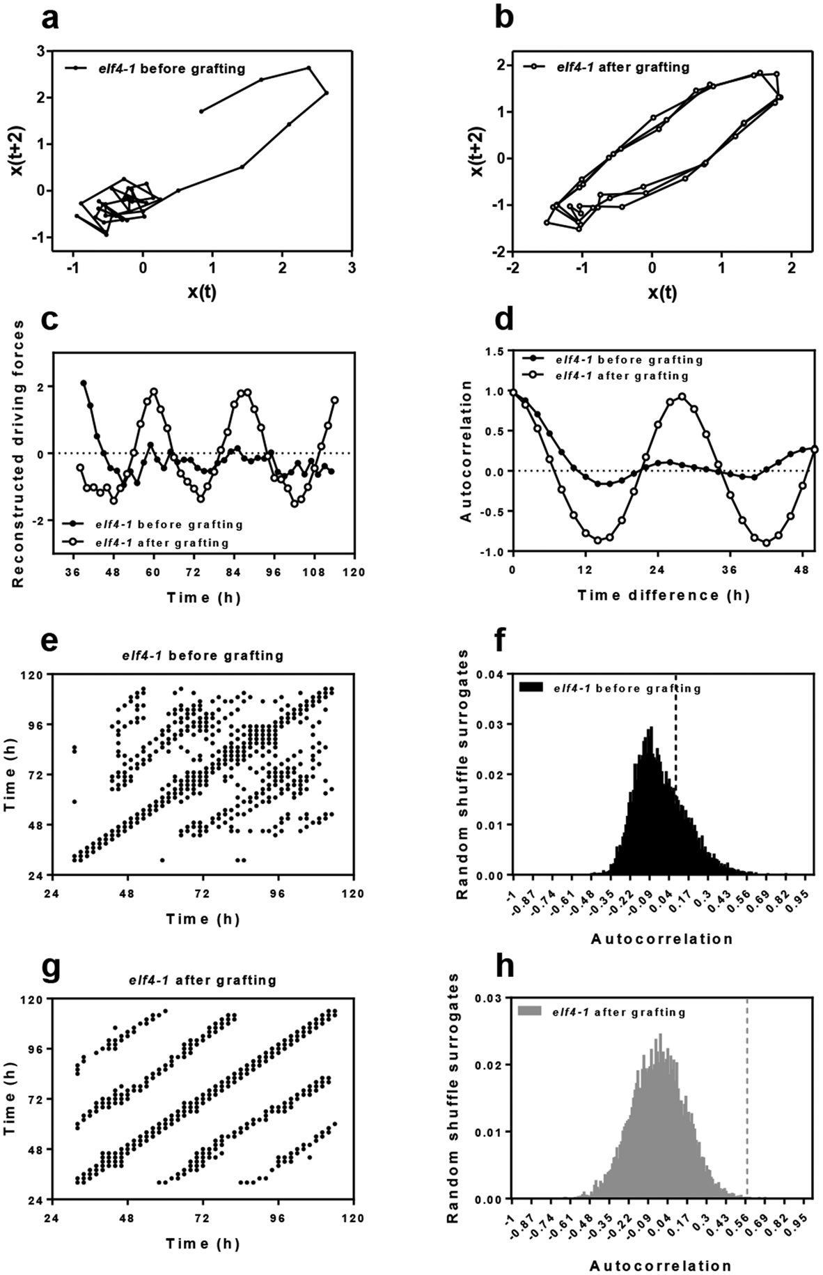
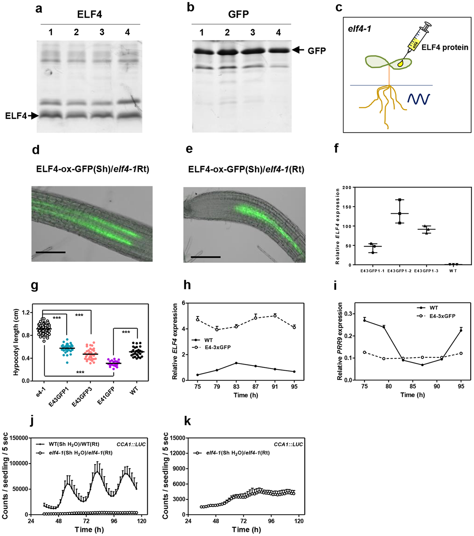
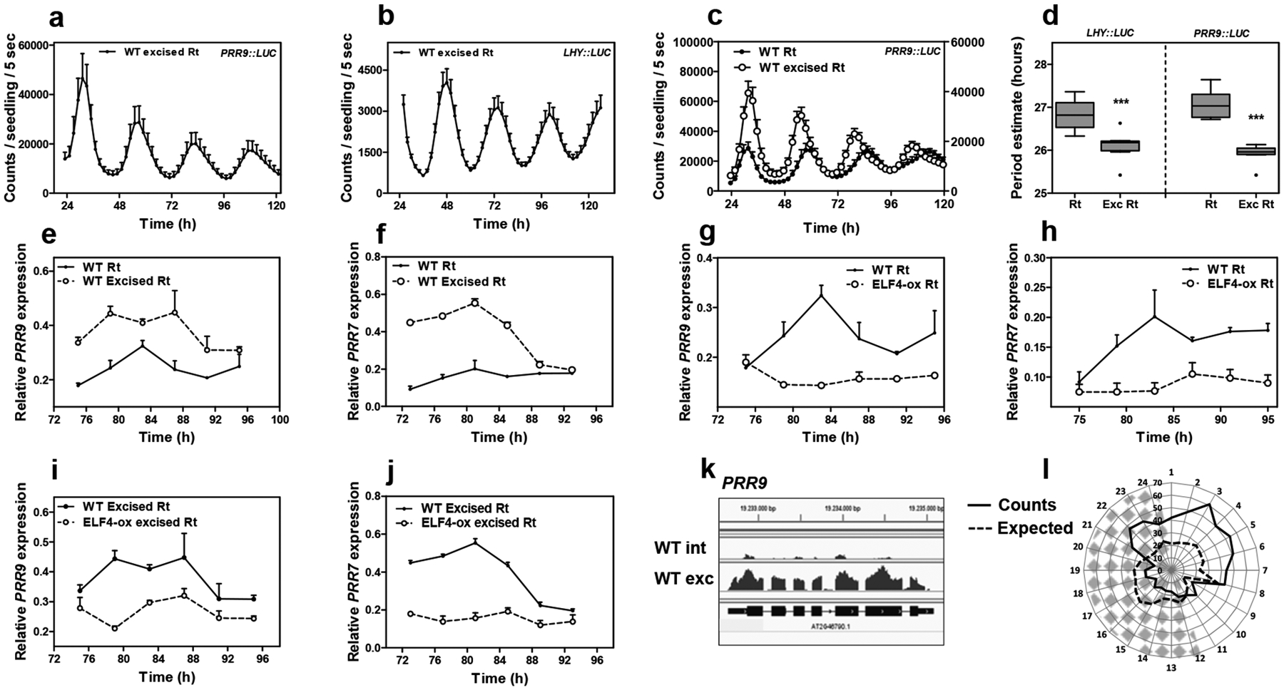
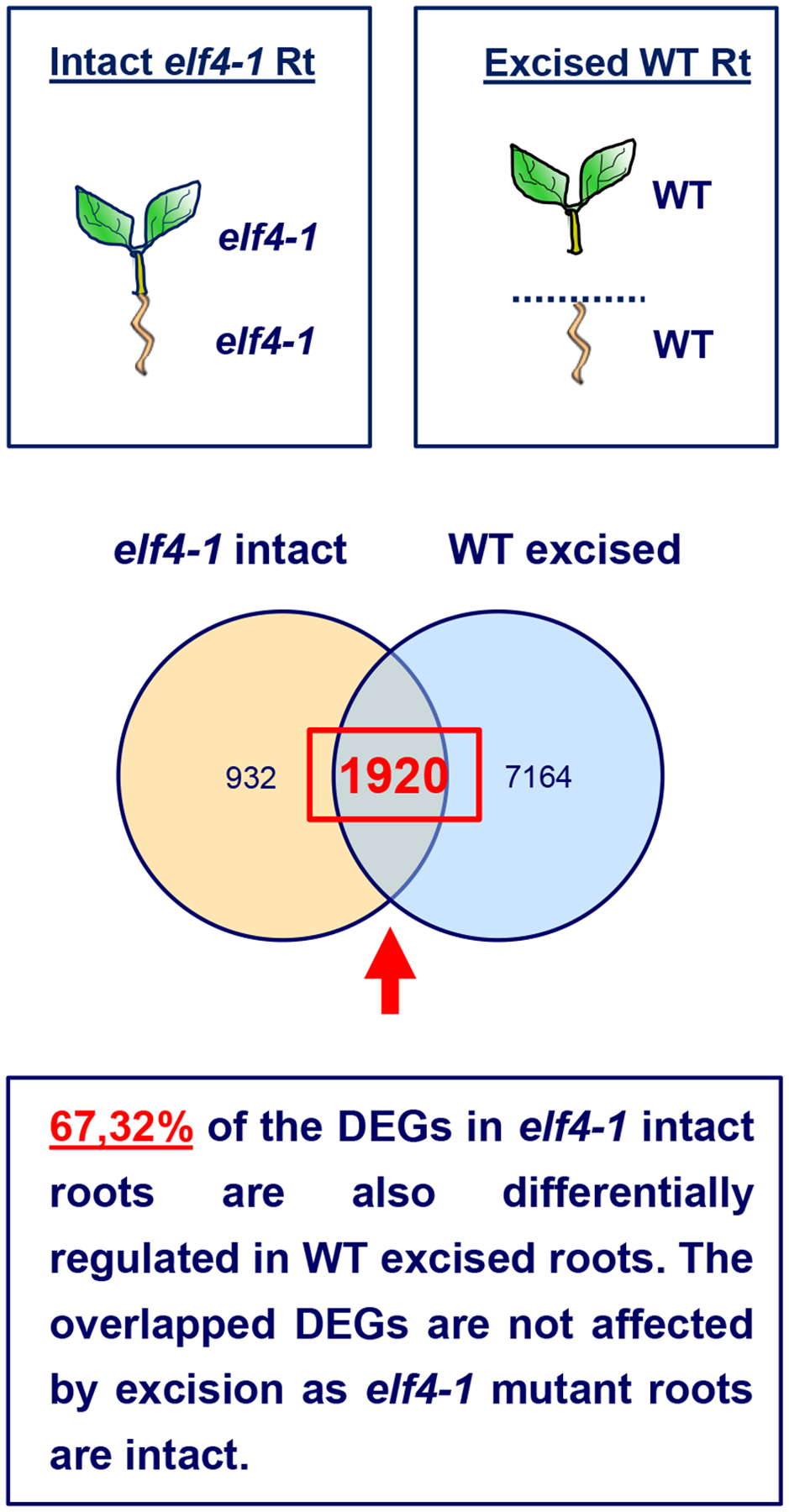
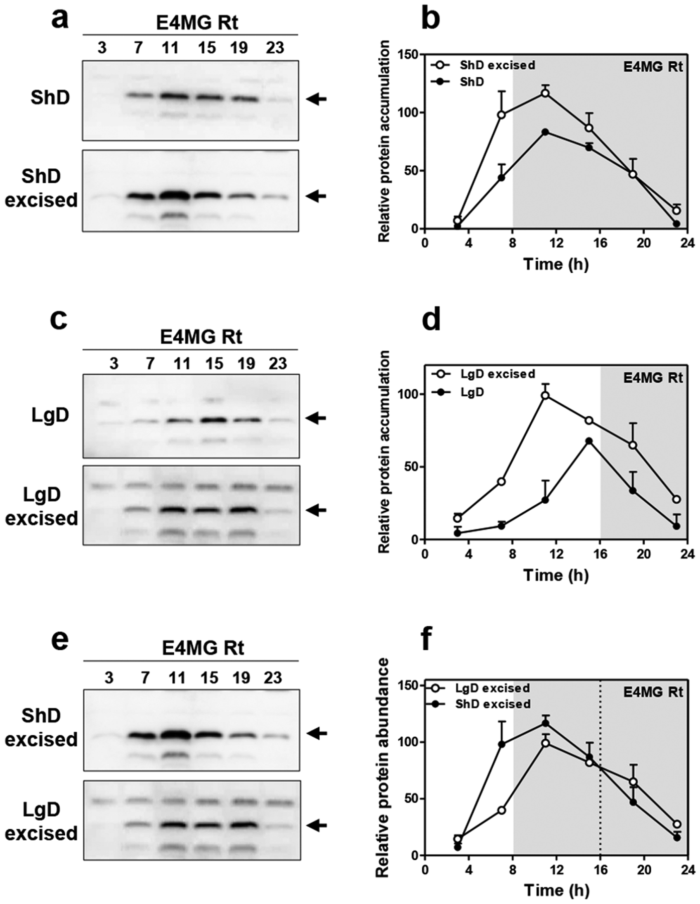
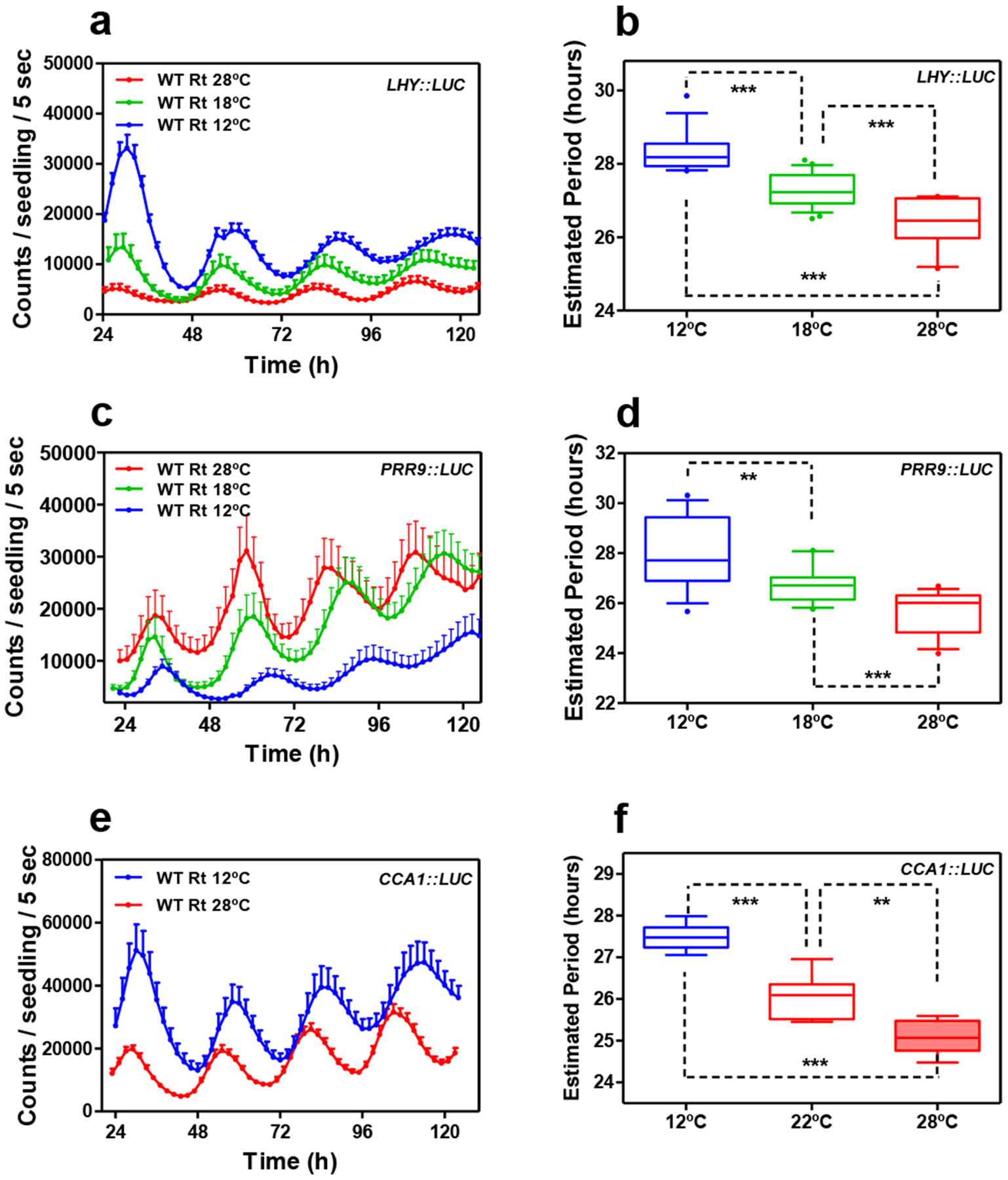
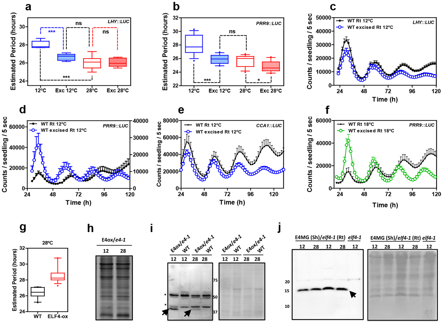
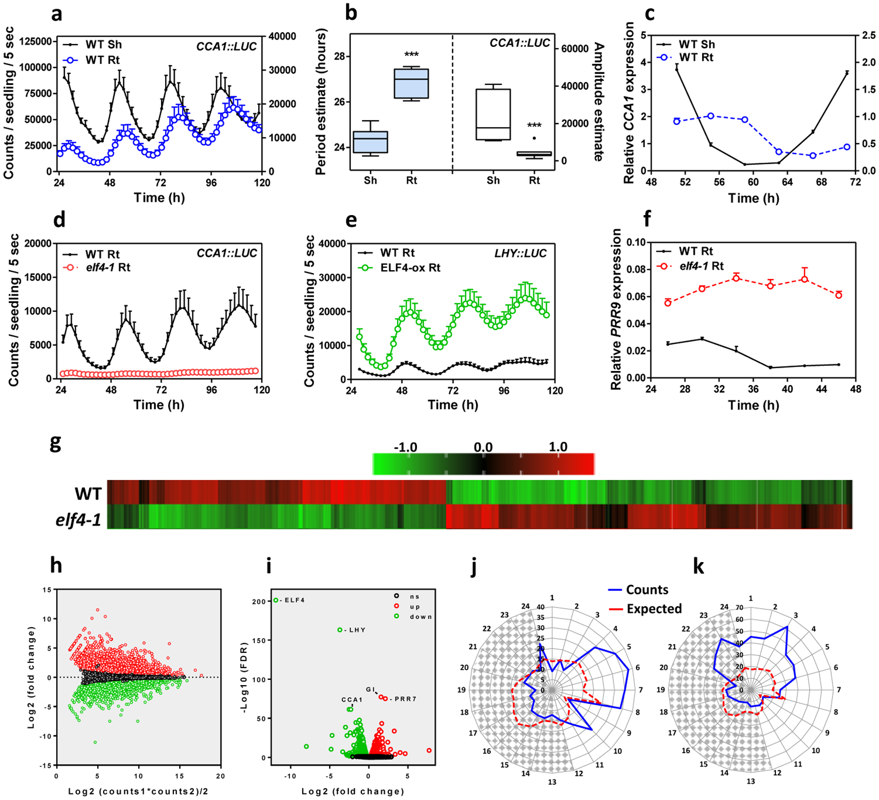
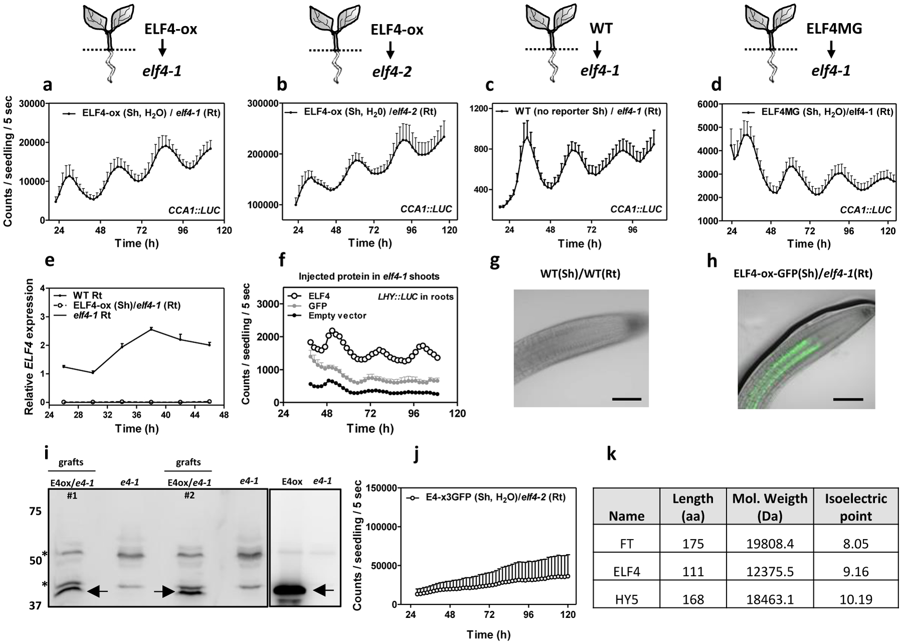
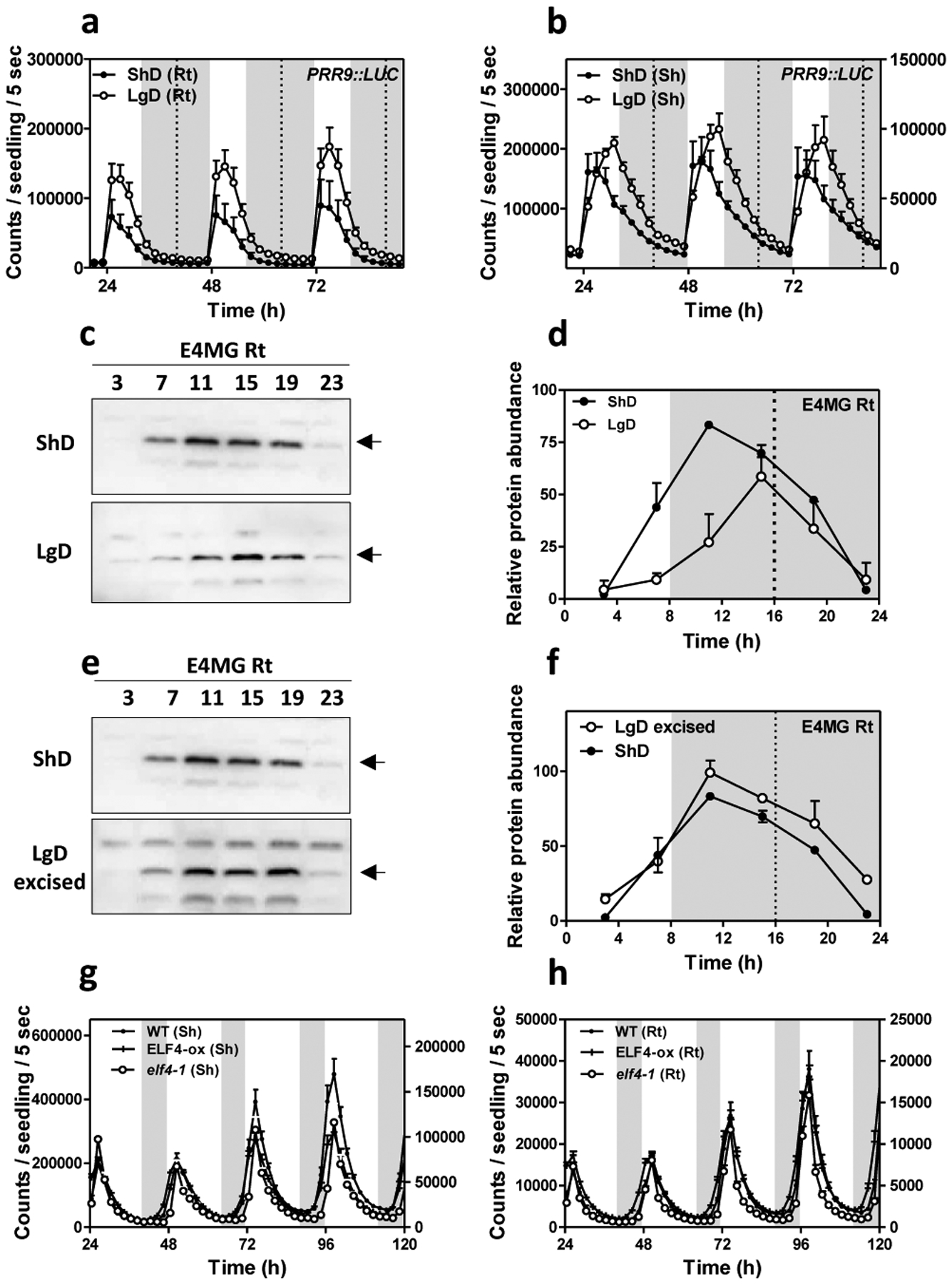
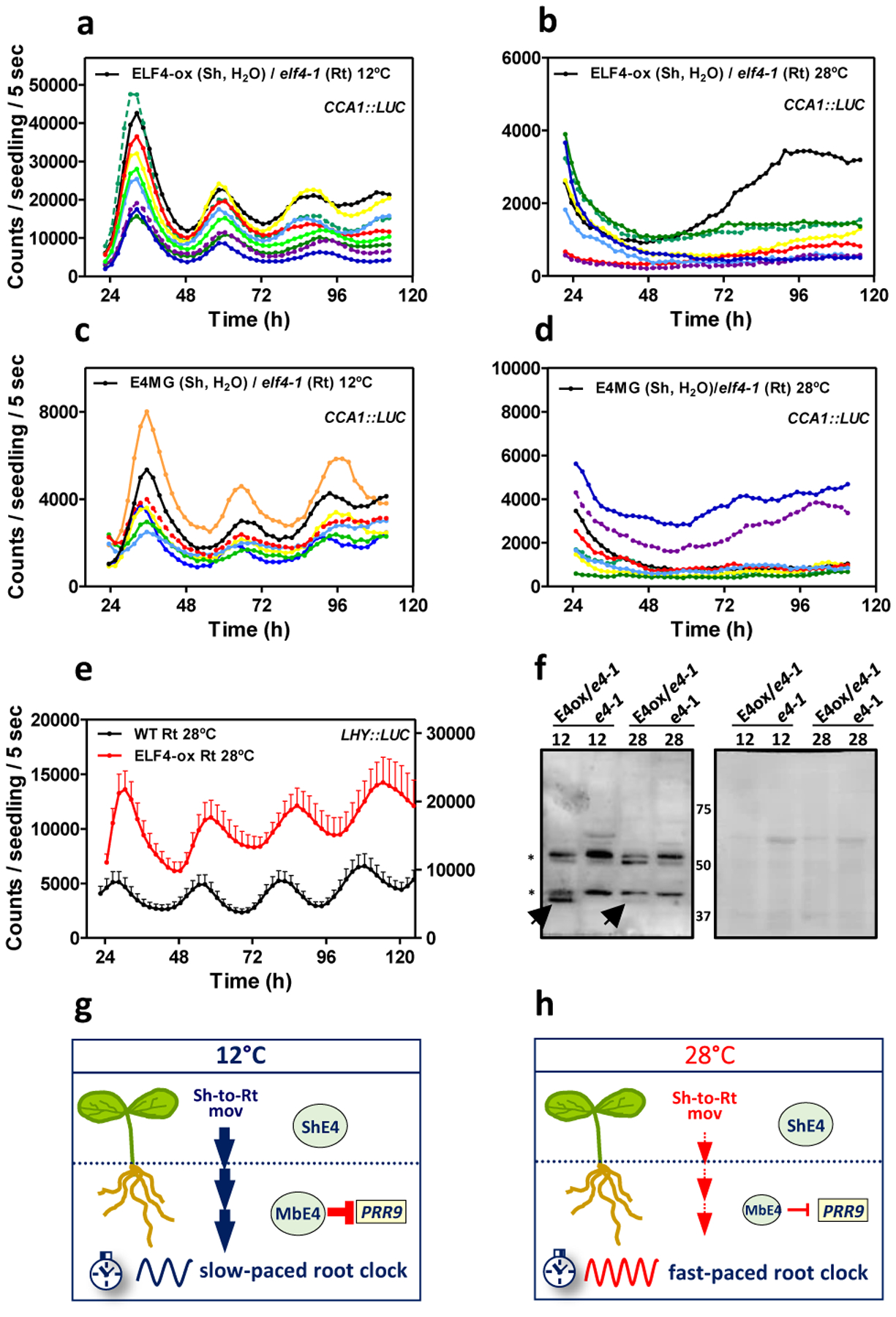
The circadian clock is synchronized by environmental cues, mostly by light and temperature. Explaining how the plant circadian clock responds to temperature oscillations is crucial to understanding plant responsiveness to the environment. Here, we found a prevalent temperature-dependent function of the Arabidopsis clock component EARLY FLOWERING 4 (ELF4) in the root clock. Although the clocks in roots are able to run in the absence of shoots, micrografting assays and mathematical analyses show that ELF4 moves from shoots to regulate rhythms in roots. ELF4 movement does not convey photoperiodic information, but trafficking is essential for controlling the period of the root clock in a temperature-dependent manner. Low temperatures favour ELF4 mobility, resulting in a slow-paced root clock, whereas high temperatures decrease movement, leading to a faster clock. Hence, the mobile ELF4 delivers temperature information and establishes a shoot-to-root dialogue that sets the pace of the clock in roots.


Conflict of interest statement

Competing interests

The authors declare no competing interests.

Figures

Extended Data Fig. 1
Extended Data Fig. 1. Comparative analyses of rhythmic circadian oscillation in shoots and roots.
Luminescence of a, LHY::LUC (n=6 for Sh, n=6 for Rt), and b, TOC1::LUC (n=16 for Sh, n=15 for Rt) rhythms simultaneously measured in shoots (Sh) and roots (Rt). Root luminescence signals in a, are represented in the right Y-axis. c, Circadian period (left Y-axis, n=16 for Sh, n=14 for Rt) and amplitude (right Y-axis, n=16 for Sh, n=15 for Rt) estimates of TOC1::LUC luminescence signals (data are represented as the median ± max and min; 25–75 percentile). *** p-value<0.0001; two-tailed t-tests with 95% of confidence. d, Luminescence of CCA1::LUC rhythms in elf4–1 Rt (n=8) (from Fig. 1d) showing the weak rhythms of the mutant. e, Circadian time course analyses of ELF4 mRNA expression in WT and elf4–1 mutant Rt. f, Luminescence of LHY::LUC rhythms in WT (n=6) and elf4–1 mutant Rt (n=6). g, Circadian time course analyses of LHY mRNA expression in roots of WT and elf4–1. h, Circadian time course analyses of ELF4 mRNA expression in WT and elf4–1 mutant Sh (also in Extended Data Fig. 3c). i, Luminescence of LHY::LUC (n=6) and j, CCA1::LUC (n=9) rhythms in WT and elf4–1 mutant Sh. k, Circadian period estimates of LHY::LUC in WT (n=12) and ELF4-ox (n=14) roots; data are represented as the median ± max and min; 25–75 percentile). *** p-value<0.0001; two-tailed t-tests with 95% of confidence. a-b, d-j, Data are represented as the means + SEM. The mRNA expression and promoter activity analyses were performed under constant light conditions previous synchronization of plants under LD cycles at 22°C. The “n” values refer to independent samples. a-k, Two biological replicates were performed for all experiments, with measurements taken from distinct samples grown and processed at different times.
Extended Data Fig. 2
Extended Data Fig. 2. RNA-Seq analyses of WT and elf4–1 mutant roots.
a, Quantitative analysis of DEGs in elf4–1 versus WT roots. The statistical analyses of the DEGs are detailed in Materials and Methods. b, Heatmap of the median-normalized expression (Z-scaled FPKM values) of the oscillator genes in WT and elf4–1 roots. Analyses by RT-QPCR of c, ELF4, d, PRR7, e, CCA1 and f, LUX mRNA expression in roots at CT75 after three days in LL. Visualization of RNA-seq reads by using the Integrative Genomics Viewer (IGV) browser for g, ELF4, h, PRR7, i, and CCA1. j, Number of lateral roots per cm in the Ws-2 (WT, n=86), elf4–1 (n=28) or ELF4-ox lines (n=48). Plants were grown for 12 days under short day (8h light:/16h darkness) photoperiod with a constant temperature of 22°C. Letters signify significant difference (p < 0.01) as determined by a one-way ANOVA with a Tukey HSD post hoc test. Data is represented as the median ± max and min; 25–75 percentile). Data represents an average of two independent experiments.
Extended Data Fig. 3
Extended Data Fig. 3. Analyses of circadian rhythmicity of the micrografting assays.
Luminescence of PRR9::LUC in a, shoots (n=3) and b, roots (n=3) of WT scion into WT rootstocks and its comparison with luminescence in (non-grafted) WT roots (n=4). c, Circadian time course analyses of ELF4 mRNA expression in shoots of WT, elf4–1 and ELF4-ox (also in Extended Data Fig. 1h). d Individual waveform of CCA1::LUC rhythmic recovery in roots of ELF4-ox scion and elf4–1 rootstocks. Water instead of luciferin was added to the wells containing ELF4-ox shoots. e, CCA1::LUC luminescence in shoots and roots of elf4–2 mutant plants (n=5 for each). f, Individual waveform of CCA1::LUC rhythmic recovery in roots of E4MG scion into elf4–1 rootstocks. Water instead of luciferin was added to the wells containing E4MG shoots. a-f, Data are represented as the means + SEM. The mRNA expression and promoter activity analyses were performed under constant light conditions previous synchronization of plants under LD cycles at 22°C. The “n” values refer to independent samples. a-f, Two biological replicates were performed for all experiments, with measurements taken from distinct samples grown and processed at different times.
Extended Data Fig. 4
Extended Data Fig. 4. Mathematical analyses of the micrografting effects on rhythms.
Two-dimensional plots of CCA1::LUC rhythms in roots a, before and b, after grating ELF4-ox scion into elf4–1 rootstocks. c, Reconstructed waveforms and d, autocorrelation analyses in roots before and after grafting. e, g, Recurrence plots for the driver obtained by delay coordinates and autocorrelation analyses f, before and h, after ELF4-ox scion grafting into elf4–1 rootstock. Number of surrogates for each test: 10000. Before grafting: 95% confidence interval: [-1 0.2932]; effect size: 0.0975; p-value: 0.2339. After grafting: 95% confidence interval: [-1 0.2720]; effect size: 0.5752; p-value: 0.00045.
Extended Data Fig. 5
Extended Data Fig. 5. Analyses of ELF4 movement from shoots to roots.
a, ELF4 and b, GFP proteins purified from bacteria and c, injected in shoots of elf4–1 mutant plants to examine rhythmic recovery in roots. d, e, Representative images showing fluorescence signals in roots of ELF4-ox-GFP scion and elf4–1 rootstock. Scale bars: 100 μm. f, Gene expression analyses of ELF4 mRNA expression in WT and different ELF4-x3GFPs lines. Data are represented as the median ± max and min; 25–75 percentile. g, Hypocotyl length of different lines expressing ELF4-x3GFPs (E43GFP) (E43GFP1 n=34; E43GFP3 n=35) transformed into elf4–1 mutant plants. Hypocotyl length was also assayed for WT (n=27), elf4–1 (n=48) and plants over-expressing ELF4 fused to 1 GFP (E41GFP) (n=19). *** p-value<0.0001; two-tailed t-tests with 95% of confidence. Data are represented as the median ± max and min; 25–75 percentile. Circadian time course analyses of h, ELF4, and i, PRR9 mRNA expression by RT-QPCR in shoots of WT and ELF4-x3GFPs. j, Luminescence of elf4–1 scion into elf4–1 rootstocks elf4–1 (Sh)/elf4–1(Rt) (n=4) and its comparison with luminescence in WT (Sh)/WT(Rt) roots (n=5). k, Luminescence signals of elf4–1 (Sh)/elf4–1(Rt) from j, shown in a separate graph. Water instead of luciferin was added to the wells containing WT and elf4–1 shoots. h-k, Data are represented as the means + SEM. a-k, At least two biological replicates were performed for all experiments, with measurements taken from distinct samples grown and processed at different times.
Extended Data Fig. 6
Extended Data Fig. 6. Shoot excision advances the phase and shortens the circadian period in roots.
Luminescence of a, PRR9::LUC (n=5) and b, LHY::LUC (n=8) circadian rhythms in WT excised roots. c, Comparison of PRR9::LUC circadian rhythms in WT intact (n=6) versus excised roots (n=6). d, Period estimates of LHY::LUC (left graph) (n=8) and PRR9::LUC (right graph) (n=8) rhythms in WT intact versus excised roots. Data are represented as the median ± max and min; 25–75 percentile. *** p-value<0.0001; two-tailed t-tests with 95% of confidence. Circadian time course analyses of e, PRR9 and f, PRR7 mRNA expression in WT intact versus excised roots. Circadian time course analyses of g, PRR9 and h, PRR7 mRNA expression in WT and ELF4-ox intact roots. Circadian time course analyses of i, PRR9 and j, PRR7 mRNA expression in WT excised and ELF4-ox excised roots. a-c, e-j, Data are represented as the means + SEM. k, Visualization of PRR9 RNA-seq reads by using the Integrative Genomics Viewer (IGV) browser. l, Circadian phases of overlapped DEGs in elf4–1 and WT excised roots relative to WT intact roots. Radial axis represents the subjective time (hours). White and gray areas represent subjective day and night, respectively. The mRNA expression and promoter activity analyses were performed under constant light conditions previous synchronization of plants under LD cycles at 22°C. The “n” values refer to independent samples. a-l, Two biological replicates were performed for all experiments, with measurements taken from distinct samples grown and processed at different times.
Extended Data Fig. 7
Extended Data Fig. 7. The lack of ELF4 movement affects gene expression in roots.
Schematic drawing depicting the RNA-Seq comparison of different root genotypes (elf4–1 mutant versus WT) and conditions (intact versus excised). The proportion of overlapped DEGs (67%) is highly significant (P-value < 0.0001, chi-square test for equality of proportions) compared to the overlapped proportion of genes (30%) using random gene lists with the same counts as the DEGs in WT excised roots. The overlapped genes cannot be the result of excision because the data of elf4–1 mutant roots come from intact roots.
Extended Data Fig. 8
Extended Data Fig. 8. Excision advances the phase of ELF4 protein accumulation in roots under entraining conditions.
a, Western-blot analysis and b, quantification of ELF4 protein accumulation in ELF4 Minigene (E4MG) intact and excised roots under ShD (also in Fig. 3c–d). c, Western-blot analysis and d, quantification of ELF4 protein accumulation in E4MG intact and excised roots under LgD (also in Fig. 3e–f). e, Western-blot analysis and f, quantification of ELF4 protein accumulation in E4MG excised roots under ShD and LgD (also in Fig. 3e–f). b, d, f, Data are represented as the means + SEM. a-f, Two biological replicates were performed for all experiments, with measurements taken from distinct samples grown and processed at different times.
Extended Data Fig. 9
Extended Data Fig. 9. The root clock is not temperature-compensated.
a, Luminescence waveforms of LHY::LUC rhythmic oscillation in WT roots at 28°C (n=8), 18°C (n=8) and 12°C (n=8) and b, circadian period estimates of LHY::LUC rhythmic oscillation in WT roots at 28°C (n=12), 18°C (n=23) and 12°C (n=14). Data are represented as the median ± max and min; 25–75 percentile. c, Luminescence waveforms of PRR9::LUC rhythmic oscillation in roots at 28°C (n=8), 18°C (n=16) and 12°C (n=8) and d, circadian period estimates of PRR9::LUC rhythmic oscillation in roots at 28°C (n=14), 18°C (n=16) and 12°C (n=12). Data are represented as the median ± max and min; 25–75 percentile. e, Luminescence waveforms and f, circadian period estimates of CCA1::LUC rhythmic oscillation in roots at 28°C (n=6), 22°C (n=6) and 12°C (n=6) Data are represented as the median ± max and min; 25–75 percentile. b d, f, *** p-value<0.0001; ** p-value<0.005; *** p-value<0.0001; two-tailed t-tests with 95% of confidence. a, c, e, Data are represented as the means + SEM. The “n” values refer to independent samples. a-f, Two biological replicates were performed for all experiments, with measurements taken from distinct samples grown and processed at different times.
Extended Data Fig. 10
Extended Data Fig. 10. Circadian rhythms in excised roots at various temperatures.
Circadian period estimates of a, LHY::LUC in intact (n=8) versus excised WT roots (n=7) at 12°C and 28°C (n=8 for intact and n=8 for excised) and b, PRR9::LUC in intact (n=12) versus excised WT roots (n=13) at 12°C and 28°C (n=14 for intact and n=16 for excised). Data are represented as the median ± max and min; 25–75 percentile. *** p-value<0.0001; * p-value<0.05; ns: non-significant p=0.369; two-tailed t-tests with 95% of confidence. Luminescence rhythmic oscillation in WT intact and excised roots (n=8 for each) at 12°C of c, LHY::LUC, d, PRR9::LUC (n=8 for each) and e, CCA1::LUC (n=4 for excised, n=5 for intact). f, Luminescence of PRR9::LUC rhythmic oscillation in WT intact and excised roots at 18°C (n=16 for each). c-f, Data are represented as the means + SEM. g, Circadian period estimates of LHY::LUC in WT (n=12) and ELF4-ox (n=17) at 28°C. Data are represented as the median ± max and min; 25–75 percentile. Promoter activity analyses were performed under constant light conditions previous synchronization of plants under LD cycles at 22°C. h, Coomassie Blue staining of protein extracts from roots of ELF4-ox-GFP scion (E4-ox) grafted into elf4–1 rootstock (e4–1) at 12°C and 28°C. i, Western-blot analyses of ELF4-GFP protein accumulation (arrows) in roots of ELF4-ox-GFP scion (E4-ox) grafted into elf4–1 rootstock (e4–1) at 12°C and 28°C. WT protein extracts were used as a control. Asterisks denote non-specific bands. Ponceau S staining of the membrane is shown in the right panel. j, Western-blot analyses of ELF4 protein accumulation (arrow) in shoots of ELF4 Minigene (E4MG) grafted into elf4–1 rootstock (e4–1) at 12°C and 28°C. elf4–1 protein extracts were used as a control. Ponceau S staining of the membrane is shown in the right panel. The “n” values refer to independent samples. a-j, Two biological replicates were performed for all experiments, with measurements or analyses taken from distinct samples grown and processed at different times.
Fig. 1.
Fig. 1.. Prevalent function of ELF4 sustaining circadian rhythms in roots.
a, Luminescence of CCA1::LUC (LUCIFERASE) oscillation simultaneously measured in shoots (Sh) (n=9) and roots (Rt) (n=9). Root luminescence signals are represented in the right Y-axis. b, Period (left Y-axis) estimates of CCA1::LUC rhythms in shoots and roots (n=8 for each) and amplitude (right Y-axis) estimates of CCA1::LUC rhythms in shoots (n=7) and roots (n=8); data are represented as the median ± max and min; 25–75 percentile). *** p-value<0.0001; two-tailed t-tests with 95% of confidence. c, Circadian time course analyses of CCA1 mRNA expression in WT Sh and Rt. d, Luminescence of CCA1::LUC rhythms in WT (n=9) and elf4–1 Rt (n=8). e, Luminescence of LHY::LUC rhythms in WT (n=8) and ELF4-ox Rt (n=9). f, Circadian time course analyses of PRR9 mRNA expression in roots of WT and elf4–1. Sampling was performed under constant light conditions (LL) following synchronization under light:dark cycles (LD). a, c-f, Data are represented as the means + SEM. g, Heatmap of the median-normalized expression (Z-scaled FPKM values) of DEGs following a hierarchical clustering using the Euclidean distance. h, Relationship between average expression and fold change for each gene. i, Volcano plot showing fold-change versus significance of the differential expression test. Black dots represent genes that are not differentially expressed, while red and green dots are the genes that are significantly up- and down-regulated, respectively. Circadian phases of j, up- and k, down-regulated DEG in elf4–1 roots. Radial axis represents the subjective time (hours). White and gray areas represent subjective day and night, respectively. The “n” values refer to independent samples. a-k, Data for all experiments are representative of two biological replicates, with measurements taken from distinct samples grown and processed at different times.
Fig. 2.
Fig. 2.. ELF4 moves from shoots to regulate rhythms in roots.
CCA1::LUC luminescence in roots of ELF4-ox scion into a, elf4–1 (n=4) and b, elf4–2 (n=3) rootstocks. Water instead of luciferin was added to the wells containing ELF4-ox shoots. CCA1::LUC luminescence in elf4–1 rootstocks with c, WT (n=8) and d, ELF4 Minigene (ELF4MG) (n=10) scions. WT scions do not express reporters and water instead of luciferin was added to the wells containing ELF4MG shoots. a-d, Schematic drawings depicting the different scion/rootstock combinations are shown above each graph. e, Circadian time course analyses of ELF4 mRNA expression in roots of WT, elf4–1 and ELF4-ox scion and elf4–1 rootstocks. f, Luminescence of LHY::LUC rhythms in elf4–1 roots after injection in shoots of purified ELF4 (n=4) or GFP proteins (n=8) and elf4–1 roots as a control (n=6). g, Representative image showing the lack of fluorescence signals in roots of WT scion and WT rootstock. h, Representative image showing fluorescence signals in roots of ELF4-ox scion into elf4–1 rootstock. Scale bar: 100 μm. i, Western-blot analysis of ELF4-GFP protein accumulation (arrows) in roots of ELF4-ox-GFP scion (E4ox) grafted into elf4–1 rootstock (e4–1) (two pools of independent grafting assays, #1 and #2, are shown). Asterisks denote non-specific bands. j, CCA1::LUC luminescence in elf4–2 rootstocks grafted with ELF4-x3GFP scions (n=5). Water instead of luciferin was added to the wells containing ELF4-x3GFP shoots. a-f, j, Data are represented as the means + SEM. k, Protein features of various plant mobile proteins. The “n” values refer to independent samples. a-j, Two biological replicates were performed for all experiments, with measurements taken from distinct samples grown and processed at different times.
Fig. 3.
Fig. 3.. Mobile ELF4 does not regulate the photoperiodic-dependent phase in roots.
Luminescence analyses of PRR9::LUC rhythms in a, roots (n=5 for ShD, n=6 for LgD) and b, shoots (n=6 for ShD, n=4 for LgD) of plants grown under short day (ShD) or long day (LgD) conditions. c, Western-blot analyses and d, quantification of ELF4 protein accumulation in ELF4 Minigene roots (E4MG Rt) of plants grown under ShD and LgD (also in Extended Data Fig. 8a–d). e, Western-blot analyses and f, quantification of ELF4 protein accumulation in E4MG roots of plants grown under ShD and excised roots under LgD (also in Extended Data Fig. 8a–f). Arrows indicate the ELF4 protein. Luminescence of LHY::LUC oscillation in WT, ELF4-ox and elf4–1 plants measured in g, shoots (Sh) (n=12) and h, roots (Rt) (n=12) under LgD conditions. a-b, d, f, g-h, Data are represented as the means + SEM. Dashed lines indicate dusk under LgD. The “n” values refer to independent samples. a-h, Two biological replicates were performed for all experiments, with measurements taken from distinct samples grown and processed at different times.
Fig. 4.
Fig. 4.. Mobile ELF4 sets the temperature-dependent pace of the root clock.
Individual luminescence waveforms of CCA1::LUC rhythmic oscillation in ELF4-ox scion into elf4–1 rootstocks at a, 12°C (n=10) and b, 28°C (n=8). Individual luminescence waveforms of CCA1::LUC rhythmic oscillation in E4MG scion into elf4–1 rootstocks at c, 12°C (n=7) and d, 28°C (n=8).Water instead of luciferin was added to the wells containing ELF4-ox and E4MG scions. e, Luminescence of LHY::LUC rhythmic oscillation in WT and ELF4-ox roots at 28°C (n=8 for WT, n=5 for ELF4-ox). Data are represented as the means + SEM. f, Western-blot analysis of ELF4-GFP protein accumulation (arrow) in roots of ELF4-ox-GFP scion (E4ox) grafted into elf4–1 rootstock (e4–1) at 12°C and 28°C. elf4–1 mutant protein extracts were used as a control. Asterisks denote non-specific bands. Ponceau S staining of the membrane is shown in the right panel. Schematic drawing depicting g, the increased shoot-to-root movement of ELF4 (Sh-to-Rt mov, thick blue vertical arrows), increased PRR9 repression and the slow pace of the root clock at low temperatures, and h, the decreased shoot-to-root movement (Sh-to-Rt mov, thin red vertical arrows), decreased PRR9 repression and fast-paced root clock at high temperature. The “n” values refer to independent samples. a-f, Two biological replicates were performed for all experiments, with measurements taken from distinct samples grown and processed at different times.

Comment in

  • ELF in the root.
    Kubota A, Endo M. Kubota A, et al. Nat Plants. 2020 Apr;6(4):336-337. doi: 10.1038/s41477-020-0636-0. Nat Plants. 2020. PMID: 32284548 No abstract available.

References

    1. Zhang EE & Kay SA Clocks not winding down: unravelling circadian networks. Nat. Rev. Mol. Cell. Biol 11, 764–776 (2010). - PubMed
    1. Greenham K & McClung CR Integrating circadian dynamics with physiological processes in plants. Nat. Rev. Genet 16, 598–610 (2015). - PubMed
    1. Nagel Dawn H. & Kay Steve A. Complexity in the wiring and regulation of plant circadian networks. Curr. Biol 22, R648–R657 (2012). - PMC - PubMed
    1. Oakenfull RJ & Davis SJ Shining a light on the Arabidopsis circadian clock. Plant Cell Environ 40, 2571–2585 (2017). - PubMed
    1. Hogenesch JB & Ueda HR Understanding systems-level properties: timely stories from the study of clocks. Nature Reviews Genetics 12, 407 (2011). - PubMed

Substances