. 2020 Apr 8;6(15):eaaw0158.
doi: 10.1126/sciadv.aaw0158. eCollection 2020 Apr.

Soft extracellular matrix enhances inflammatory activation of mesenchymal stromal cells to induce monocyte production and trafficking

Affiliations

Soft extracellular matrix enhances inflammatory activation of mesenchymal stromal cells to induce monocyte production and trafficking

Sing Wan Wong et al. Sci Adv. .

Abstract

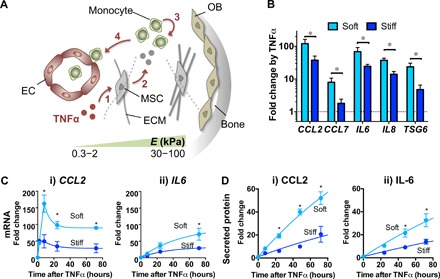
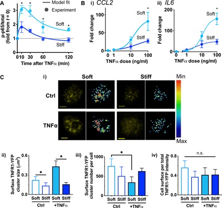
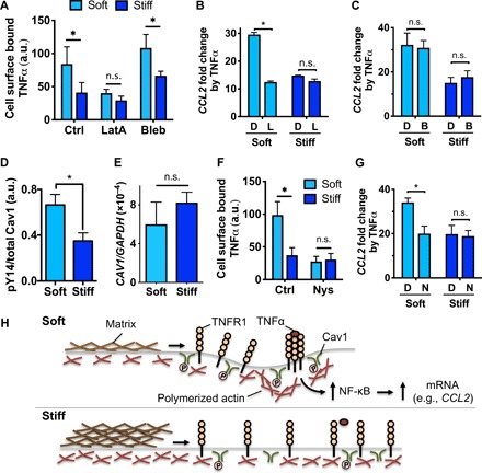
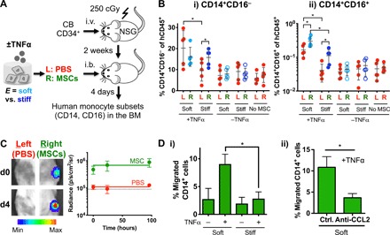
Mesenchymal stromal cells (MSCs) modulate immune cells to ameliorate multiple inflammatory pathologies. Biophysical signals that regulate this process are poorly defined. By engineering hydrogels with tunable biophysical parameters relevant to bone marrow where MSCs naturally reside, we show that soft extracellular matrix maximizes the ability of MSCs to produce paracrine factors that have been implicated in monocyte production and chemotaxis upon inflammatory stimulation by tumor necrosis factor-α (TNFα). Soft matrix increases clustering of TNF receptors, thereby enhancing NF-κB activation and downstream gene expression. Actin polymerization and lipid rafts, but not myosin-II contractility, regulate mechanosensitive activation of MSCs by TNFα. We functionally demonstrate that human MSCs primed with TNFα in soft matrix enhance production of human monocytes in marrow of xenografted mice and increase trafficking of monocytes via CCL2. The results suggest the importance of biophysical signaling in tuning inflammatory activation of stromal cells to control the innate immune system.


Figures

Fig. 1
Fig. 1. 3D matrix stiffness regulates expression of TNFα-inducible genes implicated in monocyte functions.
(A) Schematic showing that matrix stiffness in the bone marrow (BM) microenvironment can potentially influence MSC activation by TNFα to modulate monocyte functions in marrow. The extracellular matrix (ECM) in the central marrow and vascular [endothelial cell (EC)] regions is softer (Young’s modulus E = 0.3 to 2 kPa), while that near the bone surface [osteoblast (OB)] is stiffer (E = 30 to 100 kPa) (13). Upon inflammation, TNFα could activate MSCs (1) to produce secreted factors (2) that can influence monocyte production (3) and trafficking (4) before systemic distribution. (B) Effects of 3D matrix stiffness on TNFα-mediated up-regulation of gene expression in MSCs. MSCs were encapsulated in soft or stiff alginate-RGD 3D hydrogels, incubated for 1 day, and treated with TNFα (100 ng/ml) for 3 days. Transcript levels of the indicated genes were measured by qPCR and normalized against glyceraldehyde 3-phosphate dehydrogenase (GAPDH). Fold change by TNFα for each group was then calculated from the untreated control (in log scale). (C) Effects of 3D matrix stiffness on mRNA expression kinetics in response to TNFα. (i) CCL2: Data from 8 to 72 hours were fitted to standard exponential decay curves. t1/2 and plateau for soft: 5.2 hours, 90-fold; stiff: 8.9 hours, 28-fold. (ii) IL6: Data were fitted to standard one-phase association curves. t1/2 and plateau for soft: 36.5 hours, 95-fold; stiff: 23.2 hours, 33-fold. (D) Effects of 3D matrix stiffness on protein secretion kinetics in response to TNFα. Data were fitted to standard one-phase association curves. (i) CCL2: t1/2 = 126 hours for both soft and stiff; plateau for soft = 156-fold and for stiff = 61-fold. (ii) IL-6: t1/2 = 204 hours for both soft and stiff; plateau for soft = 143-fold and for stiff = 58-fold. Fold change refers to increase over untreated condition. For (B) to (D), paired t test, *P < 0.05 soft versus stiff at each time point (n = 3 donors). Error bars, ±SEM.
Fig. 2
Fig. 2. Soft matrix increases NF-κB activation by facilitating TNFR1 clustering in response to TNFα.
(A) NF-κB activation kinetics of MSCs in soft or stiff 3D gels in response to TNFα (100 ng/ml) evaluated by p65 phosphorylation at Ser536 (p-p65). The experimental results from soft and stiff alginate hydrogels are fitted to the I1-FFL–based model derived analytically (R2 > 0.8; Supplementary Materials). (B) Dose-response curves of TNFα to up-regulate mRNA expression in soft and stiff alginate-RGD hydrogels after the 3-day continuous treatment. (i) CCL2: IC50 and maximum fold increase for soft = 17 μM, 88-fold, and for stiff = 20 μM, 35-fold. (ii) IL6: IC50 and maximum fold increase for soft = 9 μM, 160-fold, and for stiff = 7 μM, 50-fold. *P < 0.05, paired t test. For (A) and (B), paired t test, *P < 0.05 soft versus stiff at each time point or dose (n = 3 experiments). Error bars, ±SEM. (C) Cell surface distribution and clustering of TNFR1-YFP in MSCs encapsulated in soft or stiff 3D gels. (i) Representative confocal images for each group showing maximum projection of TNFR1-YFP (left, yellow) and cell surface TNFR1-YFP after filtering (right, scale bar, min: 0, max: 255; fig. S2H). (ii) Cluster size of TNFR1-YFP on the cell surface. (iii) Cluster number of TNFR1-YFP per cell. (iv) Cell surface per total TNFR1-YFP intensity. P < 0.05 one-way Brown-Forsythe and Welch ANOVA for (ii) and (iii) with Dunnett T3 multiple comparisons test, *P < 0.05 (n ≥ 15 cells from two experiments). Error bars, ±SD. n.s., not significant.
Fig. 3
Fig. 3. Actin polymerization and lipid rafts mediate responsiveness of MSCs to TNFα in soft matrix.
(A) TNFα binding to MSCs in soft or stiff 3D gels in the presence of DMSO (Ctrl), latrunculin A (LatA, 0.25 μM), or blebbistatin (Bleb, 10 μM) for 3 hours. P < 0.05 two-way ANOVA with Sidak’s multiple comparison test, *P < 0.05 (n = 3 experiments). (B) Effects of inhibiting actin polymerization on TNFα-induced CCL2 mRNA expression. MSCs in soft or stiff 3D gels were preincubated with DMSO (“D”) or latrunculin A (“L”) for 1 hour, treated with or without TNFα in the presence of the drugs for 2 hours, followed by washout and incubating in media for 1 day prior to qPCR. (C) Effects of inhibiting myosin-II contractility on TNFα-induced CCL2 protein expression. Gel-encapsulated MSCs were treated with TNFα in the presence of DMSO (“D”) or blebbistatin (“B”) in the same manner as (B). (D) Phosphorylation level of caveolin-1 (Cav1) at Tyr14 in MSCs after 1 day of encapsulation in soft or stiff 3D gels. (E) Gene expression of CAV1 relative to GAPDH measured by qPCR after culturing MSCs in soft or stiff 3D gels for 1 day. (F) TNFα binding to MSCs in soft or stiff 3D gels in the presence of DMSO (Ctrl) or nystatin (Nys, 50 μM) for 2 hours, followed by washout and incubating in media for 1 day prior to qPCR. (G) Effects of inhibiting lipid rafts on TNFα-induced CCL2 mRNA expression in the presence of DMSO (“D”) or nystatin (“N”) in the same manner as (B). For (B) to (G), *P < 0.05, paired t test; n = 3 experiments; and error bars, ±SEM. (H) Model of actin polymerization and lipid rafts in mediating matrix stiffness–dependent binding of TNFα to the cell surface. In soft matrix, lipid rafts mediate redistribution of actin polymerization to facilitate clustering of TNFR1 in response to TNFα. In contrast, stiff matrix impedes this process, thereby impairing TNFR1 clustering and TNFα binding. a.u., arbitrary unit.
Fig. 4
Fig. 4. Soft matrix promotes the ability of MSCs to produce and recruit monocytes upon TNFα stimulation.
(A) Experimental scheme. Sublethally irradiated (250 cGy) NSG mice were injected intravenously (i.v.) with 20,000 human cord blood HSPCs per 20 g of mouse. After 2 weeks, MSCs that were cultured in soft or stiff gels for 1 day followed by ±TNFα treatment (100 ng/ml) for three additional days were retrieved and delivered to the right (R) tibia by an intrabone (i.b.) route, while the left (L) tibia was delivered with PBS. After 4 days, both tibias were analyzed for human monocyte subsets. (B) Percentage of (i) classical (CD14+CD16) and (ii) intermediate (CD14+CD16+) human monocytes in tibias. P < 0.05, two-way ANOVA with Sidak’s multiple comparison test, *P < 0.05. n = 5 recipients from two independent experiments. (C) MSCs remain localized in one tibia after intrabone delivery to NSG mice. Firefly luciferase–transduced MSCs were delivered in the right tibia, while PBS was injected in the left tibia, followed by IVIS imaging over 4 days. Left: Representative images at days 0 and 4 after MSC delivery. Scale bar, radiance (p/sec/cm2/sr, min: 1 M, max: 15 M). Right: Quantification of radiance signals over 4 days. The signal from the left tibia remains at the background level (~90,000, dotted line), while that from the right tibia remains ~10-fold higher over the 4 days. (D) TNFα-treated MSCs in soft matrix increase migration of human peripheral blood CD14+ monocytes via secretion of CCL2. MSCs in soft or stiff alginate hydrogel were treated with TNFα (100 ng/ml) for 1 day prior to washout and collection of the media for 1 day for the Transwell migration assay. (i) The extent of CD14+ migration under chemotactic gradient through 3-μm pores for 3 hours. P < 0.05 from one-way ANOVA with Tukey’s post hoc test, *P < 0.05 (n = 3 experiments). (ii) CCL2 is required for increased chemotaxis of CD14+ cells by the media from TNFα-treated MSCs in soft matrix. The media were treated with a neutralizing antibody against CCL2 (anti-CCL2) or isotype control (ctrl) for 3 hours prior to the Transwell assay. *P < 0.05, paired t test (n = 3 experiments). All error bars for both (i) and (ii), ±SEM.

References

    1. Yue R., Zhou B. O., Shimada I. S., Zhao Z., Morrison S. J., Leptin receptor promotes adipogenesis and reduces osteogenesis by regulating mesenchymal stromal cells in adult bone marrow. Cell Stem Cell 18, 782–796 (2016). - PubMed
    1. Méndez-Ferrer S., Michurina V., Ferraro F., Mazloom A. R., Macarthur B. D., Lira S. A., Scadden D. T., Ma’ayan A., Enikolopov G. N., Frenette P. S., Mesenchymal and haematopoietic stem cells form a unique bone marrow niche. Nature 466, 829–834 (2010). - PMC - PubMed
    1. Bartholomew A., Sturgeon C., Siatskas M., Ferrer K., McIntosh K., Patil S., Hardy W., Devine S., Ucker D., Deans R., Moseley A., Hoffman R., Mesenchymal stem cells suppress lymphocyte proliferation in vitro and prolong skin graft survival in vivo. Exp. Hematol. 30, 42–48 (2002). - PubMed
    1. Le Blanc K., Mougiakakos D., Multipotent mesenchymal stromal cells and the innate immune system. Nat. Rev. Immunol. 12, 383–396 (2012). - PubMed
    1. Parekkadan B., Milwid J. M., Mesenchymal stem cells as therapeutics. Annu. Rev. Biomed. Eng. 12, 87–117 (2010). - PMC - PubMed

Publication types

MeSH terms

LinkOut - more resources