. 2020 Apr 8;6(15):eaay1514.
doi: 10.1126/sciadv.aay1514. eCollection 2020 Apr.

Regeneration of pulpo-dentinal-like complex by a group of unique multipotent CD24a+ stem cells

Affiliations

Regeneration of pulpo-dentinal-like complex by a group of unique multipotent CD24a+ stem cells

Hong Chen et al. Sci Adv. .

Abstract

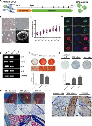
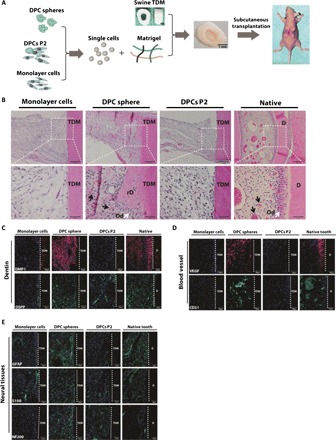
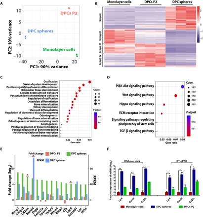
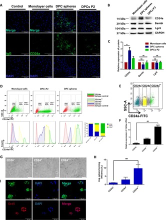
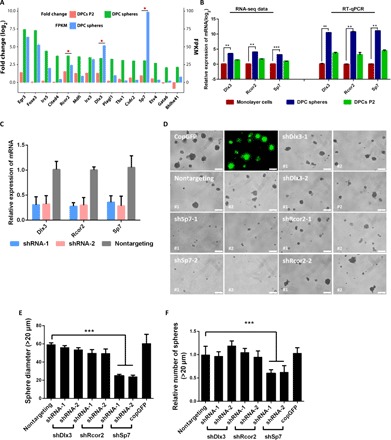
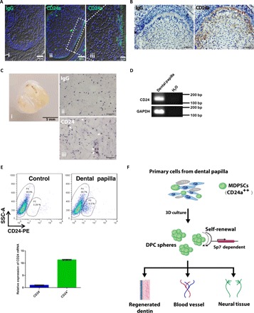
Dental pulp is critical to maintain the vitality of a tooth. Regeneration of pulpo-dentinal complex is of great interest to treat pulpitis and pulp necrosis. In this study, through three-dimensional spheroid culture, a group of unique multipotent stem cells were identified from mouse dental papilla called multipotent dental pulp regenerative stem cells (MDPSCs). MDPSCs exhibited enhanced osteogenic/odontogenic differentiation capabilities and could form regenerative dentin and neurovascular-like structures that mimicked the native teeth in vivo. Further analysis revealed that CD24a was the bona fide marker for MDPSCs, and their expansion was highly dependent on the expression of a key transcriptional factor, Sp7. Last, CD24a+ cells could be detected in primary dental papilla in mice and human, suggesting that MDPSCs resided in their native niches. Together, our study has identified a previously unidentified group of multipotent pulp regenerative stem cells with defined molecular markers for the potential treatment of pulpitis and pulp necrosis.


Figures

Fig. 1
Fig. 1. A group of multipotent sphere-forming stem cells existed in dental papilla.
(A) Schematic diagram of the 3D culture assay. DPCs were isolated from tooth germs of neonatal pups and cultured in standard DPC medium for about 1 week before further seeded in MC culture medium to initiate sphere formation. Spheres and control monolayer cells were collected at day 8 for further characterization. (B) Representative images for the typical morphology of primary mouse DPCs P2 (i) and DPC spheres on days 0 (ii) and 7 (iii). Scale bars, 200 μm. Scanning electron microscopy morphology of DPC spheres (iv). Scale bar, 20 μm. (C) 3D culture supported continuous growth of DPC spheres. The growth and diameter of DPC sphere were monitored over time in 3D culture system. (D) DPC spheres were positive for pluripotency and proliferation markers including Oct4, Sox2, Nanog, and Ki67. DPC spheres were collected at day 8 and immunostained with antibodies against Oct4, Sox2, Nanog, and Ki67. DAPI (4′,6-diamidino-2-phenylindole) was used for nuclear staining. Scale bars, 50 μm. (E) Expression of Oct4, Sox2, Nanog, and Ki67 in DPC spheres were also confirmed by RT-PCR. Complementary DNAs from mESCs (mouse embryonic stem cells) were used as the positive control and H2O as the negative control. GAPDH, glyceraldehyde-3-phosphate dehydrogenase. (F) Alizarin Red S (ARS) staining and quantification indicated DPC sphere–derived cells retained differentiation capabilities compared with 2D monolayer cultures. Monolayer cells, DPC sphere–derived cells, and DPCs at P2 were incubated in osteoinductive medium (OIM) for 10 days, respectively. Sphere-derived cells exhibited enhanced mineralization from monolayer cultures. Error bar represents two independent experiments with triplicates. ***P < 0.001 (Student’s t test). OD, optical density. (G) ALP staining and quantification indicated DPC sphere–derived cells retained differentiation capabilities compared with 2D monolayer cultures. Cells were incubated in OIM for 7 days. Error bar represents two independent experiments with triplicates. ***P < 0.001. (H) Strongly mineralized structures were formed by DPC sphere–derived cells when transplanted in renal capsules. Monolayer cells, DPC sphere–derived cells, and DPCs P2 were implanted into renal capsule of C57BL/6 mice together with Matrigel. Samples were harvested after 4 weeks. The hematoxylin and eosin (H&E) and Masson staining were used to show the mineralized structures. Scale bars, 50 μm. (I) Immunohistochemical analysis of odontogenic markers (DMP1 and DSPP) indicated that DPC sphere–derived cells retained strong potential for odontogenic differentiation. Scale bars, 20 μm.
Fig. 2
Fig. 2. DPC spheres enabled functional regeneration of the pulpo-dentinal complex-like tissue in vivo.
(A) Schematic diagram of TDM transplantation in nude mice. Single-cell suspension was prepared from DPC spheres, monolayer cells, and DPCs P2 before mixed with Matrigel. Cell-matrix mixtures were then inserted into swine TDM before subcutaneously transplanted into recipient nude mice for 4 weeks. Photo credit: Hong Chen, Sichuan University. (B) A pulpo-dentinal complex-like structure was formed only in the DPC spheres group. Tissues were harvested about 4 weeks after transplantation, and H&E staining was used to evaluate the regenerated tissue from each cell group. (C) Regenerated tissue from DPC spheres was positive for dentin-specific markers DMP1 and DSPP. rD, regenerated dentin; Od, odontoblast-like cells. Scale bars (from top to bottom), 100 μm (first lane), 20 μm (second lane), and 50 μm (the rest of the lane). (D) Regenerated tissue from DPC spheres was positive for blood vessel–specific markers VEGF and CD31. (E) Regenerated tissue from DPC spheres was positive for neural tissue–specific markers GFAP, S100, and NF200.
Fig. 3
Fig. 3. Transcriptome analysis identified the enriched genes in DPC spheres.
(A) Principal components (PC) analysis of DPC spheres indicated that these cells were significantly different from monolayer cells and DPCs P2. (B) Heat map of the differentially expressed genes in DPC spheres. RNA sequencing (RNA-seq) analysis of DPC spheres, monolayer cells, and DPCs P2 identified significantly differentially expressed genes in these populations. Cutoff line: Fold change > 2 and adjusted P value of <0.01. (C) GO analysis indicated a significant enrichment of ossification associated genes in DPC spheres. Top 20 significantly enriched GO biological processes in DPC spheres were shown. (D) Signaling pathway analysis indicated that top 2 significantly enriched signaling pathway were Wnt and phosphatidylinositol 3-kinase (PI3K) pathway in DPC spheres. TGF-β, transforming growth factor–β. (E) A group of surface markers were identified to be enriched in DPC spheres. Top 15 candidate surface markers were listed by comparing differential gene expression profile between DPC spheres and monolayer cells and overlapping with surface protein database (fold change ≥ 2). Candidates chosen for further validation were labeled with a star. FPKM, fragments per kilobase million. (F) Confirmation of surface marker candidate genes by RT-qPCR. RT-qPCR analysis was performed for selected candidate marker genes enriched in DPC spheres, including Lgr6, Bambi, and Cd24a. Error bar represents triplicate independent samples. *P < 0.05 and **P < 0.01 (Student’s t test). GAPDH was used as the house keeping control.
Fig. 4
Fig. 4. CD24a was the bona fide surface marker for sphere-initiating cells.
(A) Cells from DPC spheres were confirmed to have an enriched population of CD24a+ cells. Cells from each group were immunostained anti-mouse CD24a antibody before taking images. (B and C) Western blot and quantitative analysis of protein expression for CD24a, Bambi, and Lgr6 in DPCs P2, monolayer cells, and DPC spheres. Error bar represents data from two independent experiments. *P < 0.01 (Student’s t test). NS, not significant. (D) Cells from DPC spheres had a high percentage of CD24a++ cells. Single-cell suspension from monolayer cells, DPC spheres, and DPCs P2 was prepared and immunostained with anti-CD24a antibody labeled with fluorescein isothiocyanate (FITC). From top to bottom, scatter diagram and histogram were showed, respectively. Sphere-derived cells were CD24++ (72.9%), CD24+ (19.7%), and CD24 (2.91%). monolayer cells were CD24++ (0.179%), CD24+ (4.99%), and CD24 (93.8%). DPCs P2 were CD24++ (9.32%), CD24+ (50.8%), and CD24 (33%) as shown in the bar graph. Rat normal immunoglobulin G (IgG) was used as the negative control. SSC-A, side scatter area. (E) CD24a+ cells were collected through fluorescence-activated cell sorting (FACS). Three subpopulations of primary DPCs were sorted by FACS based on their expression of CD24a. (F) CD24a expression in the FACS-sorted cells was further confirmed by RT-qPCR. Error bar represents data from triplicate wells. (G) Only CD24a++ exhibited strong sphere-forming capability. Sorted cells were 3D cultured in MC medium for 7 days before taking images. Scale bars, 200 μm. (H) CD24a++ cells showed significantly higher sphere formation efficiency (17.04 ± 3.87‰) than CD24 cells (1.76 ± 0.24‰). **P < 0.01 (Student’s t test). (I) Spheres formed by sorted CD24a++ cells were positive for pluripotency markers Sox2 and Oct4. Spheres were collected at day 8 and immunostained with antibodies against Sox2 and Oct4. DAPI was used for nuclear staining.
Fig. 5
Fig. 5. Sp7 was the key TF to drive self-renewal of MDPSCs in vitro.
(A) A group of TFs that may be crucial for MDPSCs self-renewal were identified in DPC sphere–enriched genes. Top 15 candidate TFs were listed by comparing differential gene expression profile between DPC spheres versus monolayer cells and DPCs P2 (fold change ≥ 2). Candidates chosen for further validation were labeled with a star. (B) Expression of several enriched TFs were further confirmed by RT-qPCR. Error bar represents triplicate assays. Gapdh was used as the house keeping gene. (C) Efficient knockdown of targeted TFs by shRNAs. shRNAs targeting Dlx3, Rcor2, and Sp7 were cloned into the pLKO-puro lentiviral vector. Cell were transduced with lentivirus and harvested at day 3 for RNA extraction. RT-qPCR was then used to evaluate the knockdown efficiency of each shRNA. **P < 0.01 and ***P < 0.001 (Student’s t test). Gapdh was used as the housekeeping control. (D) Knockdown of Sp7 resulted in significant decrease of sphere formation from primary DPCs. Representative images for sphere formation in shRNAs knockdown cells were shown. Lenti-copGFP was used to monitor transduction efficiency. Nontargeting shRNA was used as the negative control. Scale bars, 100 μm. (E) Spheres from shSp7 group exhibited significant smaller diameter. Error bar represents data from three independent experiments with triplicate wells. ***P < 0.001 [one-way analysis of variance (ANOVA)]. (F) The overall number of spheres formed in shSp7-transduced cells was also significantly lower. Error bar represents data from three independent experiments with triplicate wells. ***P < 0.001 (one-way ANOVA).
Fig. 6
Fig. 6. CD24a+ cells were present in both mouse tooth germs and human dental papilla tissues.
(A) CD24a+ cells were present in the developing mouse tooth germ. Immunofluorescence staining was performed for tooth germ sections from day 1 postnatal mice. Scale bars, 50 μm (i and ii) and 20 μm (iii). (B) Immunohistochemical analysis of the developing mouse tooth germ confirmed the presence of CD24a+ cells. Scale bars, 50 μm. (C) CD24a+ cells were also present in primary samples from human dental papilla. Representative CD24a+ cells were pointed out with white arrowheads. Scale bars, 50 μm (ii and iii). Photo credit: Hong Chen, Sichuan University. (D) The expression of CD24a in human DPCs was confirmed by RT-PCR. H2O was used as the negative control. bp, base pair. (E) Human DPCs had a subpopulation of CD24a+ cells (9.37%). Single-cell suspension from human DPCs was prepared and immunostained with anti-CD24a antibody labeled with PE (phycoerythrin). PBS was used as the blank control. CD24a expression in the FACS-sorted cells was further confirmed by RT-qPCR. Error bar represents data from triplicate wells. (F) Model image for MDPSCs. MDPSCs were defined as a group of CD24a+ cells present in dental papilla, which can be expanded through a 3D culture system. Self-renewal of MDPSCs seemed to be Sp7 dependent. When transplanted in vivo, MDPSCs could give rise to regenerated dentin, blood vessel, and neural tissues, which indicated that these cells had therapeutic potential for dental pulp regeneration.

References

    1. Simon S. R. J., Berdal A., Cooper P. R., Lumley P. J., Tomson P. L., Smith A. J., Dentin-pulp complex regeneration: From lab to clinic. Adv. Dent. Res. 23, 340–345 (2011). - PubMed
    1. European Society of Endodontology , Quality guidelines for endodontic treatment: consensus report of the European Society of Endodontology. Int. Endod. J. 39, 921–930 (2006). - PubMed
    1. Sonoyama W., Liu Y., Yamaza T., Tuan R. S., Wang S., Shi S., Huang G. T.-J., Characterization of the apical papilla and its residing stem cells from human immature permanent teeth: A pilot study. J. Endod. 34, 166–171 (2008). - PMC - PubMed
    1. Miura M., Gronthos S., Zhao M., Lu B., Fisher L. W., Robey P. G., Shi S., SHED: Stem cells from human exfoliated deciduous teeth. Proc. Natl. Acad. Sci. 100, 5807–5812 (2003). - PMC - PubMed
    1. Morsczeck C., Gotz W., Schierholz J., Zeilhofer F., Kuhn U., Mohl C., Sippel C., Hoffmann K. H., Isolation of precursor cells (PCs) from human dental follicle of wisdom teeth. Matrix Biol. 24, 155–165 (2005). - PubMed

Publication types