. 2020 Apr 8:4:8.
doi: 10.1038/s41698-020-0115-0. eCollection 2020.

Accurate surgical navigation with real-time tumor tracking in cancer surgery

Affiliations

Accurate surgical navigation with real-time tumor tracking in cancer surgery

Esther N D Kok et al. NPJ Precis Oncol. .

Abstract

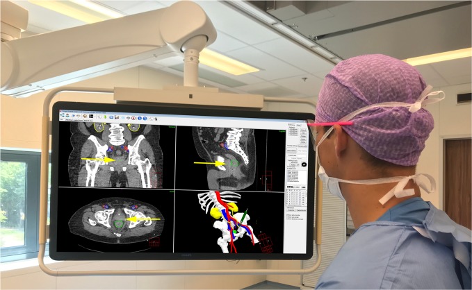
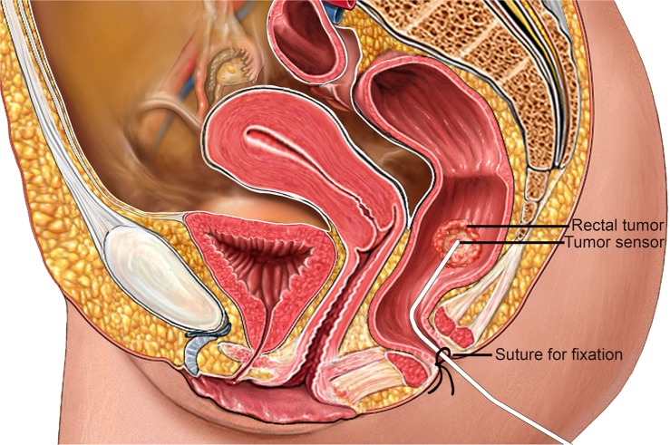
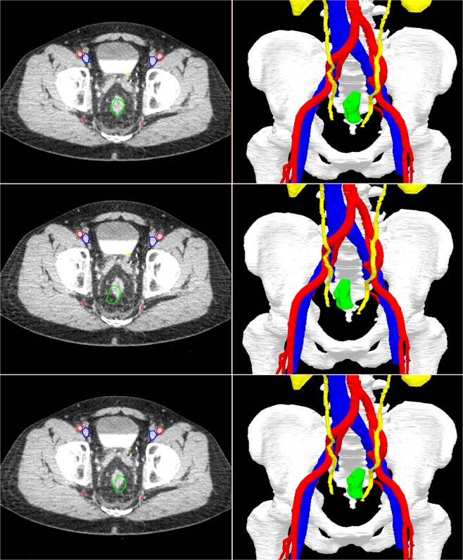
In the past decades, image-guided surgery has evolved rapidly. In procedures with a relatively fixed target area, like neurosurgery and orthopedics, this has led to improved patient outcomes. In cancer surgery, intraoperative guidance could be of great benefit to secure radical resection margins since residual disease is associated with local recurrence and poor survival. However, most tumor lesions are mobile with a constantly changing position. Here, we present an innovative technique for real-time tumor tracking in cancer surgery. In this study, we evaluated the feasibility of real-time tumor tracking during rectal cancer surgery. The application of real-time tumor tracking using an intraoperative navigation system is feasible and safe with a high median target registration accuracy of 3 mm. This technique allows oncological surgeons to obtain real-time accurate information on tumor location, as well as critical anatomical information. This study demonstrates that real-time tumor tracking is feasible and could potentially decrease positive resection margins and improve patient outcome.

Keywords: Rectal cancer; Surgical oncology.


Conflict of interest statement

Competing interestsThe authors declare no competing interests.

Figures

Fig. 1
Fig. 1. Surgical navigation user interface during surgery showing the planning CT with segmentations (top-left corner: coronal view; top-right corner: sagittal view; lower-left corner: axial view) and 3D model (lower-right corner).
Visible segmentations: bones (white), arteries (red), veins (blue), ureters/kidneys (yellow), and tumor (green). The visible slice of the CT scans is based on the location of the tip of the surgical pointer which is highlighted with yellow arrows.
Fig. 2
Fig. 2. Schematic overview of in vivo tumor sensor with the tip positioned by the surgeon at the rectal tumor.
The sensor is fixated with sutures in the perineal area (Alamy Stock Photo (https://www.alamy.com/search.html?qt=Sagittal%20View%20of%20a%20Female%20Pelvis&imgt=0), used and edited with permission).
Fig. 3
Fig. 3. An example to illustrate the real-time tumor tracking by the navigation system.
The top two images show the position of the rectal tumor in rest. In the lower four images, the rectum is moved to the right and left. Note that the green segmentation area follows the real-time tumor position in the navigation images on the right, while the CT scan on the left (still) shows the original position of the tumor. Visible segmentation: bones (white), arteries (red), veins (blue), ureters/kidneys (yellow), and tumor (green).

References

    1. Schulz C, Waldeck S, Mauer UM. Intraoperative image guidance in neurosurgery: development, current indications, and future trends. Radiol. Res. Pract. 2012;2012:197364–197364. - PMC - PubMed
    1. Claus EB, et al. Survival rates in patients with low-grade glioma after intraoperative magnetic resonance image guidance. Cancer. 2005;103:1227–1233. doi: 10.1002/cncr.20867. - DOI - PubMed
    1. Senft C, et al. Intraoperative MRI guidance and extent of resection in glioma surgery: a randomised, controlled trial. Lancet Oncol. 2011;12:997–1003. doi: 10.1016/S1470-2045(11)70196-6. - DOI - PubMed
    1. Aschendorff A, et al. Radiologically assisted navigation in cochlear implantation for X-linked deafness malformation. Cochlear Implants Int. 2009;10:14–18. doi: 10.1179/cim.2009.10.Supplement-1.14. - DOI - PubMed
    1. Tormenti MJ, et al. Intraoperative computed tomography image-guided navigation for posterior thoracolumbar spinal instrumentation in spinal deformity surgery. Neurosurgical Focus. 2010;28:E11. doi: 10.3171/2010.1.FOCUS09275. - DOI - PubMed