

Three unsuspected CT diagnoses of COVID-19
- PMID: 32285222
- PMCID: PMC7152619
- DOI: 10.1007/s10140-020-01775-4
Three unsuspected CT diagnoses of COVID-19
Abstract
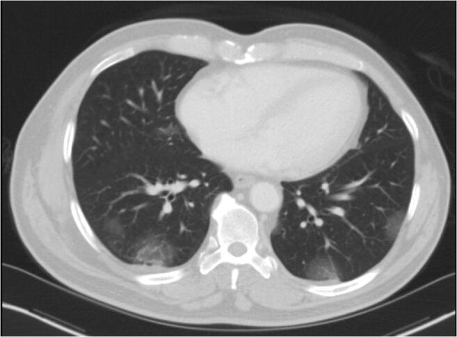
Purpose: Coronavirus disease 2019 (COVID-19) is caused by a novel strain of coronavirus named severe acute respiratory syndrome coronavirus 2 (SARS-CoV-2) that has quickly spread around the globe. Health care facilities in the USA currently do not have an adequate supply of COVID-19 tests to meet the growing demand. Imaging findings for COVID-19 are non-specific but include pulmonary parenchymal ground-glass opacities in a predominantly basal and peripheral distribution.
Methods: Three patients were imaged for non-respiratory-related symptoms with a portion of the lungs in the imaged field.
Results: Each patient had suspicious imaging findings for COVID-19, prompting the interpreting radiologist to suggest testing for COVID-19. All 3 patients turned out to be infected with COVID-19, and one patient is the first reported case of the coincident presentation of COVID-19 and an intraparenchymal hemorrhage.
Conclusion: Using imaging characteristics of COVID-19 on abdominal or neck CT when a portion of the lungs is included, patients not initially suspected of COVID-19 infection can be quarantined earlier to limit exposure to others.
Keywords: COVID-19; Ground-glass opacities; Intraparenchymal hemorrhage.
Conflict of interest statement
The authors declare that they have no conflict of interest.
Figures


References
-
- Situation Report – 55: https://www.who.int/docs/default-source/coronaviruse/situation-reports/2... [Accessed on March 15, 2020]
-
- https://www.cdc.gov/coronavirus/2019-ncov/cases-in-us.html [Accessed on March 15, 2020]
-
- https://www.cdc.gov/coronavirus/2019-ncov/lab/testing-laboratories.html [Accessed on March 12, 2020]
Publication types
MeSH terms
LinkOut - more resources
Full Text Sources
Medical
Miscellaneous
