. 2020 Apr 14;11(1):1767.
doi: 10.1038/s41467-020-15632-y.

Sex-specific innate immune selection of HIV-1 in utero is associated with increased female susceptibility to infection

Affiliations

Sex-specific innate immune selection of HIV-1 in utero is associated with increased female susceptibility to infection

Emily Adland et al. Nat Commun. .

Erratum in

Abstract

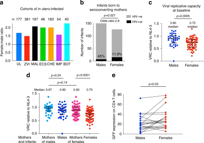
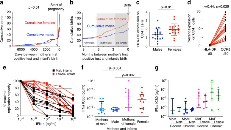
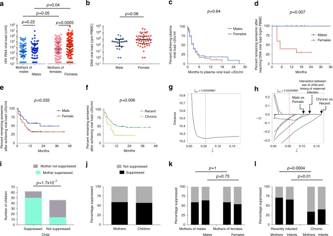
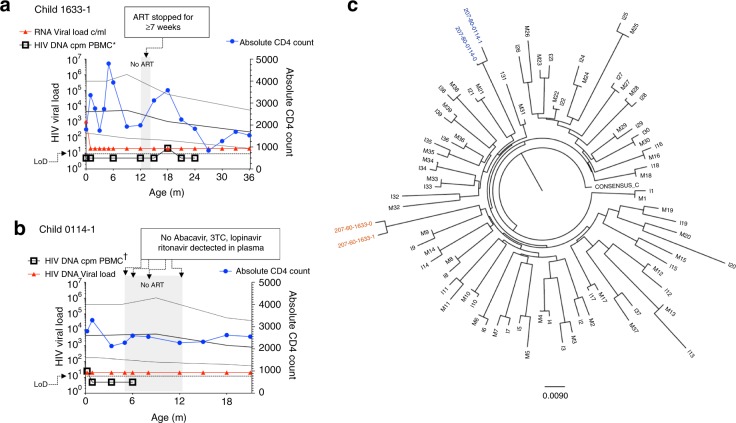
Female children and adults typically generate more efficacious immune responses to vaccines and infections than age-matched males, but also suffer greater immunopathology and autoimmune disease. We here describe, in a cohort of > 170 in utero HIV-infected infants from KwaZulu-Natal, South Africa, fetal immune sex differences resulting in a 1.5-2-fold increased female susceptibility to intrauterine HIV infection. Viruses transmitted to females have lower replicative capacity (p = 0.0005) and are more type I interferon-resistant (p = 0.007) than those transmitted to males. Cord blood cells from females of HIV-uninfected sex-discordant twins are more activated (p = 0.01) and more susceptible to HIV infection in vitro (p = 0.03). Sex differences in outcome include superior maintenance of aviraemia among males (p = 0.007) that is not explained by differential antiretroviral therapy adherence. These data demonstrate sex-specific innate immune selection of HIV associated with increased female susceptibility to in utero infection and enhanced functional cure potential among infected males.


Conflict of interest statement

The authors declare no competing interests.

Figures

Fig. 1
Fig. 1. Increased female susceptibility to in utero HIV infection compared to males.
a Female:male sex ratio in the current Ucwaningo Lwabantwana cohort of in utero HIV-infected infants and in six other similar cohorts. UL: Ucwaningo Lwabantwana; ZVI: Zvitambo; MAL: Malawi; ECS: European Collaborative Study; CHE: CHER; IMP: IMPAACT P1115; BOT: Botswana. These data refer to in utero infected infants only, except for the CHER study which included intra-partum and in utero infected infants. b Numbers of male and female infants exposed to mothers seroconverting during the pregnancy (a negative HIV antibody test followed later in the pregnancy by a positive antibody test) at Queen Nandi Hospital, Empangeni, Stanger Hospital, Stanger, and Mahatma Gandhi Memorial Hospital, between 2016 and 2018, and numbers of in utero infected infants. The numbers of exposed infants did not differ significantly between the sexes but the number of infections did (p = 0.027, 2-tailed, Fisher’s Exact test). c, d Viral replicative capacity at baseline from 101 in utero infected infants, 63 females and 38 males, and mothers. Data are presented showing medians and interquartile ranges. In panel (c), the statistical test used was the Mann–Whitney test. In panel (d) the statistical tests used were the Wilcoxon matched-pairs signed rank test (comparing males versus mothers of males), the unpaired paired t test (comparing mothers of males versus mothers of females) and the paired t test (comparing females versus mothers of females). In all cases P values were two-tailed. e In vitro HIV infection of cord blood CD4+ T-cells from 19 pairs of HIV-uninfected sex-discordant twins. The statistical test used was the Wilcoxon matched-pairs signed rank test (2-tailed). For Fig. 1c–e, source data are provided as a Source Data file.
Fig. 2
Fig. 2. Impact of timing of maternal infection in relation to sex differences in intrauterine transmission and in interferon-sensitivity of in utero transmitted viruses.
a Cumulative number of male and female in utero transmissions by time (days) between mother’s first HIV-positive test and the birth of the infant. Start of pregnancy shown as 280 days prior to delivery for simplicity (some infants were born prematurely). P value denotes sex difference in infants born to mothers whose first HIV test was prior to pregnancy (defined as ‘chronically infected’) versus those whose mothers’ first HIV test was during the pregnancy (defined as ‘recently infected’) (Fisher’s Exact test, 2-sided). b Cumulative number of male and female in utero transmissions by time (months) between mother’s first HIV-positive test and the birth of the infant in the mothers who first tested positive during the pregnancy. As in (a), start of pregnancy shown as 280 days prior to delivery. c HLA-DR expression in cord blood CD4+ T-cells in 19 sets of HIV-uninfected sex-discordant twins. Medians and interquartile ranges are shown; the statistical test used was the unpaired t test (2-sided). d Correlation between CCR5 expression on CD4 T cells on day 10 and HLA-DR expression on CD4 T cells on day 0 in females. The correlation between GFP on day 10 with baseline HLA-DR expression was r = 0.35, p = 0.07 in females. In males, correlations between HLA-DR expression on day 0 and CCR5 expression on day 10 and GFP expression on day 10 were weaker (r = 0.05 and 0.18, respectively; p = 0.41 and 0.23, respectively). In all cases, the t test used and p values were one-sided). e, f IFN-α sensitivity of Gag-Pro-NL4-3 chimeric viruses derived from baseline virus from 22 infants (10 male, 12 female). Raw data for the 22 infants are shown in panel (e) IC80 values and p values from these raw data are shown for the 22 infants and mothers of the infants in panel (f). The statistical test used in each case was the Mann–Whitney test, p values were 2-sided. Medians and interquartile ranges are shown. g Data as shown in panels (e, f) but also showing timing of maternal infection as categorised above: ‘chronic’: first HIV test prior to pregnancy; or ‘recent’: first HIV test during current pregnancy. Medians and interquartile ranges are shown. MoM: mother of male child; MoF: mother of female child. Source data are provided as a Source Data file.
Fig. 3
Fig. 3. Impact of sex differences and timing of maternal infection on outcome post-infection on plasma RNA and DNA viral loads, and viral rebound on ART.
a Baseline plasma RNA viral loads in 177 mother-child pairs. The statistical tests used were the Mann–Whitney test (comparing males versus females and comparing mothers of males versus mothers of females) and the Wilcoxon matched-pairs signed rank test (comparing males versus mothers of males and comparing females versus mothers of females). In each case, p values were 2-sided. Medians and interquartile ranges are shown. b Baseline total DNA viral loads in 69 in utero-infected infants (48 female, 21 male). The statistical test used was the Mann–Whitney test, the p value was 2-sided. cf Kaplan–Meier curves showing time to achieve plasma HIV RNA load of <20 copies/ml in males and females (panel c), time to viral rebound (>20 copies/ml) in males and females (panels d, e), time to viral rebound (>20 copies/ml) in in utero-infected children born to recently and chronically infected mothers (panel f). In panel D, data are shown from the 17 subjects who reached DNA viral loads reported as 0 cpm PBMC. In each case, the statistical tests used were the Log-rank (Mantel–Cox) test. g The cross-validation error for a grid of LASSO penalty parameters ξ in order to determine the optimal amount of penalisation. h The coefficient paths of the (standardised) regression parameter estimates versus the LASSO penalty parameter ξ; the optimal ξ is indicated by the vertical red dashed line. Re-standardised coefficients of the coefficients selected by the model were: Recent maternal infection: −0.552; Interaction between infant sex and timing of maternal infection: −0.146; Male sex of infant: −0.101; Age of infant at baseline: −0.023; plasma RNA viral load at baseline: 7.4 × 10⁻⁹. Parameters not selected were: infant baseline absolute CD4 count, CD4%, absolute CD8 count, CD8%, and CD4:CD8 ratio. i–l. Suppression of viraemia (plasma viral load <20 copies/ml) in mother-child pairs using most recent timepoint studied. Panels ij: all mother child pairs studied (n = 108); panel k: mothers of males and males (39 pairs) compared with mothers of females and females (69 pairs); panel l: recently infected mothers and children (73 pairs) compared with chronically infected mothers and children (35 pairs). In panels il, the statistical tests used was Fisher’s Exact test; p values were 2-sided. Source data are provided as a Source Data file.
Fig. 4
Fig. 4. Lack of viral rebound in two male subjects following significant periods of ART non-adherence.
a Male subject 1633-1 received no ART for at least 7 weeks at 12–14 months’ age: pharmacy records show that the aunt of the child did not collect any ART medication for 2 months following the return of the child’s mother to work 12 m after the child’s birth. ART was restarted 7 days prior to the viral load test at 15 m. Undetectable DNA viral loads are shown as 5 HIV DNA cpm PBMC. Only the 18 m timepoint was above the limit of detection (DNA load 21 cpm PBMC). b Male subject 0114-1: none of the drugs prescribed were detected in plasma tested over a 7-month period (age 5–12 months). At age 15 months and 18 months, only 1 and 2 of the 4 drugs prescribed were detected at therapeutic levels, respectively. Undetectable DNA viral loads are shown as 5 HIV DNA cpm PBMC. Only the baseline timepoint (at 7 days’ age) was above the limit of detection (DNA load 25 cpm PBMC). c Gag was amplified from plasma from the baseline sample from both these mother-child pairs and the sequences determined clustered on a phylogenetic tree, consistent with HIV infection and mother-to-child transmission in each case. Diagnosis in these cases, as in all the in utero-infected infants in the cohort, was determined by 2 or more tests from separate blood samples detecting HIV nucleic acids.

References

    1. Darwin, C. R. The Descent of Man and Selection in Relation to Sex (John Murray, London, 1871).
    1. Klein SL, Flanagan KL. Sex differences in immune responses. Nat. Rev. Immunol. 2016;16:626–638. - PubMed
    1. Flanagan KL, Fink AL, Plebanski M, Klein SL. Sex and gender differences in the outcomes of vaccination over the life course. Annu. Rev. Cell Dev. Biol. 2017;33:577–599. - PubMed
    1. Meier A, et al. Sex differences in the TLR-mediated response of pDCs to HIV-1 are associated with higher immune activation in women. Nat. Med. 2009;15:955–959. - PMC - PubMed
    1. Souyris M, et al. TLR7 escapes X chromosome inactivation in immune cells. Sci. Immunol. 2018;3:eaaqp8855. - PubMed

Publication types