. 2020 Apr 14;10(1):6384.
doi: 10.1038/s41598-020-63136-y.

Cell surface processing of the P1 adhesin of Mycoplasma pneumoniae identifies novel domains that bind host molecules

Affiliations

Cell surface processing of the P1 adhesin of Mycoplasma pneumoniae identifies novel domains that bind host molecules

Michael Widjaja et al. Sci Rep. .

Abstract

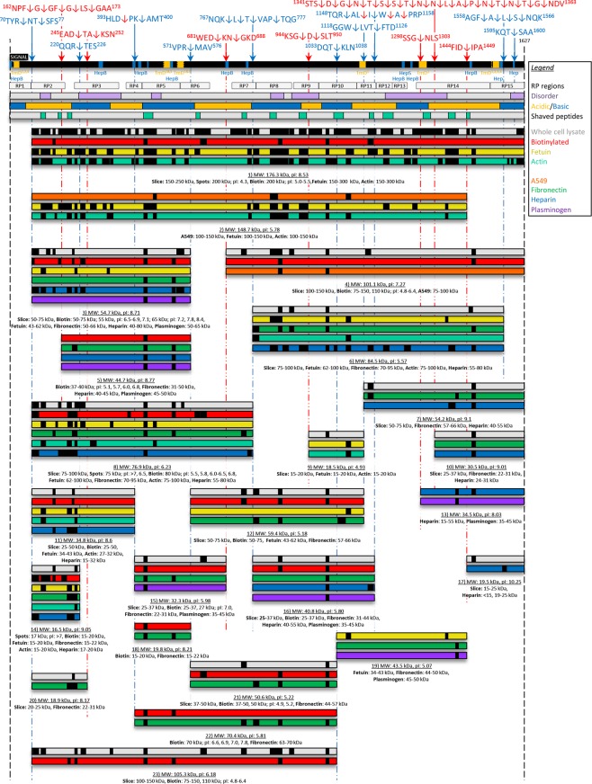
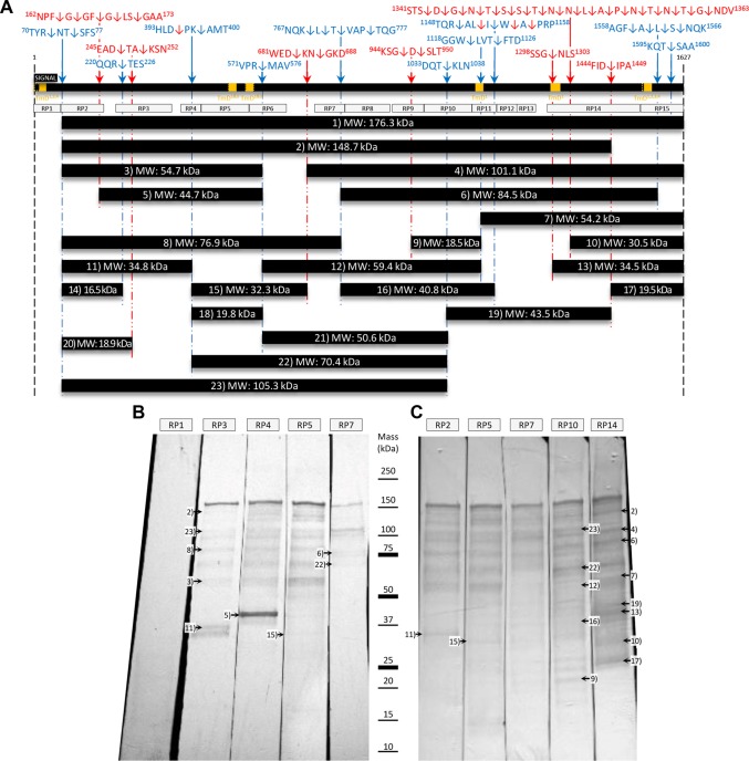
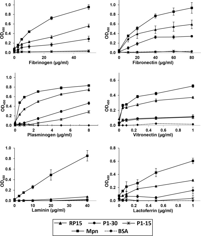
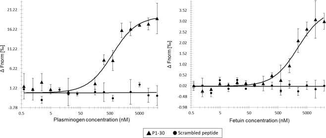
Mycoplasma pneumoniae is a genome reduced pathogen and causative agent of community acquired pneumonia. The major cellular adhesin, P1, localises to the tip of the attachment organelle forming a complex with P40 and P90, two cleavage fragments derived by processing Mpn142, and other molecules with adhesive and mobility functions. LC-MS/MS analysis of M. pneumoniae M129 proteins derived from whole cell lysates and eluents from affinity matrices coupled with chemically diverse host molecules identified 22 proteoforms of P1. Terminomics was used to characterise 17 cleavage events many of which were independently verified by the identification of semi-tryptic peptides in our proteome studies and by immunoblotting. One cleavage event released 1597TSAAKPGAPRPPVPPKPGAPKPPVQPPKKPA1627 from the C-terminus of P1 and this peptide was shown to bind to a range of host molecules. A smaller synthetic peptide comprising the C-terminal 15 amino acids, 1613PGAPKPPVQPPKKPA1627, selectively bound cytoskeletal intermediate filament proteins cytokeratin 7, cytokeratin 8, cytokeratin 18, and vimentin from a native A549 cell lysate. Collectively, our data suggests that ectodomain shedding occurs on the surface of M. pneumoniae where it may alter the functional diversity of P1, Mpn142 and other surface proteins such as elongation factor Tu via a mechanism similar to that described in Mycoplasma hyopneumoniae.


Conflict of interest statement

The authors declare no competing interests.

Figures

Figure 1
Figure 1
Cleavage map of the P1 adhesin. The full length proteoform (1627 amino acids) is shown as the black bar with cleavage sites above and fragments below this bar. Cleavage sites identified from dimethyl labelling and semi-tryptic sites are shown as the blue and red arrows, respectively. Sequences where these cleavage sites occur are also shown. Putative heparin binding sites (Hep, blue boxes, motif: X-[HRK]-[HRK]-X-[HRK]-X), heparan sulfate binding sites (HepS, blue boxes, motif: X-[HRK]-X-[HRK]-[HRK]-X), clusters of basic residues (HepB, blue boxes, motifs: X-[HRK]-X(0,2)-[HRK]-X(0,2)-[HRK]-X or X-[HRK]-X(1,3)-[HRK]-X(1,3)-[HRK]-X), and transmembrane domains (TmD, yellow boxes, TmD1 predicted by TMpred, TmD2 previously predicted, TmD3 previously predicted, and TmD4 previously predicted in a P1 paralog) are shown within the black bar. Putative transmembrane domains and the location of 15 subregions of P1 (grey ‘RP’ boxes) expressed as recombinant proteins from an earlier study are shown. Predicted disordered regions appear as purple boxes in the grey bar. Acidic and basic regions within P1 are identified as yellow and blue bars, respectively. Peptides released from surface shaving experiments and identified by mass spectrometry are shown in the light green boxes within the grey bar. Grey bars represent fragments of P1 identified during SDS-PAGE of whole cell lysates. Red bars represent fragments of P1 recovered from lysates of M. pneumoniae that have their surface proteins labelled with biotin (surface exposed fragments of P1). Peptides identified by mass spectrometry of P1 fragments isolated from affinity chromatography of fetuin (yellow bars), actin (light blue bars), A549 surface protein complexes (orange bars), fibronectin (green bars), heparin (blue bars), and plasminogen (purple bars) are shown.
Figure 2
Figure 2
Immunoblots of cell lysates of M. pneumoniae probed with sera raised against regions within P1. Sera raised against 15 different regions (‘RP’ boxes) of P1 were a gift from R. Dumke. (Top panel) Simplified cleavage map depicting the P1 adhesin, cleavage sites, and the 15 regions of P1 that have been previously cloned and expressed as recombinant fragments in E. coli. The mass of full length and smaller proteoforms of P1 as predicted by ProtParam. (Bottom panel) Immunoblots depicting M. pneumoniae cell lysate probed with the panel of anti-recombinant P1 sera. All the immunoblot lanes are part of the same blot. The membrane was blocked and then sliced to separate lanes before incubating with the described P1 sera. (Bottom right) Immunoblots with the intensity adjusted to highlight low abundant bands. Proteins migrating with masses similar to P1 proteoforms identified by LC-MS/MS have been marked on the immunoblot.
Figure 3
Figure 3
Concentration-dependent binding of the C-terminus of P1 to different human proteins. Microtitre plate binding assays were used to measure the binding abilities of RP15, P1-30, and P1-15 to human plasminogen and to different components of the human extracellular matrix. Bovine serum albumin (BSA) and whole cell lysate proteins of M. pneumoniae (Mpn) were used as a negative and positive control, respectively. Results are shown from a single experiment with a mean and standard deviation of eight replicates. The experiment was independently repeated twice.
Figure 4
Figure 4
Plasminogen and fetuin binding by P1-30 using microscale thermophoresis. Left: Thermophoretic output representing P1-30 (triangles) binding to plasminogen with a KD of 554 nM. A scrambled version of P1-30 (circles) could not be assigned a KD value. Right: Thermophoretic output representing P1-30 binding to fetuin with a KD of 2 μM. The scrambled peptide could not be assigned a KD value.
Figure 5
Figure 5
Binding of the C-terminus of P1 to immobilized A549 cells. Proteins, peptides, or A549 cells were immobilised in wells of a 96-well plate and binding was measured in a microtitre plate binding assay. ‘Without A549’ cells: protein and peptides were immobilised and detected by corresponding antisera (peptides with anti-RP15). ‘With A549 cells’: A549 cells were immobilised first before incubating with proteins and peptides. Whole antigen of M. pneumoniae (Mpn), recombinant PdhB, and RP8 served as positive and negative controls, respectively. Bars represent mean and standard deviation of eight replicates from a single experiment. The experiment was independently repeated twice.

References

    1. Krause DC, Balish MF. Structure, function, and assembly of the terminal organelle of Mycoplasma pneumoniae. FEMS Microbiol Lett. 2001;198:1–7. doi: 10.1111/j.1574-6968.2001.tb10610.x. - DOI - PubMed
    1. Baseman JB, Cole RM, Krause DC, Leith DK. Molecular basis for cytadsorption of Mycoplasma pneumoniae. J Bacteriol. 1982;151:1514–1522. doi: 10.1128/JB.151.3.1514-1522.1982. - DOI - PMC - PubMed
    1. Feldner J, Gobel U, Bredt W. Mycoplasma pneumoniae adhesin localized to tip structure by monoclonal antibody. Nature. 1982;298:765–767. doi: 10.1038/298765a0. - DOI - PubMed
    1. Krause DC. Mycoplasma pneumoniae cytadherence: unravelling the tie that binds. Mol Microbiol. 1996;20:247–253. doi: 10.1111/j.1365-2958.1996.tb02613.x. - DOI - PubMed
    1. Seto S, Layh-Schmitt G, Kenri T, Miyata M. Visualization of the attachment organelle and cytadherence proteins of Mycoplasma pneumoniae by immunofluorescence microscopy. J Bacteriol. 2001;183:1621–1630. doi: 10.1128/JB.183.5.1621-1630.2001. - DOI - PMC - PubMed

Publication types