. 2020 Apr 14;10(1):6405.
doi: 10.1038/s41598-020-63042-3.

Immunomodulatory role of Interleukin-33 in large vessel vasculitis

Affiliations

Immunomodulatory role of Interleukin-33 in large vessel vasculitis

Anne-Claire Desbois et al. Sci Rep. .

Abstract

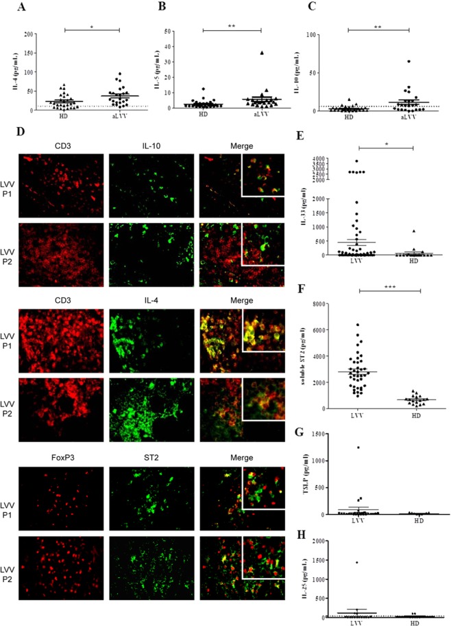
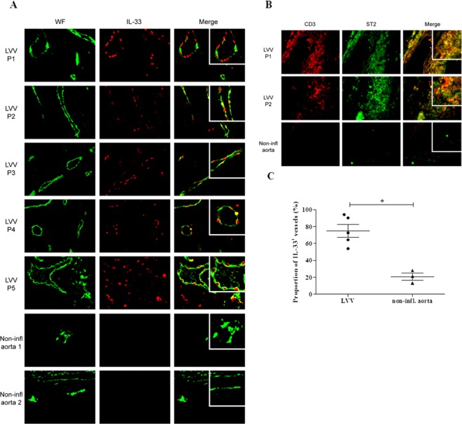
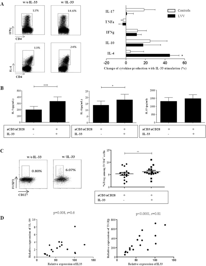
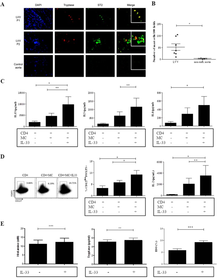
The mechanisms regulating inflammation in large vessels vasculitis (LVV) are poorly understood. Interleukin 33 (IL-33) has been shown to license innate and adaptive immunity by enhancing Th2 cytokines production. We aimed to examine the role of IL-33 in the immunomodulation of T cell activation in LVV. T cell homeostasis and cytokines production were determined in peripheral blood from 52 patients with giant cell arteritis (GCA) and 50 healthy donors (HD), using Luminex assay, flow cytometry, quantitative RT-PCR and by immunofluorescence analysis in inflammatory aorta lesions. We found increased level of IL-33 and its receptor ST2/IL-1R4 in the serum of patient with LVV. Endothelial cells were the main source of IL-33, whereas Th2 cells, Tregs and mast cells (MC) express ST2 in LVV vessels. IL-33 had a direct immunomodulatory impact by increasing Th2 and Tregs. IL-33 and MC further enhanced Th2 and regulatory responses by inducing a 6.1 fold increased proportion of Tregs (p = 0.008). Stimulation of MC by IL-33 increased indoleamine 2 3-dioxygenase (IDO) activity and IL-2 secretion. IL-33 mRNA expression was significantly correlated with the expression of IL-10 and TGF-β within aorta inflammatory lesions. To conclude, our findings suggest that IL-33 may exert a critical immunoregulatory role in promoting Tregs and Th2 cells in LVV.


Conflict of interest statement

The authors declare no competing interests.

Figures

Figure 1
Figure 1
Expression of Th2 and anti-inflammatory cytokines in LVV. (A–C) PBMC of active LVV patients (aLVV) (n = 23) and HD (n = 27) were stimulated for 4 hours with PMA-ionomycine. Expression levels of interleukin 4 (IL-4), interleukin 5 (IL-5) and interleukin 10 (IL-10) measured in supernatant are represented. The levels of IL-4, IL-5 and IL-10 are higher in LVV patients as compared to HD [p = 0.01; p = 0.002 and p = 0.003, respectively]. We used a Mann Whitney test. These data are shown as the mean ± SEM. (D) Immunofluorescence analysis of inflammatory lesions from LVV patients reveals the expression of Th2 (here IL-4) and IL-10 cytokines partially by CD3 positive cells. Expression of ST2 by Treg (defined as FOXP3 cells) by immunofluorescence analysis. (E) Expression level of IL-33 in LVV (n = 44) patients serum was higher compared to HD (n = 18), *P < 0.05. (F) Mean level of soluble ST2 was increased in LVV patient sera (n = 38) as compared to HD (n = 17), ***P < 0.001. (G) Level of TSLP in LVV (n = 16) patients serum compared to HD (n = 9). (H) Level of IL-25 in LVV (n = 16) patients serum compared to HD (n = 9).
Figure 2
Figure 2
IL-33 and its receptor ST2 were overexpressed in LVV. (A) Immunofluorescence analyses of aorta tissue specimens from LVV patients revealed that IL-33 was mainly expressed within adventitial vessels and co-localized with endothelial cells with positive von willebrand factor (WF) staining. Immunofluorescence analysis of non-inflammatory aorta did not reveal IL-33 positive staining. (B) Expression of ST2 was mainly observed within the inflammatory infiltrates in aorta tissue specimen. Immunofluorescence analyses of one non-inflammatory aorta show vessels (WF positive staining) without positive IL-33 staining and absence of ST2 staining. Immunofluorescence analysis of non-inflammatory aorta did not reveal ST2 positive staining. (C) The proportion of vessels with positive IL-33 staining was higher in aorta from LVV patients (n = 5) as compared to non-inflammatory controls (n = 3), *P < 0.05. These data are shown as the mean ± SEM.
Figure 3
Figure 3
Il-33 predominantly induced a Th2 and regulatory immune response in LVV. (A) Freshly isolated PBMC from LVV patients (with corticosteroids<15 mg/day) were cultured with anti-CD3/CD28 with or without IL-33 stimulation for 5 days. The secretion of Th1 and Th2 cytokines was assessed by flow cytometry. Left panel: Dot plots representing IFNγ and IL-4 secreting CD4+ T cells with or without IL-33 stimulation. Right panel: Changes of cytokine production in PBMC stimulated with IL-33 compared to PBMC not stimulated. The proportion of IL4-secreting CD4+ T cells was increased with IL-33 stimulation (p = 0.01) in LVV patients (n = 13) but not in HD (n = 6). These data are shown as the mean ± SEM. These results are from 13 independent experiments. (B) After 5 days of culture, quantitative determination of cytokines was performed in culture supernatants (n = 12) of LVV PBMC. IL-33 stimulation led to a significant increase of IL-5 and IL-4 secretion, *p < 0.05, ***P < 0.001. (C) Left panel: Dot plots representing CD25hiFOXP3+ and CD127lowFOXP3+ CD4+ cells are shown. On the left, PBMC of one LVV patient are cultured for 5 days without IL-33. The frequency of CD127lowFOXP3+ CD4+ cells is shown. On the right, PBMC of the same LVV patient were cultured for 5 days with IL-33. Right panel: Corresponding results of 15 LVV patients. The frequency of Tregs was increased in stimulated PBMC with IL-33 as compared to those without IL-33. These results are from 4 independent experiments. These data are shown as the mean ± SEM. **P < 0.01. The statistical test used was a Wilcoxon matched pair test. (D) We next assessed by quantitative PCR the expression of IL-33, ST2 and Th1 and Th2 cytokines within LVV aortic lesions (n = 18). Relative expression of IL-33 mRNA was significantly correlated with the expression of IL-10 mRNA [r = 0.6 (p = 0.008)]. Relative expression of IL-33 mRNA was significantly correlated with the expression of TGF-b mRNA [r = 0.8 (p < 0.0001)].
Figure 4
Figure 4
IL-33 enhanced a regulatory and Th2 immune response through MC. (A) Immunofluorescence staining of LVV inflammatory lesions of 2 LVV patients and 1 non-inflammatory aorta control for ST2 and MC. (B) The number of MC within aorta was higher in LVV patients (n = 7) than in non-inflammatory controls (n = 3) *P < 0.05. These data are shown as the mean ± SEM. (C) Quantitative determination of Th2 cytokines in culture supernatants (n = 8) of MC and CD4+ T cells. IL-33-stimulated MC led to a significant increase in IL-5, IL-13 and IL-4 secretion. *P < 0.05, **P < 0.01. These data are shown as the mean ± SEM. (D) Left panel: Dot plots representing the proportion of CD25highFOXP3+ and CD127lowFOXP3+ CD4+ cells are shown. On the left: CD4+ T cells of one LVV patient are cultured for 5 days without IL-33. The proportion of CD127lowFOXP3+ CD4+ cells is shown. On the middle: MC and stimulated CD4 T cells were cultured for 5 days without IL-33. On the right: MC and stimulated T CD4 cells were cultured for 5 days with IL-33. This patient is representative of the whole cohort (n = 8). Middle panel: The corresponding results of 8 LVV patients. MC alone and IL-33-stimulated MC promote the increase of Tregs frequency. *P < 0.05, **P < 0.01. These results are from 8 independent experiments. These data are shown as the mean ± SEM. Right panel: Quantitative determination of cytokines was performed in culture supernatants (n = 8). IL-33-stimulated MC led to a significant increase in IL-2 secretion. *P < 0.05, **P < 0.01. These data are shown as the mean ± SEM. (E) Left and middle panels: MC were incubated with LVV serum (n = 11) with or without IL-33 stimulation. IL-33 led to a discreet increase of MC degranulation (histamine and tryptase). Right panel: Indoleamine 2 3-dioxygenase (IDO) activity was dramatically increased in MC incubated with IL-33 as compared to those not stimulated with IL-33.**P < 0.01, ***P < 0.001. These data are shown as the mean ± SEM.

References

    1. Watanabe R, Zhang H, Berry G, Goronzy JJ, Weyand CM. Immune checkpoint dysfunction in large and medium vessel vasculitis. Am. J. Physiol. Heart Circ. Physiol. 2017;312:H1052–H1059. doi: 10.1152/ajpheart.00024.2017. - DOI - PMC - PubMed
    1. Saadoun, D. et al. Th1 and Th17 cytokines drive Takayasu Arteritis inflammation. Arthritis Rheumatol. Hoboken NJ, 10.1002/art.39037 (2015). - PubMed
    1. Deng J, Younge BR, Olshen RA, Goronzy JJ, Weyand CM. Th17 and Th1 T-cell responses in giant cell arteritis. Circulation. 2010;121:906–915. doi: 10.1161/CIRCULATIONAHA.109.872903. - DOI - PMC - PubMed
    1. Weyand CM, Goronzy JJ. Immune mechanisms in medium and large-vessel vasculitis. Nat. Rev. Rheumatol. 2013;9:731–740. doi: 10.1038/nrrheum.2013.161. - DOI - PMC - PubMed
    1. Terrier B, et al. Interleukin-21 modulates Th1 and Th17 responses in giant cell arteritis. Arthritis Rheum. 2012;64:2001–2011. doi: 10.1002/art.34327. - DOI - PubMed

Publication types

Substances