. 2020 Apr 14;10(1):6391.
doi: 10.1038/s41598-020-63318-8.

C-reactive protein (CRP) recognizes uric acid crystals and recruits proteases C1 and MASP1

Affiliations

C-reactive protein (CRP) recognizes uric acid crystals and recruits proteases C1 and MASP1

Anika Alberts et al. Sci Rep. .

Abstract

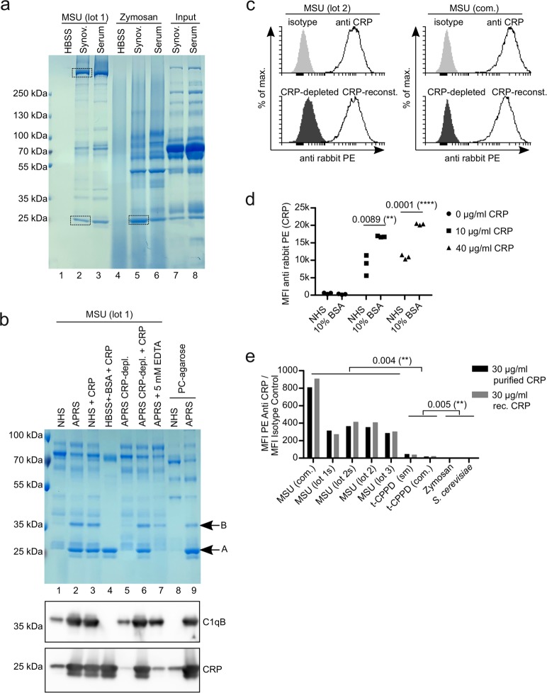
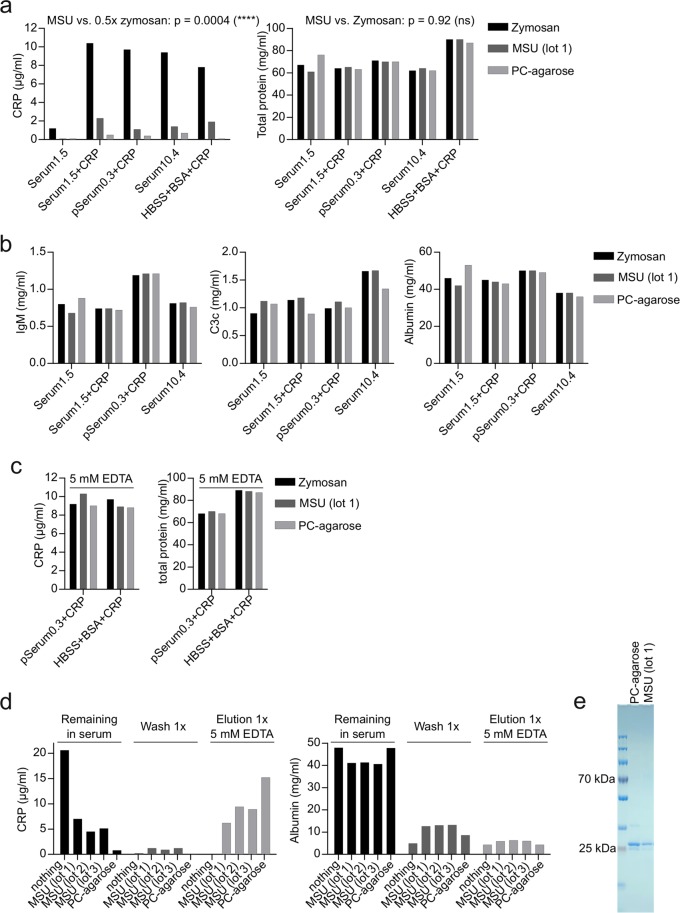
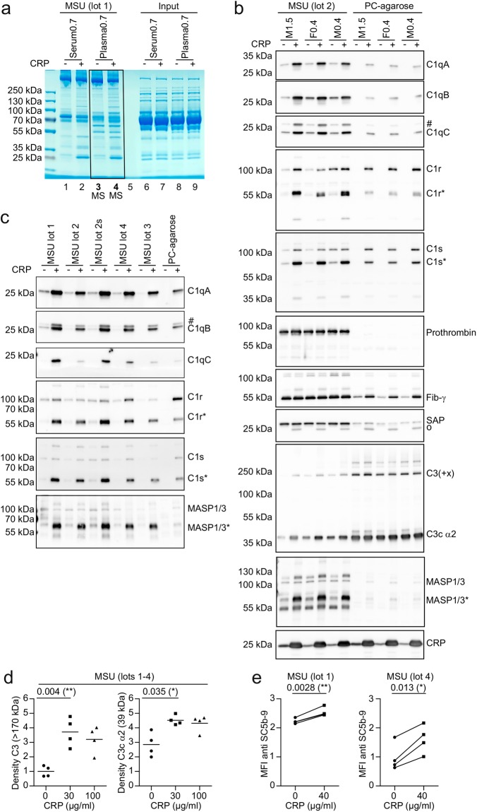
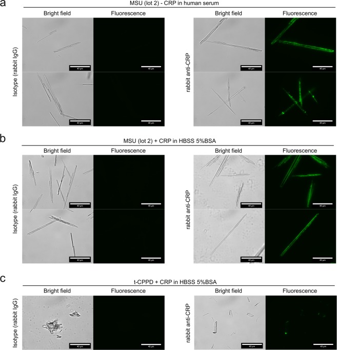
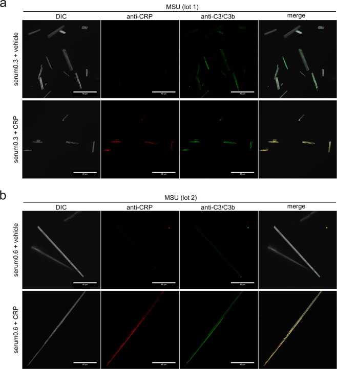
Gout is caused by crystallization of uric acid in the form of monosodium urate (MSU) crystals, which induce a sterile inflammatory response that is hardly distinguishable from microbe-induced inflammatory responses. It is unclear, if MSU crystals (like microbes) are recognized by specific pattern recognition receptors. To identify possible soluble pattern recognition molecules for MSU crystals, we purified MSU-binding proteins from human body fluids. We identified C-reactive protein (CRP) as a major MSU-binding protein. Binding of CRP was strong enough to specifically deplete CRP from human serum. We found that CRP was required for fixation of complement components C1q, C1r, C1s and MASP1. Thus, we have identified a pattern recognition molecule for MSU crystals that links to the activation of complement. Notably, CRP does not show an even binding to the complete surface of the crystals. It rather binds to edges or distinct faces of the crystals.


Conflict of interest statement

The authors declare no competing interests.

Figures

Figure 1
Figure 1
CRP binds to MSU crystals. (a) Synovial fluid or serum from a patient with pseudogout was incubated with MSU crystals (lot 2) or zymosan for 45 min at 37 °C. Unbound proteins were washed away and bound proteins were eluted and subjected to SDS-PAGE and visualized by coomassie. Bands excised for mass spectrometric analyses are indicated (rectangles). (b) Normal human serum (NHS), acute phase reaction serum (APRS; CRP around 100 µg/ml) or HBSS (always containing Ca2+) with or without depletion of CRP or addition of purified CRP to 100 µg/ml were incubated with MSU crystals or phosphorylcholine-agarose (PC-agarose) for 45 min at 37 °C. Bound proteins were eluted and subjected to SDS-PAGE and visualized by coomassie. In addition, the same samples were analyzed by Western blot analysis using CRP antibody (lower panel) or C1qB antibody (middle panel). (c) Human serum (CRP = 10.4 µg/ml) was left untreated or CRP was depleted with PC-agarose or was depleted and then reconstituted with 10 µg/ml purified CRP. All three sera were incubated with two preparations of MSU crystals (lot 1 and a commercial preparation (com.)). CRP was stained with CRP antibody and anti-rabbit-PE and analyzed using a flow cytometer. (d) Three different preparations of MSU crystals (lot 1, lot 2 and a commercial preparation) were incubated with either pool serum (NHS) (CRP < 0.3 µg/ml) or 10% BSA in HBSS, both with purified CRP added to the indicated concentrations. Binding of CRP to the crystals was analyzed as in c.  Median fluorescent intensity (MFI) is shown. (e) Four different preparations of MSU crystals (one commercial (com) and three self-made (untreated or sonicated (s)), two preparations of t-CPPD (commercial (com.) and self-made (sm)) and two preparations of S. Cerevisiae (zymosan and heat-inactivated yeast) were incubated with NHS (CRP < 0.3 µg/ml) with either 30 µg/ml purified CRP or 30 µg/ml recombinant (rec.) CRP added. Binding of CRP to the crystals and fungal particles was analyzed as in c. MFI of staining with CRP antibodies was divided by MFI of isotype controls. Uncropped images of gels and Western blot are shown in Fig. S3.
Figure 2
Figure 2
MSU crystals specifically purify CRP (a) 200 µl of serum of a single donor with 1.5 µg/ml CRP (Serum1.5) with or without addition of 10 µg/ml purified CRP, a pool serum with 0.3 µg/ml CRP (pSerum0.3) with 10 µg/ml purified CRP added, a single donor serum with 10.4 µg/ml CRP (Serum10.4) and HBSS 10%BSA with 10 µg/ml purified CRP added, were incubated with 3 mg zymosan, 5 mg MSU (lot 1) or 35 µl PC-agarose for 45 min at 37 °C. Samples were centrifuged and the supernatants were analyzed for CRP and total protein concentration. Using a one-sample t-test, the p-value of MSU samples compared to 50% of CRP concentration of the corresponding zymosan sample was calculated. For the difference of total protein in zymosan or MSU treated samples a paired t-test was used. (b) The concentration of IgM, C3c and albumin was analyzed in samples from a by turbidimetry. (c) For the indicated CRP-containing solutions the experiment was repeated in the presence of 5 mM EDTA and the supernatants were analyzed for CRP and total protein concentration. (d) 200 µl of pool serum containing 20 µg/ml purified CRP was incubated with nothing, three distinct preparations of MSU (5 mg each) or 35 µl PC-agarose for 45 min at 37 °C, washed 1x with HBSS for 5 min and then eluted with 5 mM EDTA in HBSS. Supernatants of each step were analyzed for CRP and albumin concentration by turbidimetry. (e) 100 µl of a serum containing 30 µg/ml CRP was incubated with 9 mg MSU or 35 µl PC-agarose for 45 min at 37 °C. MSU/PC-agarose was washed 5x in HBSS, and CRP was eluted by HBSS + 5 mM EDTA. Eluted proteins were applied to SDS-PAGE and proteins were visualized by coomassie staining. Uncropped image of the gel is shown in Fig. S3 (right gel). Each experiment was repeated at least once with similar results.
Figure 3
Figure 3
CRP recruits C1 and MASP1 to the surface of MSU crystals. (a) 40 µg/ml purified CRP or vehicle was added to serum and plasma (hirudin) from the same healthy male donor (CRP concentration of 0.7 µg/ml). Serum and plasma was incubated with MSU crystals (90 mg/ml) for 45 min at 37 °C. Crystals were washed extensively and bound proteins were eluted in SDS buffer, separated on a polyacrylamide gel and stained with coomassie. (b) 40 µg/ml purified CRP or vehicle was added to plasma of three donors, two male (M), one female (F). Numbers indicate original CRP concentration in µg/ml. Each plasma was incubated with MSU or PC-agarose and bound proteins were eluted as in a. Eluted proteins were subjected to Western blot analysis using the indicated antibodies. Protein names are indicated at the expected molecular weight. Cleaved/active forms of proteins are indicated with an *. Background signals due to inefficient stripping are indicated with a #. SAP antibody shows a background band at the position of CRP (o), which is likely due to cross-reactivity. (c) Five distinct preparations (4 lots, one of which untreated and sonicated (s)) of MSU crystals were incubated with pool serum containing vehicle or 40 µg/ml purified CRP. Bound proteins were eluted and analyzed as in b. (d) Purified CRP was added to NHS (originally containing 0.4 µg/ml CRP) to a concentration of 0, 30, or 100 µg/ml and was incubated with four distinct preparations of MSU crystals (lots 1-4) for 30 min at 37 °C. Bound proteins were eluted as in a and subjected to Western blot analysis using C3 antibody. Signal for full length C3 (>170 kDa) and its degradation product C3c α2 (39 kDa) were quantified by densitometry and normalized to the intensity of full length C3 in the absence of added CRP. (The corresponding Western blot is shown in Fig. S5). A paired two-tailed t-test was used to compare vehicle (0) with 30 µg/ml purified CRP. (e) Two distinct lots of MSU crystals were incubated in 4 individual human sera with 0 or 40 µg/ml purified CRP added for 30 min at 37 °C, extensively washed and stained with rabbit anti SC5b-9 plus anti rabbit PE. MSU crystals were analyzed using a flow cytometer. Median fluorescence intensity (MFI) of PE / 1000 is shown. A paired two-tailed t-test was used to compare vehicle (0) with 40 µg/ml purified CRP. Experiments from b-e are representative of at least 2 independent experiments. Uncropped images of the gel and Western blots are shown in Fig. S4 and S5.
Figure 4
Figure 4
CRP predominantly recognizes the edges or specific faces of MSU crystals. (a) MSU crystals (lot 2) were incubated for 30 min at 37 °C with human serum (CRP 0.2 µg/ml) with 30 µg/ml recombinant CRP added. CRP binding was analyzed using CRP antibody and anti-rabbit-AlexaFluor488. AlexaFluor488-fluorescence of the crystals was detected by fluorescence microscopy; scale bar = 40 µm. (b) MSU crystals (lot 2) were incubated as described in a in 30 µg/ml CRP-containing HBSS 5% BSA with 30 µg/ml recombinant CRP added. Microscopic detection of bound CRP performed as in a. (c) t-CPPD (self-made) was incubated in HBSS 5% BSA with 30 µg/ml recombinant CRP added as described in b. Microscopic detection of bound CRP performed as in a. Each experiment is representative of at least 2 independent experiments.
Figure 5
Figure 5
Co-localization of CRP and C3 on opsonized MSU crystals. (a) Confocal microscopy of MSU crystals (lot 1), which were incubated for 30 min at 37 °C with human serum (CRP 0.3 µg/ml) with or without addition of 40 µg/ml purified CRP, washed extensively and stained with rabbit anti-CRP plus anti rabbit AF568 (red) and mouse anti-C3/C3b plus anti mouse AF488 (green). DIC = digital interference contrast; scale-bar = 40 µm. (b) MSU crystals lot 2 were incubated in human serum (CRP 0.6 µg/ml) with or without addition of 40 µg/ml purified CRP, stained and microscopically detected as in a.

References

    1. Kuo CF, Grainge MJ, Zhang W, Doherty M. Global epidemiology of gout: prevalence, incidence and risk factors. Nature reviews Rheumatology. 2015;11:649–662. doi: 10.1038/nrrheum.2015.91. - DOI - PubMed
    1. Neogi T. Clinical practice. Gout. The New England journal of medicine. 2011;364:443–452. doi: 10.1056/NEJMcp1001124. - DOI - PubMed
    1. Byers PH, Ward PA, Kellermeyer RW, Naff GB. Complement as a mediator of inflammation in acute gouty arthritis. II. Biological activities generated from complement by the interaction of serum complement and sodium urate crystals. The Journal of laboratory and clinical medicine. 1973;81:761–769. - PubMed
    1. Naff GB, Byers PH. Complement as a mediator of inflammation in acute gouty arthritis. I. Studies on the reaction between human serum complement and sodium urate crystals. The Journal of laboratory and clinical medicine. 1973;81:747–760. - PubMed
    1. di Giovine FS, Malawista SE, Thornton E, Duff GW. Urate crystals stimulate production of tumor necrosis factor alpha from human blood monocytes and synovial cells. Cytokine mRNA and protein kinetics, and cellular distribution. The Journal of clinical investigation. 1991;87:1375–1381. doi: 10.1172/JCI115142. - DOI - PMC - PubMed

Publication types