Case Reports
. 2020 Apr 14;10(1):6306.
doi: 10.1038/s41598-020-63337-5.

First interchromosomal insertion in a patient with cerebral and spinal cavernous malformations

Affiliations
Case Reports

First interchromosomal insertion in a patient with cerebral and spinal cavernous malformations

Robin A Pilz et al. Sci Rep. .

Abstract

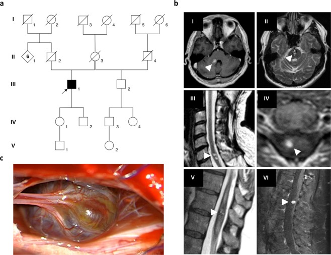
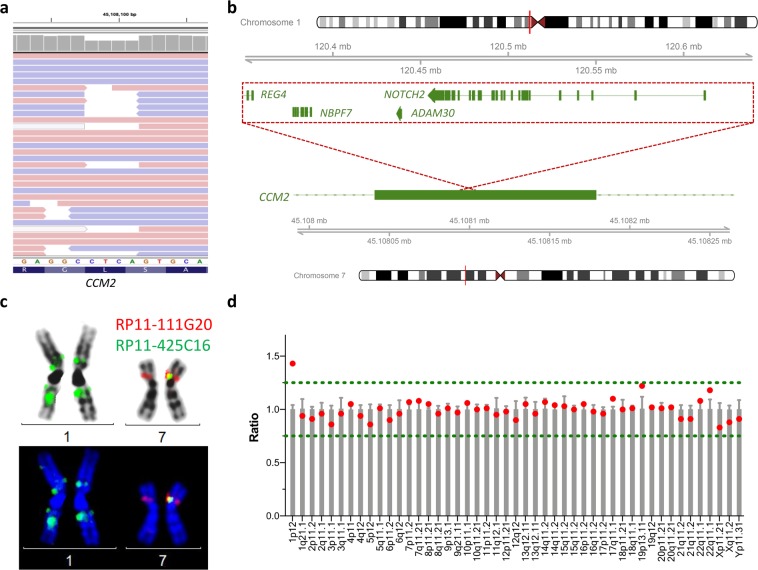
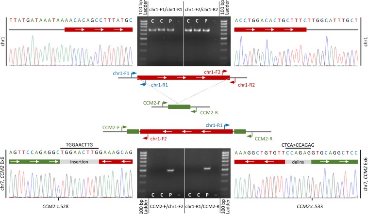
Autosomal dominant cerebral cavernous malformations (CCM) are leaky vascular lesions that can cause epileptic seizures and stroke-like symptoms. Germline mutations in either CCM1, CCM2 or CCM3 are found in the majority of patients with multiple CCMs or a positive family history. Recently, the first copy number neutral inversion in CCM2 has been identified by whole genome sequencing in an apparently mutation-negative CCM family. We here asked the question whether further structural genomic rearrangements can be detected within NGS gene panel data of unsolved CCM cases. Hybrid capture NGS data of eight index patients without a pathogenic single nucleotide, indel or copy number variant were analyzed using two bioinformatics pipelines. In a 58-year-old male with multiple CCMs in his brain and spinal cord, we identified a 294 kb insertion within the coding sequence of CCM2. Fine mapping of the breakpoints, molecular cytogenetic studies, and multiplex ligation-dependent probe amplification verified that the structural variation was an inverted unbalanced insertion that originated from 1p12-p11.2. As this rearrangement disrupts exon 6 of CCM2 on 7p13, it was classified as pathogenic. Our study demonstrates that efforts to detect structural variations in known disease genes increase the diagnostic sensitivity of genetic analyses for well-defined Mendelian disorders.


Conflict of interest statement

The authors declare no competing interests.

Figures

Figure 1
Figure 1
Multiple cerebral and spinal cavernomas in a sporadic CCM patient. (a) Pedigree of the CCM index case from pedigree 1 (III:1, arrow). (b) Repetitive magnetic resonance imaging (MRI) of the index patient’s brain and spinal cord showing progression of CCM disease with two cavernous malformations in the pons (I, II), intramedullary lesions in the cervical (III, IV) and thoracic spinal cord (V) as well as a CCM in the cauda equina (VI). MRI images were acquired between 2005 and 2019. White arrowheads indicate CCMs. (c) Intraoperative photograph of the cauda equina cavernous malformation.
Figure 2
Figure 2
Identification of an unbalanced interchromosomal insertion in CCM2. (a) Read alignment of the hybrid capture NGS data of III:1. Shown is a part of exon 6 of the CCM2 gene. The coverage plot indicated significantly reduced read depths at positions [hg19] chr7:45,108,098-45,108,101 but no reads were found that covered a deletion of 4 bp. (b) Schematic depiction of the identified interchromosomal insertion. Material from 1p12-p11.2 was found as an inverted insertion in exon 6 of the CCM2 gene on 7p13. (c) Verification of the chromosomal rearrangement [46,XY.ish ins(7;1)(p13;1p11)(RP11-425C16+,RP11-111G2 +)] by fluorescence in situ hybridization. There is a known crosshybridization of the applied probe RP11–425C16 in 1q21.1, which does not need to be considered further taking into account all other data presented here. (d) MLPA analysis of the centromere region of chromosome 1 indicated three copies of the NOTCH2 gene (1p12) which is part of the inserted fragment.
Figure 3
Figure 3
Fine mapping of the breakpoints by Sanger sequencing. Scheme of the normal and inverted insertion alleles (middle panel). PCR and sequencing primers (chr1: chr1-F1 + R1 and chr1-F2 + R2; chr7: CCM2-F + R) are depicted as blue, red, and green arrows, respectively. PCR products and chromatograms of the normal allele on chr1 (chr1-F1/R1 and chr1-F2/R2) are depicted in the upper panel. PCR products and chromatograms of the inverted insertion on chr7 (CCM2-F/chr1-F2 and chr1-R1/CCM2-R) are depicted in the lower panel. Fine mapping also revealed an additional small insertion and an indel variant at the breakpoints. C = healthy control; P = index patient; – = negative control; Ex = exon.

References

    1. Spiegler S, Rath M, Paperlein C, Felbor U. Cerebral cavernous malformations: an update on prevalence, molecular genetic analyses, and genetic counselling. Mol. Syndromol. 2018;9:60–69. doi: 10.1159/000486292. - DOI - PMC - PubMed
    1. Laberge-le Couteulx S, et al. Truncating mutations in CCM1, encoding KRIT1, cause hereditary cavernous angiomas. Nat. Genet. 1999;23:189–193. doi: 10.1038/13815. - DOI - PubMed
    1. Sahoo T, et al. Mutations in the gene encoding KRIT1, a Krev-1/rap1a binding protein, cause cerebral cavernous malformations (CCM1) Hum. Mol. Genet. 1999;8:2325–2333. doi: 10.1093/hmg/8.12.2325. - DOI - PubMed
    1. Liquori CL, et al. Mutations in a gene encoding a novel protein containing a phosphotyrosine-binding domain cause type 2 cerebral cavernous malformations. Am. J. Hum. Genet. 2003;73:1459–1464. doi: 10.1086/380314. - DOI - PMC - PubMed
    1. Denier C, et al. Mutations within the MGC4607 gene cause cerebral cavernous malformations. Am. J. Hum. Genet. 2004;74:326–337. doi: 10.1086/381718. - DOI - PMC - PubMed

Publication types

MeSH terms