Review
. 2020 May 1;44(3):351-368.
doi: 10.1093/femsre/fuaa009.

The Roles of Bacterial DNA Double-Strand Break Repair Proteins in Chromosomal DNA Replication

Affiliations
Review

The Roles of Bacterial DNA Double-Strand Break Repair Proteins in Chromosomal DNA Replication

Anurag Kumar Sinha et al. FEMS Microbiol Rev. .

Abstract

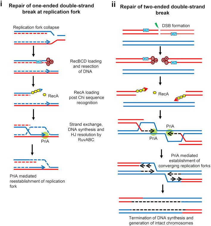
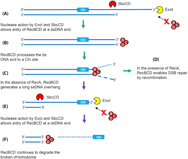
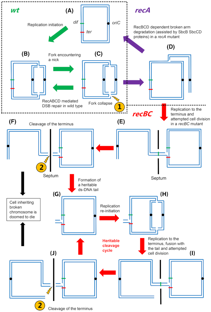
It is well established that DNA double-strand break (DSB) repair is required to underpin chromosomal DNA replication. Because DNA replication forks are prone to breakage, faithful DSB repair and correct replication fork restart are critically important. Cells, where the proteins required for DSB repair are absent or altered, display characteristic disturbances to genome replication. In this review, we analyze how bacterial DNA replication is perturbed in DSB repair mutant strains and explore the consequences of these perturbations for bacterial chromosome segregation and cell viability. Importantly, we look at how DNA replication and DSB repair processes are implicated in the striking recent observations of DNA amplification and DNA loss in the chromosome terminus of various mutant Escherichia coli strains. We also address the mutant conditions required for the remarkable ability to copy the entire E. coli genome, and to maintain cell viability, even in the absence of replication initiation from oriC, the unique origin of DNA replication in wild type cells. Furthermore, we discuss the models that have been proposed to explain these phenomena and assess how these models fit with the observed data, provide new insights and enhance our understanding of chromosomal replication and termination in bacteria.

Keywords: E. coli; DNA double-strand break repair; DNA replication; Homologous recombination.


Figures

Figure 1.
Figure 1.
RecBCD-dependent homologous recombinational repair of one-ended and two-ended DSBs. (i), A one-ended DSB is generated when a replication fork encounters a nick in a template strand, leading to replication fork collapse (a broken replication fork). (ii), Direct exposure to ionizing radiation generates a two-ended DSB. Both types of DSBs are unwound and cleaved by RecBCD using its helicase and nuclease activities. After recognizing the regulatory sequence Chi (5’-GCTGGTGG-3’), the properties of RecBCD enzyme change and a 3′-overhang terminated at a Chi sequence is generated. RecBCD then facilitates RecA loading onto this 3′-ssDNA overhang. The RecA-bound ssDNA searches for a homologous duplex DNA sequence, strand-exchange takes place to form a D-loop, followed by Holliday junction branch-migration and resolution by RuvABC proteins. PriA binds to the 3′ paired end(s), facilitates primosome assembly, and allows replication restart. Importantly, recombinational repair of a one-ended DSB leads to re-establishment of a replication fork, whereas two intact chromosomes are the products of two-ended DSB repair. Template strands and nascent strands are shown here as intact and dotted lines respectively. The leading-strand is denoted as a line with an arrowhead to mark the 3′-end.
Figure 2.
Figure 2.
Action of ExoI and SbcCD nucleases facilitates RecBCD-mediated DSB repair by recombination in recA+ cells and facilitates DNA degradation by RecBCD in recA mutant cells. RecBCD enzyme can only bind and initiate unwinding and degradation of DNA that contains a blunt or nearly blunt DSE. Exonuclease I (the product of the sbcB gene, also known as xonA) and SbcCD both contribute to the generation of such ends that can bind RecBCD. This enables productive DSB repair by recombination, in the presence of RecA (from A to D) or degradation of a broken chromosome, in the absence of RecA (from C to F).
Figure 3.
Figure 3.
Four models to explain terminus DNA amplification in a recG mutant. (i) Reverse-restart model: This model proposes concerted action of RecG and PriA in the re-establishment of a correctly oriented replication fork. According to this model, during broken replication fork repair and reassembly, the presence of RecG allows correct PriA-mediated loading of DnaB helicase on the lagging-strand template, potentiating correct replication restart (from a to d). In the absence of RecG, PriA is no longer correctly directed by RecG and can mistakenly load DnaB on the nascent lagging-strand (e). This initiates reverse-restart, via the assembly of a replication fork proceeding in the opposite direction. Meanwhile, PriA can still load DnaB in the correct location setting up a correctly oriented replication fork; (the absence of RecG does not preclude normal DnaB loading, it simply permits incorrect loading) (f). The end of the ds-DNA flap that has been generated can invade and recombine with the intact chromosome (RecA-RecBCD mediated recombination). Thus, divergent forks are established causing DNA amplification (g). If, during recombination, PriA again loads DnaB incorrectly a new flap can be generated adding to the amplification (from g to e). If new breaks are generated at one or other of the Tus/ter sites, the cycle can be re-initiated adding to amplification (from g to a). (ii) 3-flap model: This model proposes that merging of replication forks in the terminus generates a 3′-flap (from a to b). RecG binds to this 3′-flap and converts it into 5′-flap to be degraded by 5′ exonucleases to complete termination (from c to d). In the absence of RecG, the 3′-flap will be used by PriA for primosome assembly and restart of the replication fork (e). The end of the ds-DNA flap that has been generated will invade and recombine with the intact chromosome (RecA-RecBCD mediated recombination). Thus, bidirectional forks are established causing DNA amplification (f). (iii) Over-replication model: This model proposes that merging replication forks can pass each other, leading to amplification of the terminus region (from a to b). The over replicated region is processed by RecG to make it available for degradation by RecBCD and other exonucleases. In the absence of recG or exonucleases, the over replicated region persists. (iv) Fork-trap model: This model simply proposes that Tus/ter mediated trapping of multiple forks generated by stable DNA replication in a recG mutant leads to the observation of terminus DNA amplification (a-b). Yellow triangles represent the Tus/ter trap; the black circle represents the DnaB helicase. Template strands and nascent strands are shown here as intact and dotted lines respectively. The leading-strand is denoted as a line with an arrowhead representing the 3′-end.
Figure 4.
Figure 4.
Processing of a broken replication fork in wild type (wt), ΔrecA and ΔrecBCD cells. DNA replication initiates at a fixed position known as oriC (denoted by a small circle) on a circular chromosome (a) and progresses bidirectionally (b). Encounter of the replication fork with a nick on the template strand (c) leading to a broken replication fork (replication fork collapse) and generation of a DSE (one-ended DSB) at the fork (depicted by 1 in the figure). This can lead to cell division-dependent DNA cleavage of the terminus in recBC mutant cells (depicted by 2 in the figure). In wt cells, the broken fork is repaired by RecBCD-RecA mediated recombinational repair, to re-establish an intact replication fork (from c to b). In ΔrecA cells, RecBCD binds to this DSE and degrades the entire broken arm, to generate an intact single circular chromosome (from d to a). However, the degradation process of the entire broken arm by RecBCD needs assistance from exonucleases (such as ExoI, SbcCD etc.), since frequent encounter with Chi sequences by RecBCD leads to the generation of long 3′-ssDNA overhangs and disassociation of RecBCD. Owing to its ability to bind only blunt (or nearly blunt) DSEs, RecBCD is unable to bind and restart degradation from these long 3′ ssDNA overhangs. ssDNA exonucleases degrade the overhangs to make them blunt and so facilitate RecBCD loading and further degradation. In ΔrecBCD cells, the broken arm persists. The other intact replication fork continues to progress towards the terminus (e) and reaches an appropriately oriented ter site (shown here as red line). Since replication and segregation occur simultaneously in bacterial cells, the intact circular chromosome and the broken linear chromosome segregate into two halves of the cell but remain linked by the intact fork in the terminus (e). FtsK action aligns two dif sites (shown here as green lines), one on the circular and another on the linear chromosome, in the middle of the septum. Septum formation causes guillotining of the chromosome in the dif region of the terminus, separating linear and circular chromosomes into two daughter cells (f). The cell inheriting the linear chromosome does not grow further. The cell inheriting the circular chromosome has a replication fork attached to a short linear DNA tail that remains in the terminus (g). A new round of replication initiated at oriC sends forks around the chromosome (h), which merge with the previous fork in the terminus, causing the short linear tail to be enlarged to an entire broken chromosome arm (i). Attempted segregation followed by guillotining (j) makes the process heritable (from g to j and then back to g).

References

    1. Al-Deib AA, Mahdi AA, Lloyd RG. Modulation of recombination and DNA repair by the RecG and PriA helicases of Escherichia coliK-12. J Bacteriol. 1996;178:6782–9. - PMC - PubMed
    1. Anderson DG, Kowalczykowski SC. The translocating RecBCD enzyme stimulates recombination by directing RecA protein onto ssDNA in a chi-regulated manner. Cell. 1997;90:77–86. - PubMed
    1. Asai T, Bates DB, Kogoma T. DNA replication triggered by double-stranded breaks in E. coli: dependence on homologous recombination functions. Cell. 1994;78:1051–61. - PMC - PubMed
    1. Asai T, Imai M, Kogoma T. DNA damage-inducible replication of the Escherichia coli chromosome is initiated at separable sites within the minimal oriC. J Mol Biol. 1994;235:1459–69. - PubMed
    1. Asai T, Kogoma T. D-loops and R-loops: alternative mechanisms for the initiation of chromosome replication in Escherichia coli. J Bacteriol. 1994a;176:1807–12. - PMC - PubMed

Publication types

MeSH terms