. 2020 Apr 14;16(4):e1008409.
doi: 10.1371/journal.ppat.1008409. eCollection 2020 Apr.

Characterizing Emerging Canine H3 Influenza Viruses

Luis Martinez-Sobrido  1 Pilar Blanco-Lobo  1 Laura Rodriguez  1 Theresa Fitzgerald  2 Hanyuan Zhang  3   4 Phuong Nguyen  2 Christopher S Anderson  2 Jeanne Holden-Wiltse  5 Sanjukta Bandyopadhyay  5 Aitor Nogales  1 Marta L DeDiego  2 Brian R Wasik  6 Benjamin L Miller  3   4 Carole Henry  7 Patrick C Wilson  7 Mark Y Sangster  2 John J Treanor  2 David J Topham  2 Lauren Byrd-Leotis  8   9 David A Steinhauer  8 Richard D Cummings  8   9 Jasmina M Luczo  10 Stephen M Tompkins  10 Kaori Sakamoto  11 Cheryl A Jones  10 John Steel  8 Anice C Lowen  8 Shamika Danzy  8 Hui Tao  8 Ashley L Fink  12 Sabra L Klein  12 Nicholas Wohlgemuth  12 Katherine J Fenstermacher  12 Farah El Najjar  12 Andrew Pekosz  12 Lauren Sauer  13 Mitra K Lewis  13 Kathryn Shaw-Saliba  13 Richard E Rothman  13 Zhen-Ying Liu  14 Kuan-Fu Chen  14 Colin R Parrish  6 Ian E H Voorhees  6 Yoshihiro Kawaoka  15 Gabriele Neumann  15 Shiho Chiba  15 Shufang Fan  15 Masato Hatta  15 Huihui Kong  15 Gongxun Zhong  15 Guojun Wang  16   17 Melissa B Uccellini  16   17 Adolfo García-Sastre  16   17   18   19 Daniel R Perez  20 Lucas M Ferreri  20 Sander Herfst  21 Mathilde Richard  21 Ron Fouchier  21 David Burke  22 David Pattinson  22 Derek J Smith  22 Victoria Meliopoulos  23 Pamela Freiden  23 Brandi Livingston  23 Bridgett Sharp  23 Sean Cherry  23 Juan Carlos Dib  24 Guohua Yang  23 Charles J Russell  23 Subrata Barman  23 Richard J Webby  23 Scott Krauss  23 Angela Danner  23 Karlie Woodard  23 Malik Peiris  25 R A P M Perera  25 M C W Chan  25 Elena A Govorkova  23 Bindumadhav M Marathe  23 Philippe N Q Pascua  23 Gavin Smith  26 Yao-Tsun Li  26 Paul G Thomas  27 Stacey Schultz-Cherry  23
Affiliations

Characterizing Emerging Canine H3 Influenza Viruses

Luis Martinez-Sobrido et al. PLoS Pathog. .

Abstract

The continual emergence of novel influenza A strains from non-human hosts requires constant vigilance and the need for ongoing research to identify strains that may pose a human public health risk. Since 1999, canine H3 influenza A viruses (CIVs) have caused many thousands or millions of respiratory infections in dogs in the United States. While no human infections with CIVs have been reported to date, these viruses could pose a zoonotic risk. In these studies, the National Institutes of Allergy and Infectious Diseases (NIAID) Centers of Excellence for Influenza Research and Surveillance (CEIRS) network collaboratively demonstrated that CIVs replicated in some primary human cells and transmitted effectively in mammalian models. While people born after 1970 had little or no pre-existing humoral immunity against CIVs, the viruses were sensitive to existing antivirals and we identified a panel of H3 cross-reactive human monoclonal antibodies (hmAbs) that could have prophylactic and/or therapeutic value. Our data predict these CIVs posed a low risk to humans. Importantly, we showed that the CEIRS network could work together to provide basic research information important for characterizing emerging influenza viruses, although there were valuable lessons learned.


Conflict of interest statement

AG-S. is inventor of patents on influenza virus vaccines owned by the Icahn School for Medicine at Mount Sinai and licensed to Medimmune, BI Vetmedica, Vivaldi Biosciences, Zoetis and Avimex.

Figures

Fig 1
Fig 1. Replication in primary cells and explants.
(A) hNEC or (B) hBEC cultures were inoculated with Beth15, CIV-41915 or rCIV-1177 viruses at a MOI of 0.1 or MOI of 1 and incubated at 32°C or 37°C. At the indicated time, apical media was collected, and virus titers determined. Data are pooled from 2 independent experiments with n = 3 wells per virus for each experiment (n = 6 total). Two-way ANOVA was used for statistical analysis (a = p<0.05, b = p<0.001 compared to Beth15 virus). Dotted line indicates limit of detection. (C) Human bronchus explant culture was submerged in 106 TCID50/ml virus for 1 hour at 37°C, washed and placed onto a surgical sponge in a 24-well tissue culture plate filled with 1 ml/well of culture medium to create an ALI. Supernatant was collected at 1, 24, and 48 hpi and virus titer determined. Experiments were performed with tissues from 3 donors (n = 3). Two-way ANOVA was used for statistical analysis (* = p<0.03, ** = p<0.0005, *** = p<0.0001 compared to mock).
Fig 2
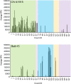
Fig 2. Glycan array binding.
Fluorescently labeled CIV-41915 and Beth15 viruses were incubated on the glycan microarray for 1 hour at 4°C, to inhibit viral neuraminidase activity, then the slide was washed to remove unbound virus and scanned using a ProScanArray microarray scanner for Alexa Fluor 488 fluorescence and results shown as RFU. Each bar represents a single glycan. Green box = α2,3; pink box = α2,3 + α2,6; blue box = α2,6; orange box = α2,8; and purple box = miscellaneous + NeuGc glycans.
Fig 3
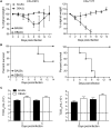
Fig 3. Pathogenicity in mice.
(A-B) Body weight and survival: Six-week-old BALB/c and DBA/2J mice (n = 5 per group) were lightly anesthetized with isoflurane and inoculated intranasally with 106 TCID50 CIV-41915 (left panel) or rCIV-1177 (right panel) in 50 μl DMEM. Mice were monitored daily for clinical disease and weighed every other day. (C) Virus loads present in whole lungs were determined 3 and 5 dpi (n = 5 mice per group). Virus titers are expressed as mean±95% CI and * = p <0.05 as compared to BALB/C.
Fig 4
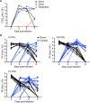
Fig 4. Transmission in ferrets and guinea pigs.
(A) 4 to 6-month-old seronegative ferrets were lightly anesthetized and intranasally inoculated with 106 TCID50 of CIV-41915 in 1.0 ml sterile PBS. At 1 dpi, naïve contacts were placed in the same cage with directly inoculated ferrets while respiratory contacts were placed in adjacent cages separated by a wire grill. Nasal washes were collected every other day for 7 dpi and viral titers determined in MDCK cells. Lines represent individual animals. (B) Guinea pigs were intranasally inoculated 10 to 103 PFU. At 1 dpi, naïve contacts were placed in the same cage with directly inoculated animals. Nasal washes were collected from anesthetized animals on days 2, 4, 6 and 8 post-inoculation and viral titers determined in MDCK cells. Lines represent individual animals.
Fig 5
Fig 5. Cartography mapping of H3, N2 and N8 influenza proteins.
Sequence-based maps of H3 (A), N2 (B) and N8 (C) viral proteins. Each point represents a single virus strain. Points are colored based on the species the virus was isolated from (A-C). Position of CIV-11613 (IL15), CIV-23 (NY09), HK68, PC73, Wy03, WI05, Vic11 and HK14 are indicated. Scale bar, 20 amino acids distance.
Fig 6
Fig 6. Pre-existing population immunity against H3N2 and H3N8 CIV in human sera.
The presence of antibodies against rCIV-11613 H3N2 (black, left), rCIV-23 H3N8 (red, middle) and WY03 H3N2 (blue, right) was examined in triplicate by (A) ELISA, (B) HAI and (C) ELLA assays using 153 human sera samples collected from healthy subjects born between 1934 and 2012 and grouped in 10 years intervals. Dotted black lines indicate the limit of detection of each of the assays. Undetectable titers were assigned a value of 10, 5 and 8 for ELISA, HAI and NAI respectively. Each dot represents the mean titer (represented as log2) of a specific human subject.
Fig 7
Fig 7. Neutralizing Ab titers to human Beth15 and CIV-41915 H3N2 viruses in patients with confirmed H3N2 infections during the 2016–17 season.
(A) Human sera collected from Johns Hopkins Health System (JHMI) and Chang Gung Memorial Hospital (CGMH) in Taipei, Taiwan were subject to two-fold serial dilutions prior to incubation with 100 TCID50 of either Beth 15 or CIV-41915. MDCK cells were then infected with the virus-sera mixture and following 24-hour incubation, cells were fixed and stained with Napthol blue black. The baseline neutralizing antibody titer was determined as the highest serum dilution that eliminated cytopathic effects in 50% of the wells. (B and C) Sera was collected from JHMI and CGMH at the time of hospital confirmation of H3N2 infection, with documentation about whether patients had or had not been vaccinated during the 2016–17 season and neutralizing antibody titers were determined as in (A). (b = p<0.001, d = p<0.0001 compared to Beth15).
Fig 8
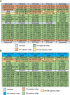
Fig 8. Layout information of the array.
(A) Array for rCIV-11613, rCIV-23, and human HK68 or (B) Wis05 H3N2 viruses. Human IgG was used as control for nonspecific IgG binding. Three anti-fluorescein (anti-FITC) solutions were uses as negative control at a concentration of 100 μg/ml (FITC-100), 200μg/ml (FITC-200) and 400μg/ml (FITC-400). Bovine IgG secondary antibodies reactive to the bovine sera used for blocking were used as positive control.
Fig 9
Fig 9. Cross-reactivity of hmAbs by AIR.
AIR images of arrays exposed to (A) rCIV-11613 H3N2, (B) rCIV-23 H3N8, (C) HK68 and (D) WI05 viruses. (E) Hierarchical cluster map of the hmAb responses. Values in this map are scaled relative to the antibody producing the strongest response for each virus. Arrows show the antibody clustering groups.
Fig 10
Fig 10. Cross-reactivity of hmAbs by immunofluorescence.
MDCK cells were infected with rCIV-11613, rCIV-23, rWY03 or HK68 viruses at a MOI of 3. At 18 hpi, cells were fixed, permeabilized and incubated with 1 μg/ml of the indicated hmAbs. After incubation with a secondary anti-human FITC-conjugated antibody, fluorescence was imaged under a fluorescent microscope. Fluorescence intensity was measured using the ImageJ 1.51s and data was displayed using a Heatmap visualization method. For each virus, the hmAb providing the highest intensity (017–10116 5B03) was considered as 100% and was used to normalize the percentage of reactivity of the rest of the hmAbs. The percentage of reactivity was used to categorized the hmAbs in 6 groups: Group 1: ≥30% reactivity against H3 CIVs and rWY03 H3N2; Group 2: ≥30% reactivity against H3 CIVs, but <30% for rWY03 H3N2; Group 3: ≥30% reactivity against H3N2 rCIV-11613 CIV but <30% for H3N8 rCIV-23 and rWY03 H3N2; Group 4: ≥30% reactivity against H3N2 rCIV-11613 and rWY03 H3N2 but <30% for H3N8 rCIV-23 CIV; Group 5: ≥30% reactivity against rWY03 H3N2 but <30% for rCIV-23 H3N8 and H3N2 rCIV-11613; and, Group 6: non-reactivity or <30% reactivity against rCIV-23 H3N8 and/or H3N2 rCIV-11613, or rWY03. Representative images of the reactivity of the hmAbs against infected cells are shown in the right. Scale represent % of recognition. Scale bars, 200 𝜇m.
Fig 11
Fig 11. Neutralizing activity of hmAbs.
MDCK cells were infected with the mCherry-expressing rCIV-23 H3N2 (black line), rCIV-23 H3N8 (red line) or rWY03 H3N2 (blue line) recombinant viruses at 0.005 MOI. After 1-hour adsorption at room temperature, cells were incubated with 2-fold serial dilutions of the indicated hmAbs (starting concentration of 0.2 mg/ml) belonging to the Groups 1 (A), 2 (B), 3 (C), 4 (D) or 6 (E). Group 5 hmAbs (≥30% reactivity against rWY03 H3N2 but <30% for rCIV-23 H3N8 and rCIV-23 H3N2) were not included in the assay. At 60 hpi, fluorescence expression was quantified using a fluorescence plate reader and sigmoidal dose response curves were used to calculate the hmAbs concentration that reduced the virus infectivity by 50% (Neutralization titer 50, NT50). Mock-infected and infected cells in the absence of hmAbs were used as controls to normalize the percentage of inhibition. Data show the mean and +/- SDs of the results determined for triplicate. An inhibition of 50% is indicated as a dotted black line.

References

    1. Garten R, Blanton L, Elal AIA, Alabi N, Barnes J, et al. (2018) Update: Influenza Activity in the United States During the 2017–18 Season and Composition of the 2018–19 Influenza Vaccine. MMWR Morb Mortal Wkly Rep 67: 634–642. 10.15585/mmwr.mm6722a4 - DOI - PMC - PubMed
    1. Lee IH, Le TB, Kim HS, Seo SH (2016) Isolation of a novel H3N2 influenza virus containing a gene of H9N2 avian influenza in a dog in South Korea in 2015. Virus Genes 52: 142–145. 10.1007/s11262-015-1272-z - DOI - PubMed
    1. Pulit-Penaloza JA, Simpson N, Yang H, Creager HM, Jones J, et al. (2017) Assessment of Molecular, Antigenic, and Pathological Features of Canine Influenza A(H3N2) Viruses That Emerged in the United States. J Infect Dis 216: S499–S507. 10.1093/infdis/jiw620 - DOI - PMC - PubMed
    1. Voorhees IEH, Glaser AL, Toohey-Kurth K, Newbury S, Dalziel BD, et al. (2017) Spread of Canine Influenza A(H3N2) Virus, United States. Emerg Infect Dis 23: 1950–1957. 10.3201/eid2312.170246 - DOI - PMC - PubMed
    1. Song D, Kang B, Lee C, Jung K, Ha G, et al. (2008) Transmission of avian influenza virus (H3N2) to dogs. Emerg Infect Dis 14: 741–746. 10.3201/eid1405.071471 - DOI - PMC - PubMed

Publication types

MeSH terms