Review
. 2016 Dec:85:36-60.
doi: 10.1016/j.trac.2016.04.004. Epub 2016 Apr 8.

Critical overview on the application of sensors and biosensors for clinical analysis

Affiliations
Review

Critical overview on the application of sensors and biosensors for clinical analysis

Celine I L Justino et al. Trends Analyt Chem. 2016 Dec.

Abstract

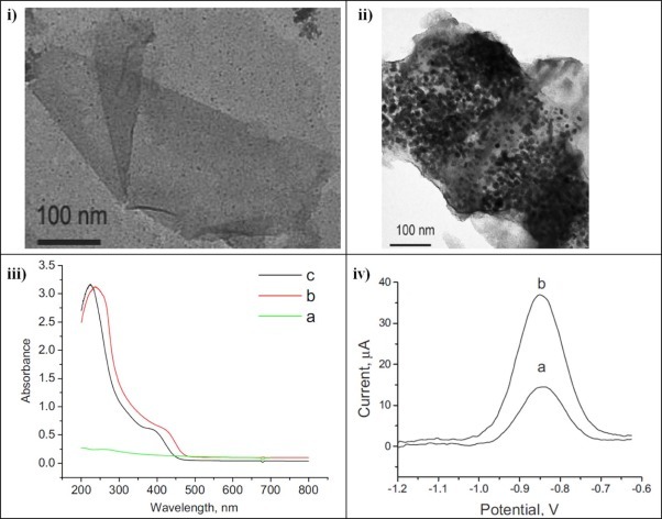
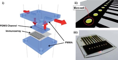
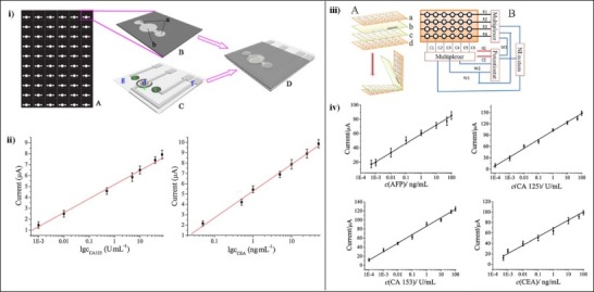
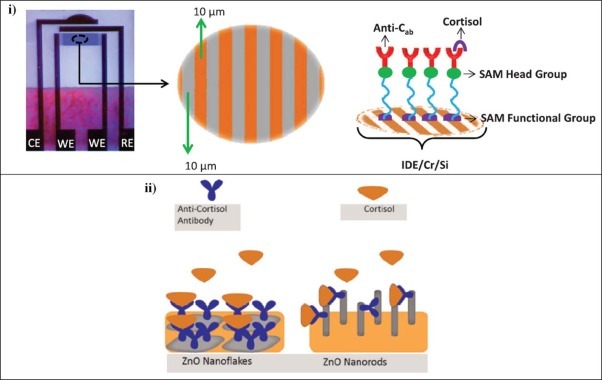
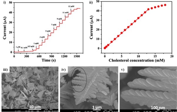
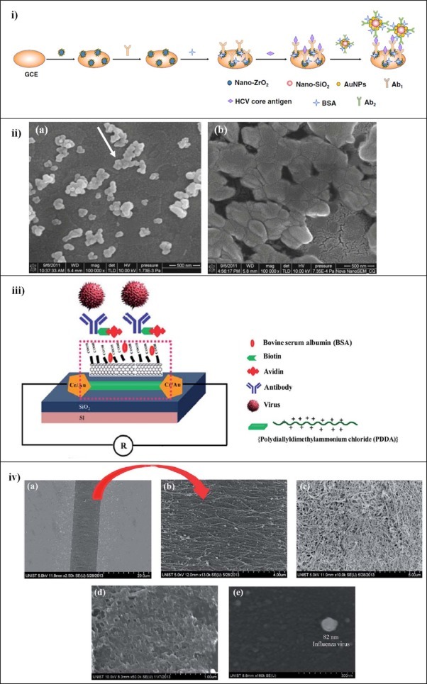
Sensors and biosensors have been increasingly used for clinical analysis due to their miniaturization and portability, allowing the construction of diagnostic devices for point-of-care testing. This paper presents an up-to-date overview and comparison of the analytical performance of sensors and biosensors recently used in clinical analysis. This includes cancer and cardiac biomarkers, hormones, biomolecules, neurotransmitters, bacteria, virus and cancer cells, along with related significant advances since 2011. Some methods of enhancing the analytical performance of sensors and biosensors through their figures of merit are also discussed.

Keywords: Analytical performance; Biosensor; Clinical analysis; Figure of merit; Sensor.


Figures

Fig. 1
Fig. 1
i) TEM image of graphene sheets; ii) TEM image of graphene sheets functionalized with quantum dots; iii) UV–Vis spectra of a) graphene sheets, b) quantum dots and c) quantum dot–graphene sheets; and iv) Square wave voltammogram response of electrodes with a) only quantum dots and b) electrodes with quantum dots in graphene sheets
Fig. 2
Fig. 2
i) Fitting the 8-electrode immunoarray into the microfluidic device. The array is sandwiched between two layers of poly(methylmethacrylate) (PMMA) and one layer of poly(dimethyl)siloxane (PDMS) acting as microfluidic channel above the sensor electrodes. The red arrows indicate the flow of buffer; ii) Computer-generated design of the gold array showing microwells around electrodes; and iii) Completed array of 8 electrodes with individual microwells containing 1 µL aqueous droplets
Fig. 3
Fig. 3
i) Schematic representation of 3D-µPED. (A): wax-patterned paper sheet; (B): 3D-µPED [a) paper working zones and b) paper auxiliary zone]; (C): screen-printed electrodes [c) carbon working electrodes, d) Ag/AgCl reference electrode, e) carbon counter electrode, f) silver conductive channel and pad, and g) transparent polyethylene terephthalate substrate]; (D): after stacking, the paper working zones and the paper auxiliary zone will be aligned to the screen-printed working electrodes, counter electrode and reference electrode (Reprinted from Wang et al. , with permission from Elsevier); ii) Calibration curves for CA 125 and CEA (eleven measurements for each point) (Reprinted from Wang et al. , with permission from Elsevier); iii) (A) Layered structure of the device; (B) Schematic diagram of the addressable electrode array detection system [a) and d) polyethylene terephthalate substrates with column electrodes (Cn) and row electrodes (Rn); b) reference electrode (RE), c) sensing sites with column electrodes (Wn) and counter electrode (CE)] (Reproduced from Ge et al. with permission of The Royal Society of Chemistry); iv) Calibration curves for immunoassay of tumour markers
Fig. 4
Fig. 4
i) Schematic representation of IDE, where the binding of cortisol with antibody (Anti-Cab) blocks the electron transport from the medium to IDE and fabrication of immunosensor proposed by Pasha et al. (Reproduced from Pasha et al. by permission of The Electrochemical Society). CE: counter electrode; WE: working electrode; SAM: self-assembled monolayer; ii) Illustration of ZnO nanorods and ZnO nanoflakes along with immobilization of monoclonal anti-cortisol antibody to fabricate electrochemical cortisol immunosensor proposed by Vabbina et al.
Fig. 5
Fig. 5
i) Amperometric response to different cholesterol concentrations in 0.10 PBS at +0.38 V and ii) Calibration curve (Reprinted from Ahmad et al. , with permission from Elsevier). FESEM images of as-synthesized α-Fe2O3 micro-pine-shaped hierarchical structures at iii) low magnification, iv) and v) high resolution
Fig. 6
Fig. 6
i) Fabrication and measurement procedures for the immunosensor proposed by Ma et al. and ii) Sizes and morphologies of nanocomposites [(a) SEM images of ZrO2 nanoparticle–chitosan and (b) antibody–Au nanoparticle–ZrO2 nanoparticle–chitosan] (With kind permission from Springer Science+Business Media: Microchimica Acta, Ma et al. [141]); iii) Schematic illustration of the SWCNT immunosensor for H1N1 virus detection proposed by Singh et al. and iv) SEM images of (a) PDDA–SWCNT, (b) PDDA–SWCNT at higher magnification, (c) SWCNT deposited by sedimentation, (d) H1N1 antibody immobilized on PDDA–SWCNT and (e) H1N1 antibody immobilized on PDDA–SWCNT after capturing a single influenza virus

References

    1. Justino C.I.L., Rocha-Santos T.A.P., Duarte A.C. Advances in point-of-care technologies with biosensors based on carbon nanotubes. Trends Anal. Chem. 2013;45:24–36.
    1. Giri B., Pandey B., Neupane B., Ligler F.S. Signal amplification strategies for microfluidic immunoassays. Trends Anal. Chem. 2015;79:326–334.
    1. Justino C.I.L., Rocha-Santos T.A.P., Duarte A.C. Review of analytical figures of merit of sensors and biosensors in clinical applications. Trends Anal. Chem. 2010;29:1172–1183.
    1. Justino C.I.L., Duarte A.C., Rocha-Santos T.A.P. Immunosensors in clinical laboratory diagnostics. Adv. Clin. Chem. 2016;73:65–108. - PubMed
    1. Thévenot D.R., Toth K., Durst R.A., Wilson G.S. Electrochemical biosensors: recommended definitions and classification. Biosens. Bioelectron. 2001;16:121–131. - PubMed

LinkOut - more resources