. 2004 Aug 15;26(16):121-128.
doi: 10.1016/j.clinmicnews.2004.08.001. Epub 2004 Aug 11.

Life beyond PCR: alternative target amplification technologies for the diagnosis of infectious diseases, part I

Affiliations

Life beyond PCR: alternative target amplification technologies for the diagnosis of infectious diseases, part I

Christine C Ginocchio. Clin Microbiol Newsl. .

Abstract

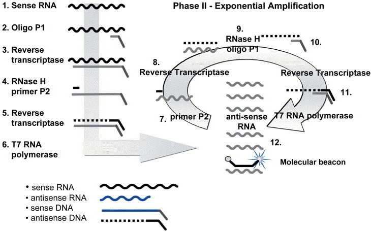
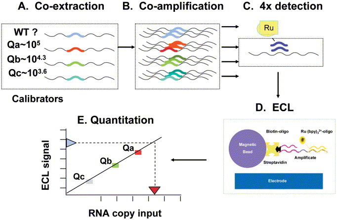
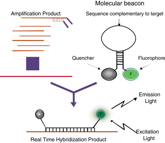
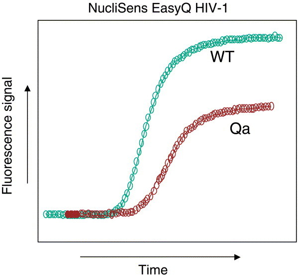
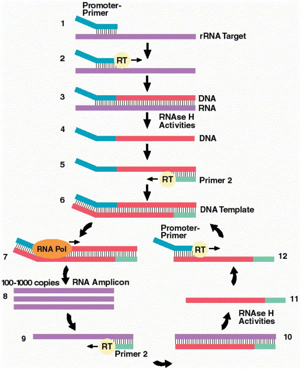
Non-PCR-based target amplification technologies, including transcription-mediated amplification (TMA), nucleic acid sequence-based amplification (NASBA), and strand displacement amplification (SDA), are currently the basis for a broad range of clinical infectious-disease molecular diagnostics. These amplification technologies are very sensitive and specific and can be used in combination with traditional end-point or "real-time" detection formats. For several nucleic acid targets, TMA, NASBA, and SDA have certain advantages over PCR-based applications. This two-part article will review the molecular basis of each technology and how the technology has been applied to clinical diagnostic systems. The articles will describe the current testing platforms available, U.S. Food and Drug Administration (FDA)- and non-FDA-approved assays, and availability of analyte-specific reagents. In addition, an open-platform system is described that utilizes standardized reagents and methods and allows the user to develop in-house protocols. Finally, applications for the future are discussed.


Figures

Figure 1
Figure 1
Nucleic acid sequenced based amplification (NASBA). An example of an RNA target is shown. Included in the amplification reaction are reverse transcriptase (RT), RNase H, T7 RNA polymerase, nucleotides, primers, and molecular beacons if performing real-time detection. Phase I. Template amplification 1. Single-stranded sense RNA. 2. Oligonucleotide P1, containing the T7 promoter recognition sequence, binds to the complementary sequence on the target. 3. RT makes a DNA copy of the RNA template. 4. RNase H removes the RNA from the duplex, and P2 binds the antisense DNA strand. 5. RT copies the antisense DNA to form a double-stranded DNA complex. 6. T7 RNA polymerase recognizes the double-stranded T7 promoter sequence and initiates transcription, making hundreds of antisense RNA copies. Phase II. Exponential amplification 7. P2 binds the complementary sequence on the antisense RNA strands. 8. RT makes a DNA copy of the RNA template. 9. RNase H removes the RNA from the duplex. 10. P2 binds the antisense DNA strand. 11. RT creates a double-stranded T7 RNA polymerase promoter. 12. Additional rounds of transcription occur, resulting in 108 to 1010 copies of antisense RNA that are templates both for more rounds of amplification and for detection by using probes and electrochemiluminescence or in real time with molecular beacons incorporated into the amplification reaction, as shown. (Figure reprinted with permission of bioMérieux, Inc., Durham, NC.)
Figure 2
Figure 2
Schematic of HIV-1 QT assay. (A) Three internal calibrators, each with a unique altered 20-nucleotide internal sequence and of known RNA concentration, are added to lysis buffer with patient sample, and total nucleic acids are co-isolated. (B) Calibrators and wild-type patient HIV-1 RNA are co-amplified by NASBA in a single tube. (C) Four separate ECL detections are performed using specific ruthenium-labeled probes targeted to the altered internal calibrator sequences or the wild-type HIV-1 sequence. (D) Streptavidin paramagnetic beads with a bound HIV-1-specific biotinylated oligonucleotide capture the amplified targets. Voltage is applied, and ECL signals are read by the NucliSens Analyzer. (E) Wild-type HIV-1 RNA quantitation is based on unique calibration curves generated by the calibrators Qa, Qb, and Qc for each sample. (Figure reprinted with permission of bioMérieux, Inc., Durham, NC.)
Figure 3
Figure 3
Real-time amplification and detection using molecular beacons. Added to the amplification reaction are molecular beacons specific for the target of interest. When the target is present, beacons open and bind to the complementary target sequence separating the fluorophore and the quencher. Upon excitation, light is emitted and measured. (Figure reprinted with permission of bioMérieux, Inc., Durham, NC.)
Figure 4
Figure 4
HIV-1 EasyQ quantitation using molecular beacons. Real-time HIV-1 quantitation is based on the presence of a single calibrator (Qa) of known RNA concentration, which is co-extracted and co-amplified with the patient sample. Algorithms based on multiple parameters (including internal calibrator values, kinetics of the assay, molecular-beacon binding, time to detection, and fluorescence units) determine the number of wild-type HIV-1 RNA copies in the patient sample. (Figure reprinted with permission of bioMérieux, Inc., Durham, NC.)
Figure 5
Figure 5
Transcription-mediated amplification. (Step 1) Promoter-primer binds to rRNA target. (Step 2) Reverse transcriptase (RT) creates DNA copy of rRNA target. (Step 3) RNA-DNA duplex. (Step 4) RNase H activities of RT degrades the rRNA. (Step 5) Primer 2 binds to the DNA, and RT creates a new DNA copy. (Step 6) Double-stranded DNA template with a promoter sequence. (Step 7) RNA polymerase (RNA Pol) initiates transcription of RNA from DNA template. (Step 8) 100 to 1,000 copies of RNA amplicon are produced. (Step 9) Primer 2 binds to each RNA amplicon and RT creates a DNA copy. (Step 10) RNA-DNA duplex. (Step 11) RNase H activities of RT degrade the rRNA. (Step 12) Promoter-primer binds to the newly synthesized DNA. RT creates a double-stranded DNA, and the autocatalytic cycle repeats, resulting in a billion-fold amplification. (Figure reprinted with permission of Gen-Probe Inc., San Diego, CA.)

References

    1. Urdea M.S. Branched amp-lification multimers for the sensitive, direct detection of human hepatitis virus. Nucleic Acids Symp. Ser. 1991;24:197–200. - PubMed
    1. Barany F. The ligase chain reaction (LCR) in a PCR world. PCR Methods Appl. 1991;1:5–16. - PubMed
    1. Wharam S.D. Specific detection of DNA and RNA targets using a novel isothermal nucleic acid amplification assay based on the formation of a three-way junction. Nucleic Acids Res. 2001;29:e54. - PMC - PubMed
    1. Zhang D.Y. Detection of rare DNA targets by isothermal ramification amplification. Gene. 2001;274:209–216. - PubMed
    1. Kwiatkowski R.W. Clinical, genetic, and pharmacogenetic applications of the Invader Assay. Mol. Diagn. 1999;4:353–364. - PubMed

LinkOut - more resources