. 2014 Mar 31:193:334-339.
doi: 10.1016/j.snb.2013.11.087. Epub 2013 Dec 1.

Investigation of C-terminal domain of SARS nucleocapsid protein-Duplex DNA interaction using transistors and binding-site models

Affiliations

Investigation of C-terminal domain of SARS nucleocapsid protein-Duplex DNA interaction using transistors and binding-site models

You-Ren Hsu et al. Sens Actuators B Chem. .

Abstract

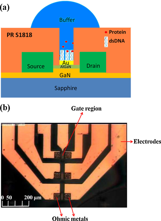
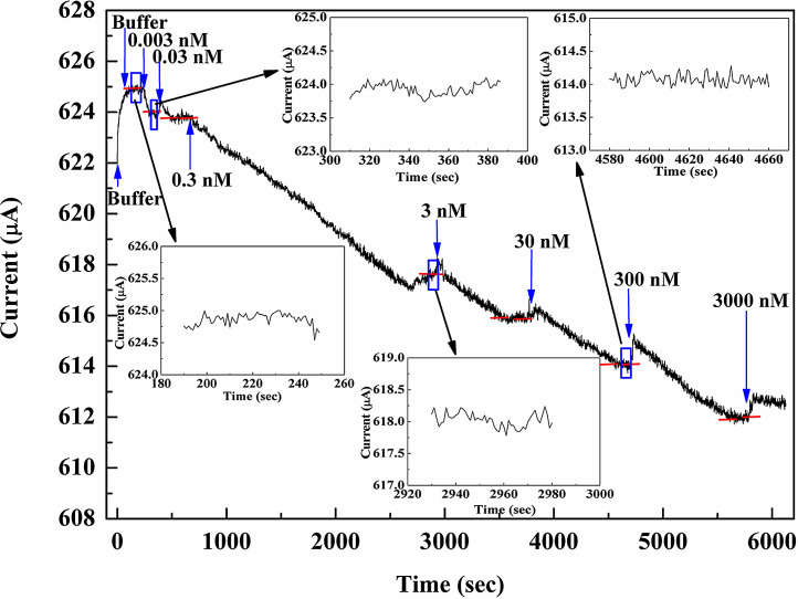
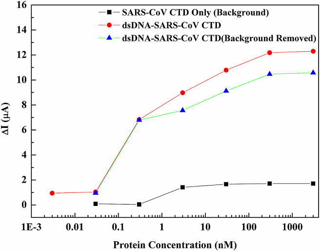
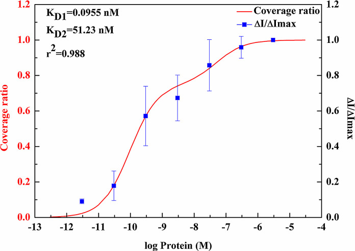
AlGaN/GaN high electron mobility transistors (HEMTs) were used to sense the binding between double stranded DNA (dsDNA) and the severe acute respiratory syndrome coronavirus (SARS-CoV) nucleocapsid protein (N protein). The sensing signals were the drain current change of the HEMTs induced by the protein-dsDNA binding. Binding-site models using surface coverage ratios were utilized to analyze the signals from the HEMT-based sensors to extract the dissociation constants and predict the number of binding sites. Two dissociation constants, K D1 = 0.0955 nM, K D2 = 51.23 nM, were obtained by fitting the experimental results into the two-binding-site model. The result shows that this technique is more competitive than isotope-labeling electrophoretic mobility shift assay (EMSA). We demonstrated that AlGaN/GaN HEMTs were highly potential in constructing a semiconductor-based-sensor binding assay to extract the dissociation constants of nucleotide-protein interaction.

Keywords: Binding sites; Dissociation constants; GaN; HEMTs; SARS; Sensors.


Figures

Fig. 1
Fig. 1
(a) Schematics of the N protein sensor. (b) Plan-view photography of the sensor.
Fig. 2
Fig. 2
Real-time detection of the N protein from 0.003 nM to 3000 nM at constant bias of 350 mV. (For interpretation of the references to colour in this figure legend, the reader is referred to the web version of this article.)
Fig. 3
Fig. 3
Current changes at different concentrations of N protein CTD in log scale. The red line and the black line represent the current changes from the HEMT with and without DNA immobilized, respectively. The green line is the net current by subtracting the black line from the red line. (For interpretation of the references to colour in this figure legend, the reader is referred to the web version of this article.)
Fig. 4
Fig. 4
(a) Dissociation constant extracted from the linear regression of Langmuir isotherm equation (b) Surface coverage ratio as a function of the protein concentration calculated with the dissociation constant extracted from (a). The ΔIImax from the experiment are shown as dots versus protein concentration.
Fig. 5
Fig. 5
The curve-fitting of the two-binding-site model with surface coverage ratio at different SARS-N protein-CTD concentrations and the two dissociation constants extracted from the model.

References

    1. Stadler K., Masignani V., Eickmann M., Becker S., Abrignani S., Klenk H.-D. SARS-beginning to understand a new virus. Nature Reviews Microbiology. 2003;1:209–218. - PMC - PubMed
    1. Saikatendu K.S., Joseph J.S., Subramanian V., Neuman B.W., Buchmeier M.J., Stevens R.C. Ribonucleocapsid formation of severe acute respiratory syndrome coronavirus through molecular action of the N-terminal domain of N protein. Journal of Virology. 2007;81:3913–3921. - PMC - PubMed
    1. Chang C.-k., Hsu Y.l., Chang Y.h., Chao F.a., Wu M.C., Huang Y.s. Multiple nucleic acid binding sites and intrinsic disorder of SARS coronavirus nucleocapsid protein-implication for ribonucleocapsid protein packaging. Journal of Virology. 2008 JVI.02001-08. - PMC - PubMed
    1. Takeda M., Chang C.-k., Ikeya T., Güntert P., Chang Y.-h., Hsu Y.-l. Solution structure of the C-terminal dimerization domain of SARS coronavirus nucleocapsid protein solved by the SAIL-NMR method. Journal of Molecular Biology. 2008;380:608–622. - PMC - PubMed
    1. Chen C.-Y., Chang C.-k., Chang Y.-W., Sue S.-C., Bai H.-I., Riang L. Structure of the SARS coronavirus nucleocapsid protein RNA-binding dimerization domain suggests a mechanism for helical packaging of viral RNA. Journal of Molecular Biology. 2007;368:1075–1086. - PMC - PubMed