

COVID-19 pneumonia manifestations at the admission on chest ultrasound, radiographs, and CT: single-center study and comprehensive radiologic literature review
- PMID: 32289051
- PMCID: PMC7129441
- DOI: 10.1016/j.ejro.2020.100231
COVID-19 pneumonia manifestations at the admission on chest ultrasound, radiographs, and CT: single-center study and comprehensive radiologic literature review
Abstract
Purpose: To investigate the imaging features of emerging COVID-19 pneumonia on chest ultrasound (US), radiographs (CXR) and computed tomography (CT) examinations performed at admission and to provide a comprehensive radiological literature review on ongoing radiological data from recent publications.
Materials and methods: In this retrospective single-center study, we enrolled consecutive patients from February 15, 2020, to March 15, 2020, with laboratory-confirmed SARS-CoV-2 hospitalized in Valduce Hospital (Como, Italy). Multi-modality imaging findings were evaluated and compared. Literature research was conducted through a methodical search on Pubmed and Embase databases.
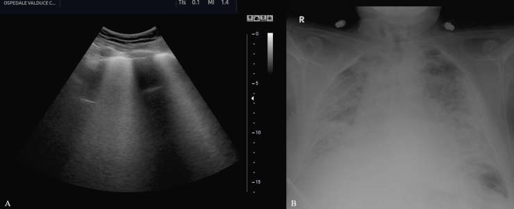
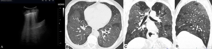
Results: Fifty-eight patients (36 men, 22 women; age range, 18-98 years) were included in the study. Among these, chest US, CXR, and CT were performed respectively in twenty-two, thirty-two and forty-two patients. Lung US findings were consistent with diffuse B lines (100%) and subpleural consolidations (27.3%). CXR showed prevalent manifestations of consolidations (46.9%) and hazy increased opacities (37.5%). Typical CT features included bilateral and multilobar ground-glass opacities (GGO) with (59.5%) and without (35.7%) consolidations having a predominantly peripheral distribution (64.3%). Other imaging features included crazy paving pattern (57.1%), fibrous stripes (50%), subpleural lines (35.7%), architectural distortion (28.6%), air bronchogram sign (26.2%), vascular thickening (23.8%) and nodules (2.4%). Also, enlarged lymph nodes (14.3 %) and pleural effusion (7.1%) were observed. The literature review identified twenty-six original studies supporting our imaging chest findings.
Conclusion: The spectrum of chest imaging manifestations of COVID-19 pneumonia upon admission includes B-lines and consolidations on US, consolidations and hazy increased opacities on CXR, and multifocal GGO with consolidations on CT.
Keywords: COVID-19; Computed Tomography (CT); SARS-CoV-2; coronavirus disease; pneumonia; radiographic chest examination (CXR); ultrasound (US).
© 2020 The Author(s).
Conflict of interest statement
The authors declare that they have no conflict of interest.
Figures


References
-
- Guan W.-J., Ni Z.-Y., Hu Y., Liang W.-H., Ou C.-Q., He J.-X., Liu L., Shan H., Lei C.-L., Hui D.S.C., Du B., Li L.-J., Zeng G., Yuen K.-Y., Chen R.-C., Tang C.-L., Wang T., Chen P.-Y., Xiang J., Li S.-Y., Wang J.-L., Liang Z.-J., Peng Y.-X., Wei L., Liu Y., Hu Y.-H., Peng P., Wang J.-M., Liu J.-Y., Chen Z., Li G., Zheng Z.-J., Qiu S.-Q., Luo J., Ye C.-J., Zhu S.-Y., Zhong N.-S. China Medical Treatment Expert Group for Covid-19, Clinical Characteristics of Coronavirus Disease 2019 in China. N. Engl. J. Med. 2020 - PMC - PubMed
-
- Hansell D.M., Bankier A.A., MacMahon H., McLoud T.C., Müller N.L., Remy J. Fleischner Society: Glossary of Terms for Thoracic Imaging. Radiology. 2008;246:697–722. - PubMed
-
- Huang C., Wang Y., Li X., Ren L., Zhao J., Hu Y., Zhang L., Fan G., Xu J., Gu X., Cheng Z., Yu T., Xia J., Wei Y., Wu W., Xie X., Yin W., Li H., Liu M., Xiao Y., Gao H., Guo L., Xie J., Wang G., Jiang R., Gao Z., Jin Q., Wang J., Cao B. Clinical features of patients infected with 2019 novel coronavirus in Wuhan, China. Lancet. 2020;395:497–506. - PMC - PubMed
LinkOut - more resources
Full Text Sources
Medical
Miscellaneous
