. 2020 May 4;219(5):e201909094.
doi: 10.1083/jcb.201909094.

Nek2-mediated GAS2L1 phosphorylation and centrosome-linker disassembly induce centrosome disjunction

Affiliations

Nek2-mediated GAS2L1 phosphorylation and centrosome-linker disassembly induce centrosome disjunction

Franco K C Au et al. J Cell Biol. .

Abstract

Centrosome disjunction occurs in late G2 to facilitate bipolar spindle formation and is mediated by the NIMA-related kinase Nek2. Here, we show that GAS2L1, a microtubule- and F-actin-binding protein required for centrosome disjunction, undergoes Nek2-mediated phosphorylation at Ser352 in G2/M. The phosphorylation is essential for centrosome disjunction in late G2 and for proper spindle assembly and faithful chromosome segregation in mitosis. GAS2L1 contains a calponin-homology (CH) domain and a GAS2-related (GAR) domain, which bind to F-actin and microtubules, respectively. Notably, the CH and GAR domains bind to each other to inhibit the functions of both domains, and Ser352 phosphorylation disrupts the interaction between the two domains and relieves the autoinhibition. We dissected the roles of the GAS2L1 phosphorylation and of centrosome-linker disassembly, which is another Nek2-mediated event, and found that these events together trigger centrosome disjunction. Therefore, our findings demonstrate the concerted Nek2 actions that split the centrosomes in late G2.


Figures

Figure 1.
Figure 1.
GAS2L1 undergoes cell cycle-dependent phosphorylation. (A) HeLa cells were released from double-thymidine block into medium containing 5 µM STLC. Whole-cell extracts were collected at indicated times and immunoblotted with antibodies against GAS2L1, cyclin B1, and β-actin. (B) Following double-thymidine release for 12 h, HeLa cells were lysed and incubated at 30°C with or without calf intestinal alkaline phosphatase for 15 min; subsequently, cell extracts were immunoblotted with antibodies against GAS2L1 and β-actin. (C) Left: Preparation of GAS2L1 samples for identification of phosphorylation sites by using mass spectrometry. Right: Coomassie Blue–stained gel of GAS2L1 proteins immunoprecipitated (using the attached tag) from interphase and mitotic cells. (D) Left: GAS2L1 residues identified to undergo mitotically induced phosphorylation. Right: Schematic structure of GAS2L1 indicating the location of five clustered Ser-phosphorylation sites. (E) Sequence alignment of GAS2L1 from various vertebrate species. Asterisks: identified phosphorylation sites within the region.
Figure 2.
Figure 2.
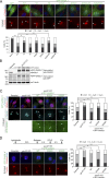
Phosphorylation at Ser352 is required for GAS2L1 function in centrosome separation. (A) Asynchronous RPE-1 cells were transfected with GFP or the following GFP-tagged GAS2L1 constructs: WT and mutants 5A, 5D, S352A, S352D, S352A/4D, and S352D/4A. Centrosomes were labeled with anti-γ-tubulin antibody. Centrosome distance was measured in the transfected cells from three independent experiments: n = 96 (GFP), 110 (WT), 120 (5A), 114 (5D), 112 (S352A), 110 (S352D), 100 (S352A/4D), and 100 (S352D/4A). Arrowheads: centrosomes. (B) GAS2L1 expression was examined in RPE-1 parental cells and sublines. gas2l1 KO, gas2l1-knockout line; GFP-GAS2L1 (WT) or mutants (S352A and S352D) were stably expressed in the knockout line. Anti-GAS2L1, anti-GFP, and anti-α-tubulin immunoblotting was performed. (C) RPE-1 parental cells and sublines were stained for CENP-F and γ-tubulin. Centrosome distance during late G2 was measured from CENP-F–positive cells in three independent experiments: n = 161 (parental), 170 (gas2l1 KO), 150 (WT), 162 (S352A), and 149 (S352D). Arrowheads: centrosomes. (D) RPE-1 lines as in C were arrested with aphidicolin and then released and treated with STLC. Centrosomes were labeled through anti-centrin staining. Centrosome distance was measured in mitotic cells (arrested in prometaphase by STLC) from three independent experiments: n = 100 (parental), 100 (gas2l1 KO), 120 (WT), 123 (S352A), and 121 (S352D). (A, C, and D) Arrowheads, centrosomes. Scale bars, 10 µm in cell micrographs and 5 µm in enlarged images. χ2 test; **, P < 0.001; ***, P < 0.0001; n.s., not significant.
Figure S1.
Figure S1.
Ser-to-Ala substitution at position 355, 357, 358, or 360 does not affect centrosome-separating activity of GAS2L1. RPE-1 cells were transfected with GFP-tagged GAS2L1 WT or mutants (S355A, S357A, S358A, and S360A), and stained with an anti-γ-tubulin antibody. Centrosome distance was measured from three independent experiments: n = 110 (WT), 114 (S355A), 121 (S357A), 98 (S358A), and 104 (S360A).
Figure 3.
Figure 3.
GAS2L1 and its Ser352 phosphorylation regulate centrosome separation and facilitate mitotic progression. (A) Representative time-lapse sequences of parental RPE-1 cells and the indicated sublines before and during mitosis. Images are labeled with times relative to NEBD; t = 0, time of NEBD; onset times of centrosome disjunction are shown in yellow. Boxed areas are enlarged. Arrowheads: centrosomes; arrows in gas2l1−/− cells and S352A-expressing gas2l1−/− cells: chromosome bridges. Scale bar, 5 µm. (B) Intercentrosomal distances measured before NEBD from cells in A; t = 0, time of NEBD. (C) Dot plot of times of centrosome disjunction (d > 2 µm) occurrence. Each dot represents a single cell with data collected. Midline: mean; upper and lower lines: SD. (D) Durations of mitosis and different mitotic stages presented as means ± SEM. (C and D) Data were quantified from three different experiments: n = 122 (parental), 107 (gas2l1 KO), 118 (WT), 96 (S352A), and 121 (S352D). One-way ANOVA; *, P < 0.01; **, P < 0.001; ***, P < 0.0001; n.s., not significant.
Figure 4.
Figure 4.
GAS2L1 and its Ser352 phosphorylation are required for proper spindle organization and chromosome segregation. (A) RPE-1 parental cells and sublines were stained for microtubules (anti-α-tubulin) and centrosomes (anti-γ-tubulin). The sublines examined were gas2l1-knockout line (gas2l1 KO) and the knockout line stably expressing GFP-GAS2L1 (WT) or mutants (S352A and S352D). Angles between the spindle pole axis and the metaphase plate axis were determined from three repeats and are presented in a dot plot: n = 143 (parental), 123 (gas2l1 KO), 131 (WT), 135 (S352A), and 136 (S352D). Midline: mean; upper and low lines: SD. One-way ANOVA; **, P < 0.001; n.s., not significant. (B) Time-lapse imaging of RPE-1 parental cells and sublines after incubation with 0.1 µg/ml Hoechst 33342. Incidence of chromosome-segregation defects (bridging and lagging chromosomes) was quantified from three independent experiments: n = 113 (parental), 151 (gas2l1 KO), 125 (WT), 118 (S352A), and 124 (S352D). Data are presented as means ± SD of three experimental repeats. χ2 test; *, P < 0.01; **, P < 0.001; n.s., not significant. Scale bars, 5 µm. (C) Times of centrosome disjunction occurrence were determined from cells exhibiting normal segregation or mis-segregation of chromosomes. Data are presented as means ± SEM. One-way ANOVA; **, P < 0.001.
Figure S2.
Figure S2.
gas2l1 knockout or the disruption of GAS2L1 Ser352 phosphorylation does not affect centrosome maturation. (A and B) RPE-1 parental cells and sublines were stained for (A) γ-tubulin or (B) pericentrin. DNA was labeled with Hoechst 33258. Prophase cells were identified based on DNA patterns and analyzed for centrosomal intensities of γ-tubulin and pericentrin; data were collected from three independent experiments. Arrowheads: centrosomes. γ-Tubulin: n = 52 (Parental), 58 (gas2l1 KO), 61 (gas2l1 KO + WT), 55 (gas2l1 KO + S352A), and 50 (gas2l1 KO + S352D). Pericentrin: n = 48 (Parental), 51 (gas2l1 KO), 44 (gas2l1 KO + WT), 49 (gas2l1 KO + S352A), and 56 (gas2l1 KO + S352D). (C) Microtubule regrowth assay was performed on RPE-1 parental cells and sublines. Microtubules were visualized through anti-α-tubulin labeling, and DNA was stained with Hoechst 33258. Centrosome-based microtubule asters were quantified in prophase cells from three independent experiments: n = 50 (Parental), 50 (gas2l1 KO), 51 (gas2l1 KO + WT), 55 (gas2l1 KO + S352A), and 53 (gas2l1 KO + S352D). Boxed regions are enlarged to show microtubule asters. Scale bars, 10 µm.
Figure 5.
Figure 5.
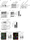
GAS2L1 CH and GAR domains form an autoinhibitory module. (A) Schematic of GAS2L1 fragments prepared for binding and sedimentation assays. (B and C) GAS2L1 constructs (FLAG-tagged) were transiently expressed in HEK293T cells for anti-FLAG immunoprecipitation. Samples of immunoprecipitates (50%) and cell-lysate inputs (1%) were immunoblotted (WB) with antibodies against FLAG and β-actin. IP, immunoprecipitation; WB, Western blotting. (D) Recombinant GAS2L1 proteins (5 µM; His6-FLAG tagged) were incubated with polymerized F-actin. After sedimentation of F-actin, pellets and supernatants were collected for anti-His6-tag and anti-β-actin immunoblotting. Sup, supernatant. To test the inhibitory effect of GAR protein, His6-FLAG-CH (5 µM) was preincubated with His6-FLAG-GAR (25 µM) for 30 min before binding with F-actin. Proportions of GAS2L1 proteins in pellets were quantified and are presented as means ± SD of three independent assays. One-way ANOVA; **, P < 0.001. (E) His6-FLAG-tagged GAS2L1 proteins (10 µM) were incubated with taxol-stabilized microtubules in the sedimentation assay, and the distribution of the proteins into the microtubule pellets and the supernatants was probed by means of anti-His6-tag and anti-α-tubulin immunoblotting. To test the effect of CH protein, His6-FLAG-CH (50 µM) was preincubated with His6-FLAG-GAR (10 µM) for 30 min. GAS2L1 proteins that cosedimented with microtubules were quantified, and the data are presented as means ± SD of three independent experiments. One-way ANOVA; **, P < 0.001. (F) GAS2L1 fragments (FLAG-tagged) were transiently coexpressed in HEK293T cells with the CH domain (GFP-tagged) for anti-FLAG immunoprecipitation. Aliquots of immunoprecipitates (50%) and cell-lysate inputs (1%) were immunoblotted (WB) with antibodies against FLAG and GFP. (G) Recombinant proteins of CH domain (His6 tagged; 2 µM) and GAR domain (His6-FLAG tagged; 1 µM) were incubated, and the mixtures were then subject to anti-FLAG immunoprecipitation. Immunoprecipitates (50%) and inputs (5%) were analyzed through anti-His6 immunoblotting.
Figure S3.
Figure S3.
Analysis of purified bacterially expressed GAS2L1 fragments. (A) GAS2L1 fragments harboring a His6-FLAG or His6 tag were expressed in bacteria and purified through the His6 tag. Purified proteins were analyzed using SDS-PAGE and Coomassie Blue staining. Arrowheads: recombinant proteins. (B) Recombinant GAS2L1 fragments (His6-tagged) were used in F-actin or microtubule sedimentation assays in the absence of F-actin or microtubules, respectively, in actin polymerization buffer (F-actin buffer) or BRB80 buffer (microtubule buffer). Pellets and supernatants were collected after sedimentation for anti-His6 immunoblotting.
Figure 6.
Figure 6.
Ser352 phosphorylation relieves CH-GAR autoinhibition. (A) S352A or S352D mutant of GAR-Tail construct (197–681; FLAG-tagged) was transiently coexpressed in HEK293T cells with GAS2L1 CH domain (GFP-tagged) for anti-FLAG immunoprecipitation. Aliquots of immunoprecipitates (50%) and cell-lysate inputs (1%) were immunoblotted with anti-FLAG and anti-GFP. IP, immunoprecipitation. Amounts of coimmunoprecipitated GFP-CH were quantified and are presented as means ± SD from three independent experiments. Unpaired Student’s t test (two-tailed); **, P < 0.001. (B) GAS2L1 constructs (Bio-GFP–tagged) were transiently expressed in HEK293T cells for streptavidin pull-downs. WT, WT GAS2L1; S352A and S352D, GAS2L1 mutants. To disrupt F-actin, cells were treated with latrunculin B (Lat B) for 30 min before harvesting. Samples of pull-downs (50%) and cell extracts (1%) were analyzed by immunoblotting. F-actin was quantified from pull-downs performed without latrunculin B treatment, and the data are presented as means ± SD from three independent experiments. One-way ANOVA; *, P < 0.01; **, P < 0.001. (C) RPE-1 gas2l1−/− lines stably expressing GFP-GAS2L1 (WT) or mutants (S352A and S352D) were stained for Nedd1, cyclin B1, and F-actin. Boxed areas are enlarged. Centrosome-associated F-actin was quantified from G2 cells from three independent experiments, and the data are presented as means ± SD of three experimental repeats: n = 77 (WT), 79 (S352A), and 88 (S352D). One-way ANOVA; *, P < 0.01. (D) RPE-1 cells were detached for F-actin staining. G2 cells were identified through anti-cyclin B1 staining, and centrosomes were labeled with anti-Nedd1 antibody. Boxed areas are enlarged. Intensity of centrosome-associated F-actin was determined and is presented as means ± SD from three independent experiments: n = 91 (cyclin B1–positive) and 99 (cyclin B1–negative). Unpaired Student’s t test; *, P < 0.01. Scale bars, 5 µm in cell micrographs and 2.5 µm in enlarged images.
Figure S4.
Figure S4.
Binding of Ser352 mutants of GAS2L1 to EB1. Bio-GFP-tagged GAS2L1 constructs were transiently expressed in HEK293T cells and pulled down using streptavidin beads. Aliquots of the pull-downs (50%) and lysate inputs (2%) were immunoblotted for GFP and EB1. WT, GAS2L1 WT.
Figure 7.
Figure 7.
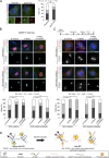
Nek2A mediates G2/M phosphorylation of GAS2L1. (A) GAS2L1 (Bio-FLAG–tagged) was coexpressed with a kinase-dead mutant of GFP-Nek2A (K37R) in HEK293T cells. After pull-down of ectopically expressed GAS2L1, pull-down samples (50%) and cell-extract inputs (5%) were immunoblotted with anti-FLAG and anti-GFP. (B) FLAG-GAS2L1 was transiently expressed with GFP-Nek2A or the K37R mutant in HEK293T cells, and then cell extracts were immunoblotted. An aliquot of the extract from cells coexpressing FLAG-GAS2L1 and GFP-Nek2A was treated with calf intestinal alkaline phosphatase before immunoblotting. (C) Purified GFP-GAS2L1 and S352A mutant proteins were phosphorylated in vitro with GST-Nek2A and then immunoblotted for GAS2L1, pSer352-GAS2L1, and Nek2A. (D) RPE-1 cells were synchronized with aphidicolin and then released into STLC-containing medium. Cells were collected at indicated time points and analyzed by immunoblotting. (E) RPE-1 cells transfected with control siRNA (si-Control) or nek2-targeting siRNA (si-Nek2) were arrested with aphidicolin and then released into STLC-containing medium for 12 h. Cell extracts were immunoblotted with indicated antibodies. Intensity of Ser352-phosphorylated GAS2L1 was measured in GAS2L1 doublets and normalized and is presented as means ± SD from three independent experiments. Unpaired Student’s t test; **, P < 0.001. (F) HEK293T cells were transfected with GAS2L1 constructs and Nek2A WT or kinase-dead mutant K37R. After anti-FLAG immunoprecipitation, immunoprecipitates (50%) and lysate inputs (1%) were immunoblotted with anti-FLAG and anti-GFP antibodies. Anti-pSer352-GAS2L1 immunoblotting was also performed on lysate inputs. IP, immunoprecipitation. The coimmunoprecipitated CH domain was quantified and normalized and is presented as means ± SD from three independent experiments. Unpaired Student’s t test; ***, P < 0.0001. (G) GFP-GAS2L1 or the S352D mutant was overexpressed in asynchronous RPE-1 cells transfected with si-Control or si-Nek2. Centrosomes were labeled through anti-γ-tubulin staining. Boxed areas are enlarged; arrowheads: centrosomes. Centrosome distance was measured in GFP-GAS2L1- and S352D-expressing cells from three independent experiments: n = 157 (si-Control + GFP-GAS2L1 WT), 148 (si-Nek2 + GFP-GAS2L1 WT), 144 (si-Control + GFP-GAS2L1 S352D), and 161 (si-Nek2 + GFP-GAS2L1 S352D). χ2 test; **, P < 0.001; n.s., not significant. Scale bars, 10 µm in cell micrographs and 5 µm in enlarged centrosome images.
Figure S5.
Figure S5.
GAS2L1 and its S352A mutant prepared from HEK293T cells expressing the proteins. GFP-tagged GAS2L1 WT and S352A mutant, both also containing a biotinylation tag, were expressed in HEK293T cells, purified using streptavidin-coupled beads, resolved on SDS-PAGE gels, and analyzed through Coomassie Blue staining and anti-GFP immunoblotting. Arrowheads: GFP-GAS2L1 full-length protein; asterisks: fragment of the protein.
Figure 8.
Figure 8.
Centrosome disjunction is mediated by GAS2L1 phosphorylation and centrosome-linker disassembly. (A) RPE-1 parental cells and gas2l1-knockout line (GAS2L1 KO) were transfected with GFP-Nek2A and stained with anti-γ-tubulin. Boxed areas are enlarged; arrowheads: centrosomes. Centrosome distance was measured in asynchronous cells transfected with GFP-Nek2A from three independent experiments: n = 112 (parental) and 109 (GAS2L1 KO). Scale bars, 10 µm in cell micrographs and 5 µm in enlarged centrosome images. (B) RPE-1 gas2l1−/− cells stably expressing GFP-GAS2L1 or the S352D mutant were transfected with indicated siRNAs. Centrosomes were labeled through anti-γ-tubulin staining; G2 cells were identified based on positive CENP-F staining. Centrosome distance was measured in G2 cells (CENP-F–positive cells) from three independent experiments: n = 198 (si-Control), 210 (si-Nek2), and 204 (si-Nek2 + si-rootletin). Boxed areas are enlarged. Scale bars, 10 µm in cell micrographs and 5 µm in enlarged centrosome images. (C) RPE-1 cells used as in B were synchronized with aphidicolin, released for 6 h, and treated with STLC for 4 h. Cells were stained for centrin and rootletin. Centrosome distance was measured in mitotic cells (arrested in prometaphase by STLC) from three independent experiments: GFP-GAS2L1 (WT) stable cells: n = 125 (si-Control), 136 (si-Nek2), and 119 (si-Nek2 + si-rootletin); GFP-GAS2L1 (S352D) stable cells: n = 127 (si-Control), 135 (si-Nek2), and 123 (si-Nek2 + si-rootletin). Arrowheads: centrosomes. Scale bars, 5 µm in cell micrographs and 2.5 µm in enlarged centrosome images. (D) Model depicting molecular events underlying centrosome disjunction. The disjunction is driven by GAS2L1 phosphorylation at Ser352 plus centrosome-linker disassembly, both of which are mediated by Nek2A. (A–C) χ2 test; *, P < 0.01; **, P < 0.001; n.s., not significant.

References

    1. Agircan F.G., Schiebel E., and Mardin B.R.. 2014. Separate to operate: control of centrosome positioning and separation. Philos. Trans. R. Soc. Lond. B Biol. Sci. 369:20130461 10.1098/rstb.2013.0461 - DOI - PMC - PubMed
    1. Algeciras-Schimnich A., and Peter M.E.. 2003. Actin dependent CD95 internalization is specific for Type I cells. FEBS Lett. 546:185–188. 10.1016/S0014-5793(03)00558-1 - DOI - PubMed
    1. Applewhite D.A., Grode K.D., Duncan M.C., and Rogers S.L.. 2013. The actin-microtubule cross-linking activity of Drosophila Short stop is regulated by intramolecular inhibition. Mol. Biol. Cell. 24:2885–2893. 10.1091/mbc.e12-11-0798 - DOI - PMC - PubMed
    1. Au F.K.C., Jia Y., Jiang K., Grigoriev I., Hau B.K.T., Shen Y., Du S., Akhmanova A., and Qi R.Z.. 2017. GAS2L1 Is a Centriole-Associated Protein Required for Centrosome Dynamics and Disjunction. Dev. Cell. 40:81–94. 10.1016/j.devcel.2016.11.019 - DOI - PubMed
    1. Bahe S., Stierhof Y.-D., Wilkinson C.J., Leiss F., and Nigg E.A.. 2005. Rootletin forms centriole-associated filaments and functions in centrosome cohesion. J. Cell Biol. 171:27–33. 10.1083/jcb.200504107 - DOI - PMC - PubMed

Publication types