. 2020 Jun 1;217(6):e20190634.
doi: 10.1084/jem.20190634.

ACKR4 restrains antitumor immunity by regulating CCL21

Affiliations

ACKR4 restrains antitumor immunity by regulating CCL21

Carly E Whyte et al. J Exp Med. .

Expression of concern in

Abstract

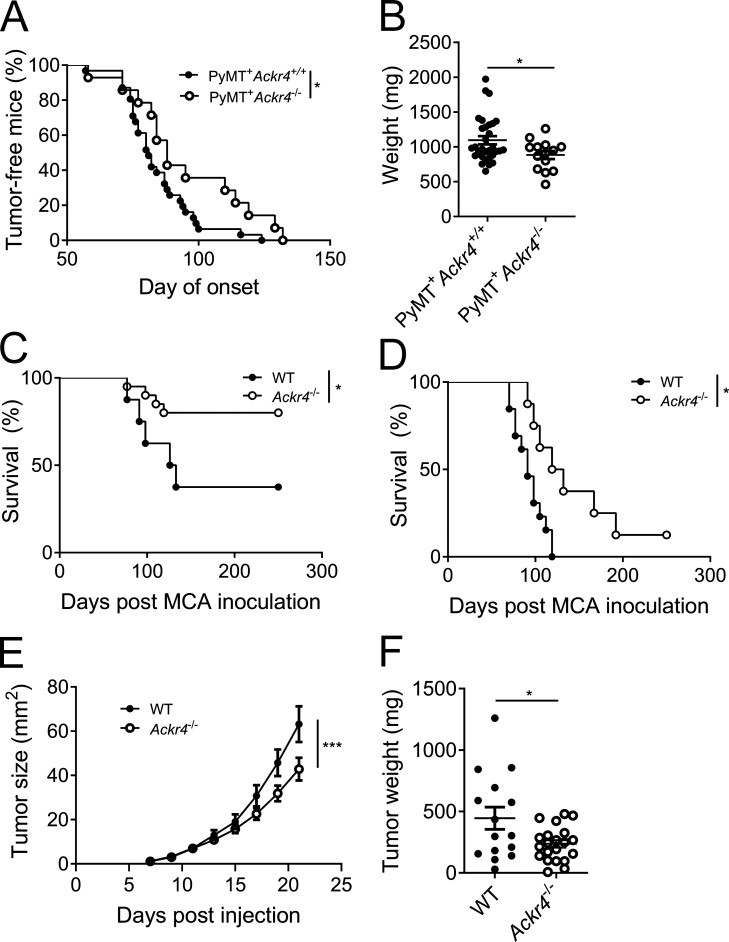
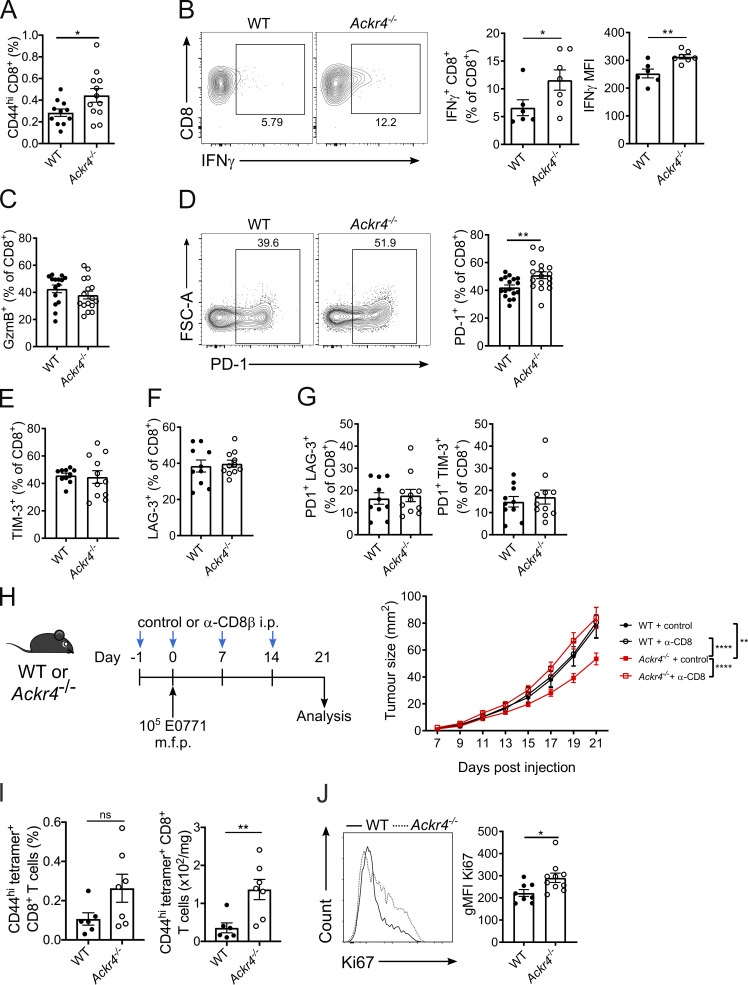
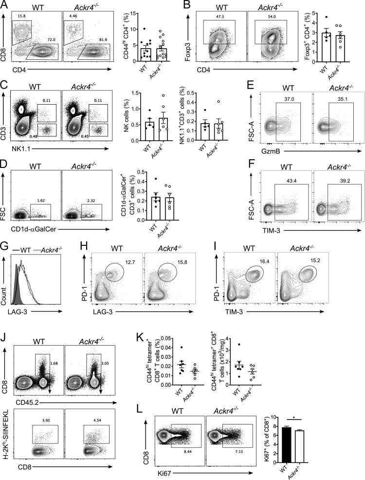
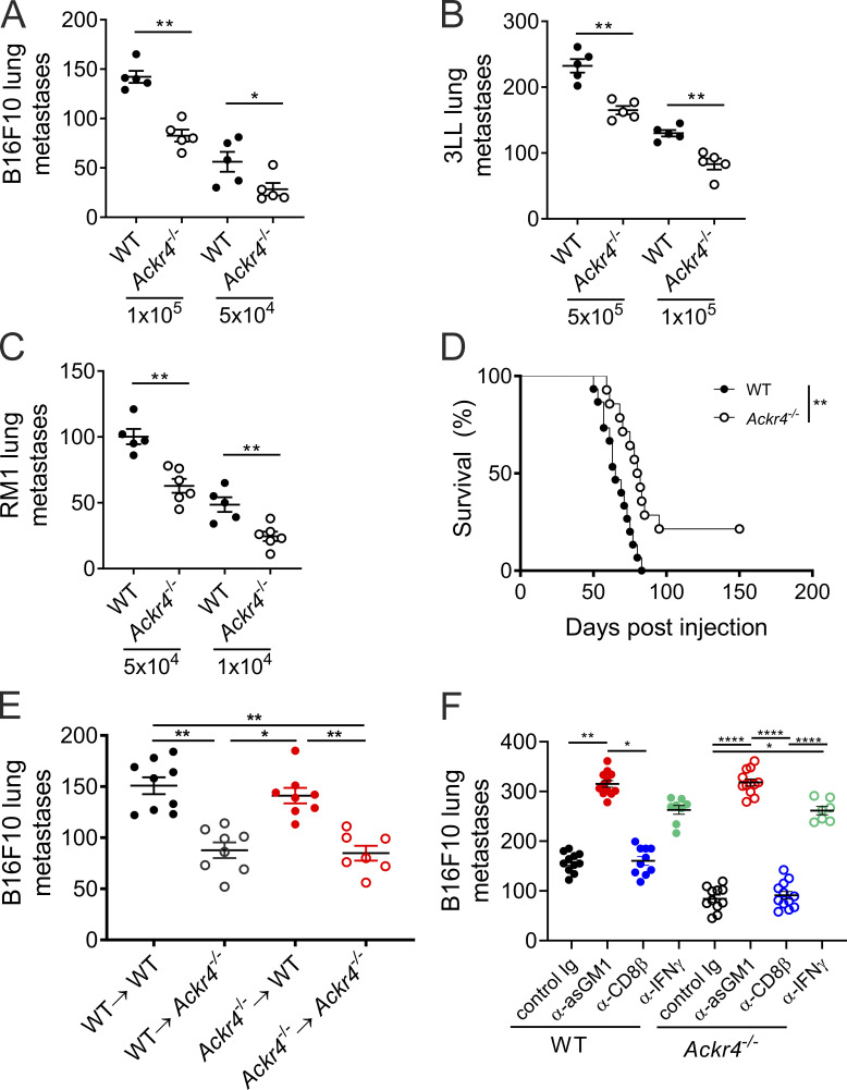
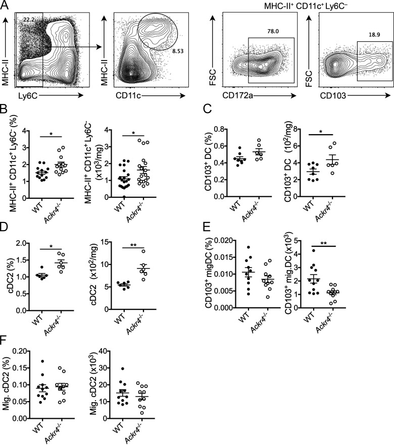
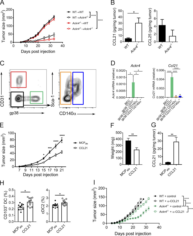
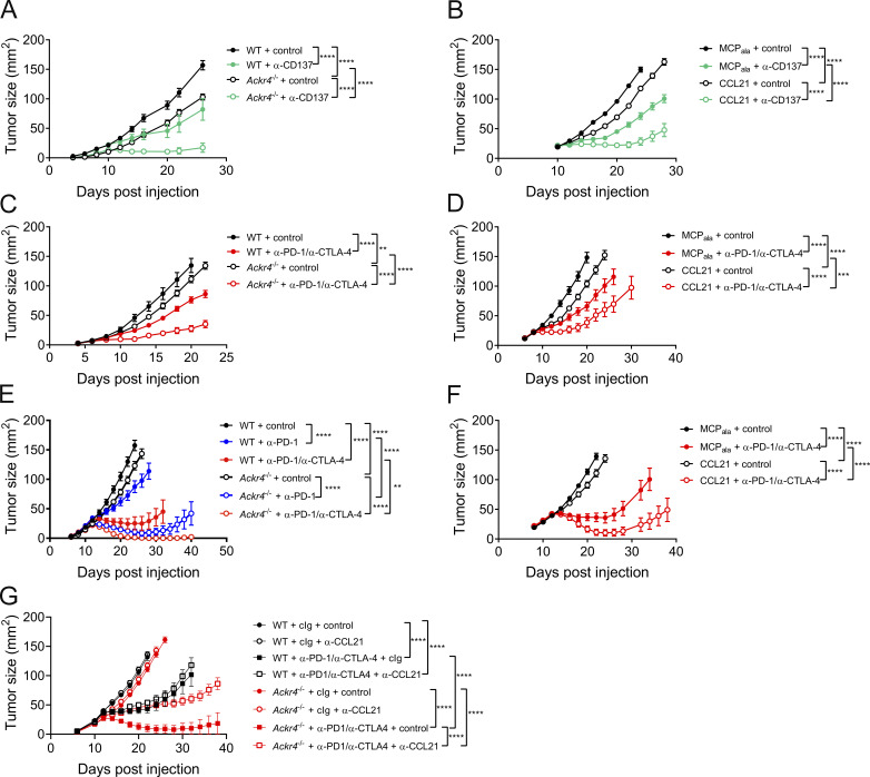
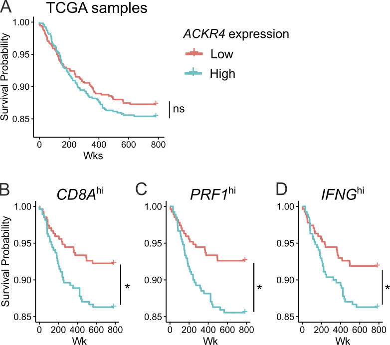
Current immunotherapies involving CD8+ T cell responses show remarkable promise, but their efficacy in many solid tumors is limited, in part due to the low frequency of tumor-specific T cells in the tumor microenvironment (TME). Here, we identified a role for host atypical chemokine receptor 4 (ACKR4) in controlling intratumor T cell accumulation and activation. In the absence of ACKR4, an increase in intratumor CD8+ T cells inhibited tumor growth, and nonhematopoietic ACKR4 expression was critical. We show that ACKR4 inhibited CD103+ dendritic cell retention in tumors through regulation of the intratumor abundance of CCL21. In addition, preclinical studies indicate that ACKR4 and CCL21 are potential therapeutic targets to enhance responsiveness to immune checkpoint blockade or T cell costimulation.


Conflict of interest statement

Disclosures: Dr. Harata-Lee reported a patent number 2012903874 with royalties paid, "Tizona Therapeutics." Dr. Comerford reported a patent number 2012903874 with royalties paid, "Tizona Therapeutics." Dr. Smyth reported grants from Bristol Meyers Squibb, grants from Tizona Therapeutics, grants from Aduro Biotech, personal fees from Tizona Therapeutics, and personal fees from Compass Therapeutics outside the submitted work. In addition, Dr. Smyth had a patent number 2012903874 issued. Dr. McColl reported grants from Tizona Therapeutics, personal fees from Tizona Therapeutics, and grants from Carina Biotech outside the submitted work. In addition, Dr. McColl had a patent number 2012903874 with royalties paid, "Tizona Therapeutics." No other disclosures were reported.

Figures

Figure 1.
Figure 1.
Loss of ACKR4 inhibits tumor growth in vivo. (A) Age of tumor onset for MMTV-PyMT B6 (Ackr4+/+) and Ackr4−/− mice; n = 31 (Ackr4+/+), 14 (Ackr4−/−), Mantel–Cox test. (B) Total weight of mammary glands in MMTV-PyMT B6 and Ackr4−/−at 20 wk of age; n = 31 (Ackr4+/+), 14 (Ackr4−/−), unpaired t test. (C and D) Survival curve of WT and Ackr4−/− mice inoculated with 25 µg (C) or 300 µg (D) MCA in the hind flank (C: n = 8, 20; D: n = 13, 8, Mantel-Cox test). (E and F) Growth curves (E) and weights (F) of WT or Ackr4−/− mice injected with 105 E0771 mammary carcinoma cells. Data in E and F are pooled from three independent experiments; n = 16–21 (E, two-way ANOVA; F, unpaired t test). Data are presented as mean ± SEM. *, P ≤ 0.05; ***, P ≤ 0.001.
Figure 2.
Figure 2.
ACKR4-deficient mice mount an enhanced antitumor CD8+ T cell response. (A–G) WT or Ackr4−/− mice were injected with 105 E0771 cells and analyzed 18–21 d after injection. (A) Frequency of CD44hi CD8+ T cells (of total viable cells) in tumors; n = 11–13, unpaired t test. MFI, mean fluorescence intensity. (B) Frequency of IFN-γ expression in intratumor CD8+ T cells and MFI of IFN-γ+ CD8+ T cells; n = 6–7, unpaired t test. (C and D) Frequency of granzyme B (C) or PD-1 expression (D) in intratumor CD8+ T cells; n = 17, unpaired t test. FSC-A, forward scatter area. (E–G) Frequency of TIM-3 (E), LAG-3 (F), PD-1+ TIM-3+ (G), or PD-1+ LAG-3+ expression in intratumor CD8+ T cells; n = 10–11. (H) WT or Ackr4−/− mice were injected with E0771 cells and treated with 100 µg anti-CD8β depleting antibody or isotype control. Tumor growth curve; n = 9–14, two-way ANOVA. m.f.p., mammary fat pad. (I) Frequency (of total viable cells) and number of OVA-specific CD8+ T cells in E0771-OVA tumors; n = 6–7 mice, unpaired t test. (J) Representative flow cytometry and geometric mean fluorescence intensity (gMFI) of Ki67 on intratumor CD8+ T cells from mice injected with 105 E0771 cells; n = 10, unpaired t test. Data are pooled (A, C, G, and H) or representative (B, I, and J) from at least two independent experiments (experiments shown in E–G were performed once). Data represent mean ± SEM; *, P ≤ 0.05; **, P ≤ 0.01; ****, P ≤ 0.0001; ns, P > 0.05.
Figure S1.
Figure S1.
Tumor-infiltrating lymphocytes and CD8+ T cell priming in mice bearing E0771 tumors. (A–G) WT or Ackr4−/− mice were injected with 105 E0771 cells into the fourth mammary gland and analyzed 18–21 d after injection. (A) Representative gating and frequency (of total viable cells) of intratumor CD44hi CD4+ T cells; n = 11–13. (B) Representative gating and frequency of intratumor Foxp3+ regulatory t cells. n = 5–6. (C) Representative gating and frequency of intratumor NK cells and NK1.1+ CD3+ NKT cells. n = 5–6. (D) Representative gating and frequency of type I invariant NKT cells. n = 5–6. FSC, forward scatter. (E–H) Representative gating strategy of intratumor CD8+ T cells for (E) granzyme B, (F) TIM-3, (G) LAG-3, (H) PD-1+ LAG-3+, and (I) PD-1+ TIM-3+ (data in Fig. 2, C and E–G). (J) Representative gating strategy of OVA-specific CD8+ T cells in E0771-OVA tumors (data in Fig. 2 F). (K) Frequency and number of OVA-specific CD44hi CD8+ T cells in dLNs of mice injected with 5 × 10⁵ E0771-OVA cells; n = 6–7. (L) Representative gating and frequency of Ki67 expression on CD8+ T cells in the dLNs of mice injected with 105 E0771 cells. n = 10, unpaired t test. Data representative of at least two independent experiments. Data are presented as mean ± SEM. *, P ≤ 0.05.
Figure S2.
Figure S2.
Loss of nonhematopoietic ACKR4 inhibits tumor metastasis. (A–C) WT or Ackr4−/− mice were intravenously injected with (A) B16F10 melanoma, (B) 3LL lung carcinoma, or (C) RM1 prostate cells, and the number of lung metastases were counted (n = 5–6 mice, Mann–Whitney test). (D) Survival curve of WT or Ackr4−/− mice injected with 2 × 10⁴ E0771 cells, with primary tumors resected at day 15 (n = 14–15, Mantel–Cox test). (E) Lung metastasis in BM chimeras of WT and Ackr4−/− mice (donor BM→host). Mice were injected with 105 B16F10 cells, lungs were harvested 14 d after inoculation, and colonies were counted (n = 7–9, Kruskal–Wallis test). (F) WT or Ackr4−/− mice were injected with 105 B16F10 cells and treated with depleting antibodies against CD8β (100 µg), asialoGM1 (50 µg), or IFNγ (250 µg) on days −1, 0, and 7 (n = 7–12, Kruskal–Wallis test). Data are pooled from two independent experiments and presented as mean ± SEM. *, P ≤ 0.05; **, P ≤ 0.01; ****, P ≤ 0.0001.
Figure 3.
Figure 3.
Loss of ACKR4 promotes intratumor DC accumulation. WT or Ackr4−/− mice were injected with E0771 cells, and tumors were analyzed 18 d later. (A) Representative gating strategy for intratumoral DCs pregated on live CD45+ cells. FSC, forward scatter. (B–D) Frequency (of total viable cells) and number of intratumor (B) DCs (MHC-II+ CD11c+ Ly6C), (C) CD103+ DCs, and (D) CD172a+ cDC2s. (E and F) Frequency and number of migratory (E) CD103+ DCs and (F) CD172a+ cDC2s in dLNs. Data are pooled from two independent experiments (B) or representative of at least two independent experiments (C–F); n = 6–10 mice, unpaired t test. Data are presented as mean ± SEM. *, P ≤ 0.05; **, P ≤ 0.01.
Figure 4.
Figure 4.
ACKR4 regulates CCL21 availability and enhances intratumor DC retention. (A) E0771 tumor growth in BM chimeras of WT and Ackr4−/− mice (donor BM→host). Mice were injected with 5 × 10⁵ E0771 cells at least 8 wk after reconstitution; n = 9, two-way ANOVA. (B) CCL21 and CCL25 abundance in endpoint E0771 tumors; n = 6, unpaired t test. (C and D) Ackr4 and Ccl21 expression in stromal cells sorted from d13 E0771-mCherry tumors. (C) Representative gating strategy, pregated on CD45 mCherry cells. (D) Relative expression of Ackr4 and Ccl21 by qPCR; n = 7, one-way ANOVA. (E–H) WT mice were injected with 105 E0771 cells into contralateral sides of the inguinal mammary glands. MCPala or CCL21 (3 µg) was injected into contralateral glands every 3 d from the day of E0771 injection; n = 19 mice. (E) Tumor growth curves; two-way ANOVA. (F) Tumor weights at day 21 after injection; paired t test. (G) Intratumor CCL21 abundance; paired t test. (H) Frequency of intratumor CD103+ and CD172a+ cDC2s (of total viable cells); n = 10, paired t test. (I) E0771 tumor growth in WT and Ackr4−/− mice treated with control or anti-CCL21 (100 µg) every 6 d from day −1; n = 6, two-way ANOVA. Data are representative (A, B, H, and I) or pooled (C−G) from at least two independent experiments. Data are presented as mean ± SEM. *, P ≤ 0.05; **, P ≤ 0.01; ***, P ≤ 0.001; ****, P ≤ 0.0001.
Figure 5.
Figure 5.
ACKR4 deficiency enhances responsiveness to immunotherapy. (A) Tumor growth in WT or Ackr4−/− mice injected with 105 E0771 cells and administered 100 µg anti-CD137 (clone 3H3) or rat IgG every 3 d from days 10 to 19; n = 7–9. (B) Tumor growth in WT mice injected with 105 E0771 cells and administered MCPala or CCL21 (3 µg intratumorally) plus 100 µg anti-CD137 or rat IgG every 3 d from days 10 to 19; n = 5. (C) Tumor growth in WT or Ackr4−/− mice injected with 105 B16F10 cells and administered 250 µg anti–PD-1 (clone RMP1-14) and 250 µg anti–CTLA-4 (clone UC10-4F10) or 500 µg control rat/hamster Ig every 3 d from days 6 to 15; n = 5–6 mice. (D) Tumor growth in WT mice injected with 105 B16F10 cells and administered MCPala or CCL21 (3 µg intratumorally) plus 250 µg anti–PD-1 and 250 µg anti–CTLA-4 or 500 µg control rat/hamster Ig every 3 d from days 6 to 15; n = 6. (E) Tumor growth curves from WT or Ackr4−/− mice injected subcutaneously with 105 MC38 cells and administered 250 µg anti–PD-1, 250 µg anti–PD-1/250 µg anti–CTLA-4, or 500 µg rat/hamster Ig every 3 d from days 12 to 21; n = 7–8 mice. (F) Tumor growth curves from WT mice injected subcutaneously with 105 MC38 cells and administered MCPala or CCL21 (3 µg, intratumorally) plus 250 µg anti–PD-1 and 250 µg anti–CTLA-4 or 500 µg rat/hamster Ig every 3 d from days 12 to 21; n = 10 mice. (G) Tumor growth curves from WT or Ackr4−/− mice injected subcutaneously with 105 MC38 cells and administered 250 µg anti-PD-1 (clone RMP1-14) and 250 µg anti–CTLA-4 (clone UC10-4F10) or 500 µg rat/hamster Ig every 3 d from days 12 to 21 plus control or anti-CCL21 (100 µg) every 6 d from day −1; n = 6–7 mice. Data are representative from two independent experiments (A–F) or are from one experiment (G). All statistical analyses were performed using a two-way ANOVA, and data are presented as mean ± SEM. **, P ≤ 0.01; ***, P ≤ 0.001; ****, P ≤ 0.0001.
Figure S3.
Figure S3.
Low ACKR4 expression correlates with increased survival in stratified patient groups. (A) Survival curves from TCGA analyses of patients with breast-invasive carcinoma stratified by ACKR4 expression, with the median log counts per million value across all samples as the boundary between low and high expression. (B–D) Survival curves from patients with high expression of (B) CD8A, (C) PRF1, or (D) IFNG stratified by ACKR4 expression; n = 1,085, pairwise comparisons by log-rank test, adjusted with Benjamini–Hochberg method. *, P ≤ 0.05; ns, P > 0.05.

References

    1. Ahern, E., Smyth M.J., Dougall W.C., and Teng M.W.L.. 2018. Roles of the RANKL-RANK axis in antitumour immunity - implications for therapy. Nat. Rev. Clin. Oncol. 15:676–693. 10.1038/s41571-018-0095-y - DOI - PubMed
    1. Binnewies, M., Mujal A.M., Pollack J.L., Combes A.J., Hardison E.A., Barry K.C., Tsui J., Ruhland M.K., Kersten K., Abushawish M.A., et al. . 2019. Unleashing Type-2 Dendritic Cells to Drive Protective Antitumor CD4+ T Cell Immunity. Cell. 177:556–571.e16. 10.1016/j.cell.2019.02.005 - DOI - PMC - PubMed
    1. Boyle, S.T., Ingman W.V., Poltavets V., Faulkner J.W., Whitfield R.J., McColl S.R., and Kochetkova M.. 2016. The chemokine receptor CCR7 promotes mammary tumorigenesis through amplification of stem-like cells. Oncogene. 35:105–115. 10.1038/onc.2015.66 - DOI - PubMed
    1. Broz, M.L., Binnewies M., Boldajipour B., Nelson A.E., Pollack J.L., Erle D.J., Barczak A., Rosenblum M.D., Daud A., Barber D.L., et al. . 2014. Dissecting the tumor myeloid compartment reveals rare activating antigen-presenting cells critical for T cell immunity. Cancer Cell. 26:638–652. 10.1016/j.ccell.2014.09.007 - DOI - PMC - PubMed
    1. Bryce, S.A., Wilson R.A., Tiplady E.M., Asquith D.L., Bromley S.K., Luster A.D., Graham G.J., and Nibbs R.J.. 2016. ACKR4 on Stromal Cells Scavenges CCL19 To Enable CCR7-Dependent Trafficking of APCs from Inflamed Skin to Lymph Nodes. J. Immunol. 196:3341–3353. 10.4049/jimmunol.1501542 - DOI - PubMed

Publication types