. 2020 May 13;27(5):841-848.e3.
doi: 10.1016/j.chom.2020.04.004. Epub 2020 Apr 13.

An Infectious cDNA Clone of SARS-CoV-2

Affiliations

An Infectious cDNA Clone of SARS-CoV-2

Xuping Xie et al. Cell Host Microbe. .

Abstract

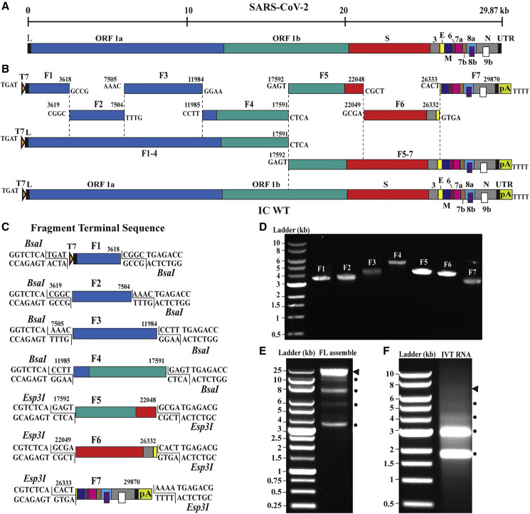
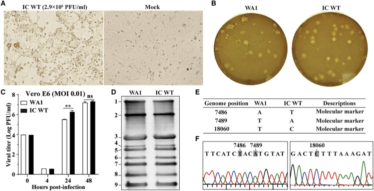
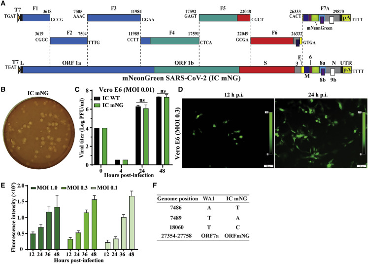
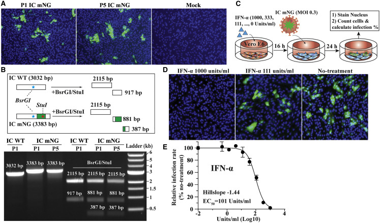
The ongoing pandemic of COVID-19, caused by severe acute respiratory syndrome coronavirus 2 (SARS-CoV-2), underscores the urgency to develop experimental systems for studying this virus and identifying countermeasures. We report a reverse genetic system for SARS-CoV-2. Seven complimentary DNA (cDNA) fragments spanning the SARS-CoV-2 genome were assembled into a full-genome cDNA. RNA transcribed from the full-genome cDNA was highly infectious after electroporation into cells, producing 2.9 × 10⁶ plaque-forming unit (PFU)/mL of virus. Compared with a clinical isolate, the infectious-clone-derived SARS-CoV-2 (icSARS-CoV-2) exhibited similar plaque morphology, viral RNA profile, and replication kinetics. Additionally, icSARS-CoV-2 retained engineered molecular markers and did not acquire other mutations. We generated a stable mNeonGreen SARS-CoV-2 (icSARS-CoV-2-mNG) by introducing this reporter gene into ORF7 of the viral genome. icSARS-CoV-2-mNG was successfully used to evaluate the antiviral activities of interferon (IFN). Collectively, the reverse genetic system and reporter virus provide key reagents to study SARS-CoV-2 and develop countermeasures.

Keywords: COVID-19; SARS-CoV; SARS-CoV-2; antiviral; coronavirus; vaccine.


Conflict of interest statement

Declaration of Interests X.X., V.D.M, and P.-Y.S. have filed a provisional patent on the reverse genetic system of SARS-CoV-2. Other authors have no conflicts of interest to declare.

Figures

None
Graphical abstract
Figure 1
Figure 1
Assembly of a Full-Length SARS-CoV-2 Infection cDNA Clone (A) Genome structure SARS-CoV-2. The open reading frames (ORFs) from the full genome are indicated. (B) Strategy for in vitro assembly of an infectious cDNA clone of SARS-CoV-2. The nucleotide sequences and genome locations of the cohesive overhangs are indicated. The WT full-length (FL) cDNA of SARS-CoV-2 (IC WT) was directionally assembled using in vitro ligation. (C) Diagram of the terminal sequences of each cDNA fragment recognized by BsaI and Esp3I. (D) Gel analysis of the seven purified cDNA fragments. Individual fragments (F1–F7) were digested from corresponding plasmid clones and gel purified. Seven purified cDNA fragments (50–100 ng) were analyzed on a 0.6% native agarose gel. The 1-kb DNA ladders are indicated. (E) Gel analysis of cDNA ligation products. About 400 ng of purified ligation product was analyzed on a 0.6% native agarose gel. Triangle indicates the FL cDNA product. Circles indicate the intermediate cDNA products. (F) Gel analysis of RNA transcripts. About 1 μg of in-vitro-transcribed (IVT) RNAs were analyzed on a 0.6% native agarose gel. DNA ladders are indicated. Because this is a native agarose gel, the DNA size is not directly corelated to the RNA size. Triangle indicates the genome-length RNA transcript. Circles show the shorter RNA transcripts.
Figure 2
Figure 2
Characterization of the IC WT (A) Bright-field images of the Vero E6 cells electroporated with RNA transcripts. CPEs appeared in the IC-WT-RNA-transfected cells on day 4 after transfection. The titer of the P0 virus (directly from the transfected cells) is shown in PFUs per ml. (B) Plaque morphology of the original clinical isolate (WA1 = 2019-nCoV/USA_WA1/2020) and the recombinant P1 IC WT virus. Plaques were developed in Vero E6 cells on day 2 after infection. (C) Replication kinetics. Vero E6 cells were infected with the clinical isolate or recombinant P1 IC WT virus at MOI 0.01. Viruses in culture fluids were quantified by plaque assay. Results from triplicate experiments were presented with error bars indicating standard deviations. (D) Northern blot analysis of FL and subgenomic RNAs. Numbers indicated the FL (band 1) and eight subgenomic RNAs (bands 2–9). (E) Sequence differences between the original clinical isolate WA1 and the recombinant P1 IC WT. The three silent nucleotide changes were engineered as molecular markers. (F) Chromatograms of Sanger sequencing results. The engineered molecular maker mutations are indicated.
Figure 3
Figure 3
Generation of a mNeonGreen SARS-CoV-2 (A) Assembly of the FL mNG SARS-CoV-2 cDNA. The mNG gene was placed downstream of the regulatory sequence of ORF7 to replace the ORF7 sequence (Sims et al., 2005) in the subclone F7. (B) Plaque morphology of the P1 IC mNG virus. Plaques were developed in Vero E6 cells on day 2 after infection. (C) Replication kinetics. Vero E6 cells were infected with the IC WT or reporter icSARS-CoV-2-mNG (IC mNG) at MOI of 0.01. Viruses in culture medium were quantified by plaque assay. (D) Fluorescence microscopy analysis of P1-mNG-virus-infected cells. Vero E6 cells were infected with P1 mNG viruses at MOI of 0.3. Representative mNG-positive (green) images are shown. (E) Kinetics of fluorescence intensity. Vero E6 cells were infected with MOIs of 1.0, 0.3, or 0.1. After background signal subtraction, the fluorescence intensities from 12 to 48 h after infection are shown. Results from triplicate experiments were presented with bars representing standard deviations. (F) Summary of full-genome sequence of mNG virus (P1 IC mNG). Nucleotides different from the original clinical isolate (WA1) are indicated.
Figure 4
Figure 4
Stability and Application of mNeonGreen Virus The stability of mNG virus was analyzed by comparing the fluorescent signals between the cells infected with P1 and P5 reporter viruses. The presence of mNG gene in the P1 and P5 reporter viruses was also verified using RT-PCR. The application of mNG virus was examined by testing the antiviral activity of IFN-α treatment. (A) Fluorescence microscopy analysis of the P1- and P5-mNG-virus-infected cells. Vero E6 cells were infected with P1 or P5 virus at an MOI of 0.3. The cells were monitored for mNG-positive signals at 24 h after infection. Color representations are as follows: green, mNG; blue, nucleus. (B) Gel analysis of mNG virus stability. The top graphic depicts the theoretical results of RT-PCR followed by restriction enzyme digestion. The bottom graphic shows the gel analysis of the RT-PCR products before (lanes 1–3) and after BsrGI/StuI digestion (lanes 4–6). About 100 ng DNA samples were analyzed on a 0.6% agarose gel. The DNA sizes are indicated. (C) Schematic diagram of IFN-α treatment. (D) Representative fluorescence images of reporter-virus-infected cells after IFN-α treatment. The doses of IFN-α treatment are indicated. (E) Dose response curve of mNG signal inhibited by IFN-α. The Hillslope and EC50 values are indicated. Results from triplicate experiments were presented with bars representing standard deviations.

References

    1. Almazán F., Sola I., Zuñiga S., Marquez-Jurado S., Morales L., Becares M., Enjuanes L. Coronavirus reverse genetic systems: infectious clones and replicons. Virus Res. 2014;189:262–270. - PMC - PubMed
    1. Assiri A., Al-Tawfiq J.A., Al-Rabeeah A.A., Al-Rabiah F.A., Al-Hajjar S., Al-Barrak A., Flemban H., Al-Nassir W.N., Balkhy H.H., Al-Hakeem R.F. Epidemiological, demographic, and clinical characteristics of 47 cases of Middle East respiratory syndrome coronavirus disease from Saudi Arabia: a descriptive study. Lancet Infect. Dis. 2013;13:752–761. - PMC - PubMed
    1. Coutard B., Valle C., de Lamballerie X., Canard B., Seidah N.G., Decroly E. The spike glycoprotein of the new coronavirus 2019-nCoV contains a furin-like cleavage site absent in CoV of the same clade. Antiviral Res. 2020;176:104742. - PMC - PubMed
    1. Curtis K.M., Yount B., Baric R.S. Heterologous gene expression from transmissible gastroenteritis virus replicon particles. J. Virol. 2002;76:1422–1434. - PMC - PubMed
    1. de Wit E., van Doremalen N., Falzarano D., Munster V.J. SARS and MERS: recent insights into emerging coronaviruses. Nat. Rev. Microbiol. 2016;14:523–534. - PMC - PubMed

Publication types

MeSH terms