Review
. 2020 Jul:329:113307.
doi: 10.1016/j.expneurol.2020.113307. Epub 2020 Apr 11.

Mitochondrial damage & lipid signaling in traumatic brain injury

Affiliations
Review

Mitochondrial damage & lipid signaling in traumatic brain injury

Andrew M Lamade et al. Exp Neurol. 2020 Jul.

Abstract

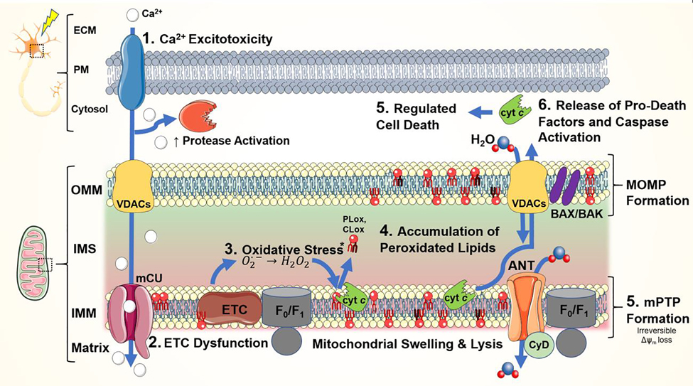
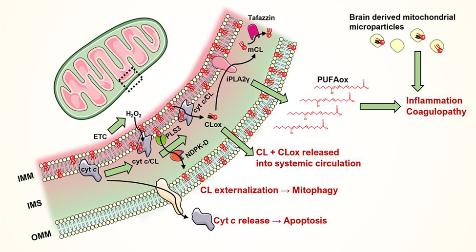
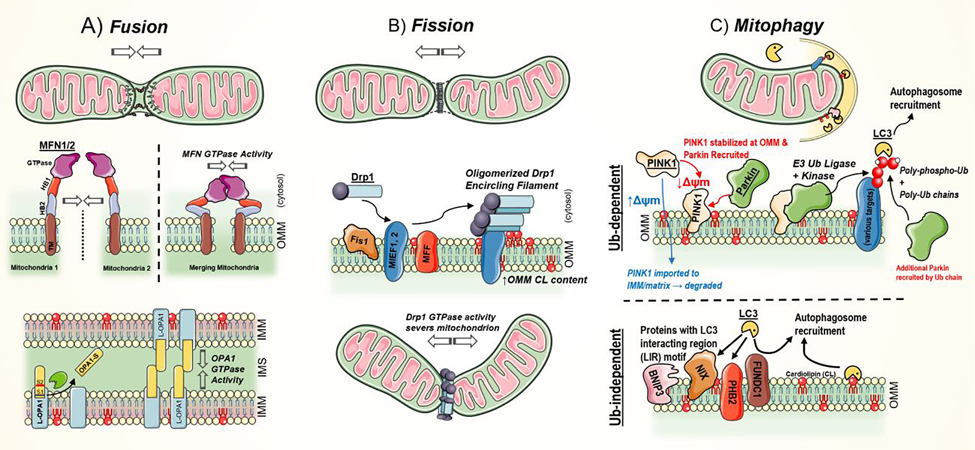
Mitochondria are essential for neuronal function because they serve not only to sustain energy and redox homeostasis but also are harbingers of death. A dysregulated mitochondrial network can cascade until function is irreparably lost, dooming cells. TBI is most prevalent in the young and comes at significant personal and societal costs. Traumatic brain injury (TBI) triggers a biphasic and mechanistically heterogenous response and this mechanistic heterogeneity has made the development of standardized treatments challenging. The secondary phase of TBI injury evolves over hours and days after the initial insult, providing a window of opportunity for intervention. However, no FDA approved treatment for neuroprotection after TBI currently exists. With recent advances in detection techniques, there has been increasing recognition of the significance and roles of mitochondrial redox lipid signaling in both acute and chronic central nervous system (CNS) pathologies. Oxidized lipids and their downstream products result from and contribute to TBI pathogenesis. Therapies targeting the mitochondrial lipid composition and redox state show promise in experimental TBI and warrant further exploration. In this review, we provide 1) an overview for mitochondrial redox homeostasis with emphasis on glutathione metabolism, 2) the key mechanisms of TBI mitochondrial injury, 3) the pathways of mitochondria specific phospholipid cardiolipin oxidation, and 4) review the mechanisms of mitochondria quality control in TBI with consideration of the roles lipids play in this process.

Keywords: Lipid peroxidation; Mitochondria; Mitochondria quality control; Mitophagy; Redox lipidomics; Traumatic brain injury.


Conflict of interest statement

Declaration of Interests

The authors declare no competing interests.

Figures

Figure 1.
Figure 1.. Mitochondria-focused mechanisms of neuronal injury following TBI.
1) Widespread neuronal depolarization results in the unregulated release of excitatory neurotransmitters and the activation of their corresponding ion channels. Increased cytosolic Ca2+ is buffered, in part, by the mitochondria leading to disruption of the mitochondrial membrane potential (Δψm). Ensuing 2) electron transport chain (ETC) dysfunction causes protracted increases in 3) ROS & RNS production. Injury becomes self-perpetuating as endogenous oxidant stress response systems are overwhelmed. 4) Mitochondrial lipids, especially cardiolipin (CL), are important sources of oxidized free fatty acid inflammatory mediators. Both non-enzymatic and cyt c/CL peroxidase-mediated enzymatic CL peroxidation occurs in the setting of high ROS levels. 5) Permeabilization of the mitochondrial membranes and oxidation of CL prompts the release of pro-death factors – ultimately leading to neuronal death. (OMM, outer mitochondrial membrane; IMS, intermembrane space; IMM, inner mitochondrial membrane; ANT, adenine nucleotide translocator; BAK/BAX, Bcl-2-associated X protein; CyD, cyclophilin D; F0/F1, ATP synthase; mCU, mitochondrial calcium uniporter, VDAC, voltage-dependent anion channel)
Figure 2.
Figure 2.. Mitochondrial CL signaling in brain injury.
The inner leaflet of mitochondrial inner membrane (IMM) is enriched in cardiolipins (CL). Upon injury, CLs are translocated to the outer leaflet of IMM, where it forms a complex with cytochrome c. The cytochrome c/cardiolipin (cyt c/CL) complex is a potent peroxidase that oxidizes the polyunsaturated acyl chains of CL (or other anionic lipids, to a lesser extent). Oxidation of CL (CLox) leads to the translocation of cyt c to the cytoplasm and subsequent apoptosis. IMM-localized non-oxidized CL are translocated to the outer membrane by the phospholipid scramblase 3 (PLS3) and mitochondrial nucleoside diphosphate kinase (NDPK-D). Externalized CL serve as a “eat me” signal for mitophagy. CLs previously oxidized by the cyt c/CL complex undergo hydrolysis by calcium-independent phospholipase A2 - gamma (iPLA2γ) to release oxidized polyunsaturated fatty acids (PUFAox). These PUFAox serve as lipid mediators that regulate inflammation and can lead to coagulopathy. Intact tetraacylated CLs can be regenerated from monolyso-CL (mCL) by phospholipid-lysophospholipid transacylases (e.g. tafazzin). CLs present on the brain-derived mitochondrial microparticles also regulates the injury-related inflammation and coagulation.
Figure 3.
Figure 3.. Mitochondrial quality control system and mitophagy.
A) Mitochondrial fusion leads to the joining of two mitochondrion. As fusion-destined mitochondria approach, outer mitochondrial membrane (OMM)-anchored mitofusins 1 and 2 (MFN1/2) dimerize and bridge the (~20 nm) gap between membranes. Subsequent MFN GTPase activity pulls the two organelles into proximity. The HB domains then participate in membrane destabilization and integration. This process is most common at sites of lipid packing detects – due to presence of bulky or conical lipids, like cardiolipin. B) Mitochondrial fusion leads to increased network fragmentation. The large GTPase, dynamin-related protein 1 (Drp1), drives fission by mechanically constricting and severing both the OMM and inner mitochondrial membrane (IMM). Soluble Drp1 is initially recruited to the OMM with assistance by OMM-bound adaptors: mitochondrial fission 1 (Fis1), mitochondrial fission factor (MFF), and mitochondrial elongation factors 1 & 2 (MIEF1, MIEF2). Increased OMM cardiolipin and phosphatidic acid content are linked to efficient fission activity. C) Mitophagy is a selective autophagy of mitochondria. Broadly, it can proceed in a ubiquitin (Ub)-dependent and -independent manner. In the Ub-dependent pathway, phosphatase and tensin homologue (PTEN)-induced punitive kinase 1 (PINK1) accumulates in the OMM when mitochondrial membrane potential (Δψm) is depleted. PINK1 recruits Parkin, an E3 Ub ligase. The PINK1/Parkin complex then proceeds to poly-phospho-ubiquitinate a range of mitochondrial proteins (e.g. translocase of the OMM, Miro, MFNs) and itself in feed-forward mechanism. By direct engagement or assistance of several adaptor proteins (OPTN, NDP52, p62, TAX1Bp1, and NBR1), LC3 (LC3-II) binds these poly-Ub and poly-phospho-ubiquitinated proteins, leading to autophagosomal engulfment. Ub-independent mechanisms similarly proceed in an LC3-dependent manner. However, in this case, LC3 directly engages various OMM protein (contain an LC3 interacting motif, e.g. BNIP3, NIX, PHB2, and FUNDC1) or select lipids, like cardiolipin (CL). This leads to autophagosome recruitment.

References

    1. Adachi Y, Itoh K, Yamada T, Cerveny KL, Suzuki TL, Macdonald P, Frohman MA, Ramachandran R, Iijima M, Sesaki H, 2016. Coincident Phosphatidic Acid Interaction Restrains Drp1 in Mitochondrial Division. Mol Cell 63, 1034–1043. - PMC - PubMed
    1. Adam-Vizi V, Tretter L, 2013. The role of mitochondrial dehydrogenases in the generation of oxidative stress. Neurochemistryinternational 62, 757–763. - PubMed
    1. Aguirre JD, Dunkerley KM, Mercier P, Shaw GS, 2017. Structure of phosphorylated UBL domain and insights into PINK1-orchestrated parkin activation. Proc Natl Acad Sci U S A 114, 298–303. - PMC - PubMed
    1. Alavi MV, Fuhrmann N, 2013. Dominant optic atrophy, OPA1, and mitochondrial quality control: understanding mitochondrial network dynamics. Mol Neurodegener 8, 32. - PMC - PubMed
    1. Anthonymuthu TS, Kenny EM, Amoscato AA, Lewis J, Kochanek PM, Kagan VE, Bayır H, 2017. Global assessment of oxidized free fatty acids in brain reveals an enzymatic predominance to oxidative signaling after trauma. Biochimica et Biophysica Acta (BBA) - Molecular Basis of Disease 1863, 2601–2613. - PMC - PubMed

Publication types