

Molecular Mechanism of Evolution and Human Infection with SARS-CoV-2
- PMID: 32290077
- PMCID: PMC7232534
- DOI: 10.3390/v12040428
Molecular Mechanism of Evolution and Human Infection with SARS-CoV-2
Abstract
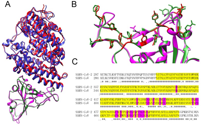
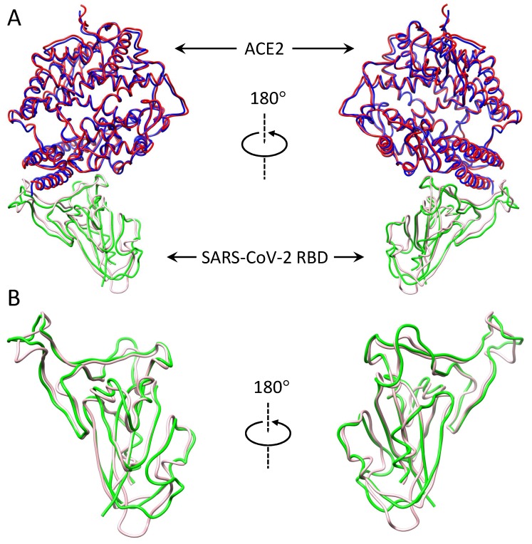
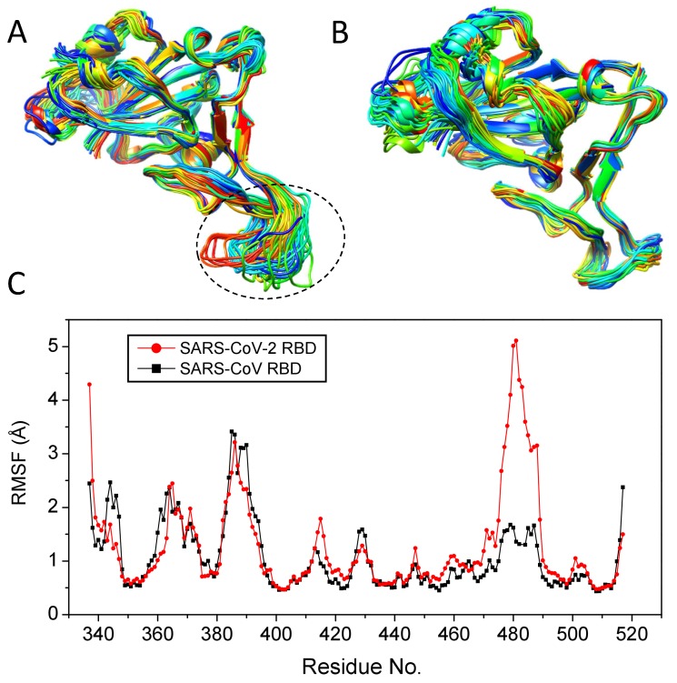
The outbreak of a novel coronavirus, which was later formally named the severe acute respiratory coronavirus 2 (SARS-CoV-2), has caused a worldwide public health crisis. Previous studies showed that SARS-CoV-2 is highly homologous to SARS-CoV and infects humans through the binding of the spike protein to ACE2. Here, we have systematically studied the molecular mechanisms of human infection with SARS-CoV-2 and SARS-CoV by protein-protein docking and MD simulations. It was found that SARS-CoV-2 binds ACE2 with a higher affinity than SARS-CoV, which may partly explain that SARS-CoV-2 is much more infectious than SARS-CoV. In addition, the spike protein of SARS-CoV-2 has a significantly lower free energy than that of SARS-CoV, suggesting that SARS-CoV-2 is more stable and may survive a higher temperature than SARS-CoV. This provides insights into the evolution of SARS-CoV-2 because SARS-like coronaviruses have originated in bats. Our computation also suggested that the RBD-ACE2 binding for SARS-CoV-2 is much more temperature-sensitive than that for SARS-CoV. Thus, it is expected that SARS-CoV-2 would decrease its infection ability much faster than SARS-CoV when the temperature rises. These findings would be beneficial for the disease prevention and drug/vaccine development of SARS-CoV-2.
Keywords: MD simulations; SARS-CoV; SARS-CoV-2; coronaviruses; human infection; molecular mechanism; protein docking.
Conflict of interest statement
The authors declare no conflict of interest.
Figures
References
-
- Zhong N.S., Zheng B.J., Li Y.M., Poon L.L., Xie Z.H., Chan K.H., Li P.H., Tan S.Y., Chang Q., Xie J.P., et al. Epidemiology and cause of severe acute respiratory syndrome (SARS) in Guangdong, People’s Republic of China, in February, 2003. Lancet. 2003;362:1353–1358. doi: 10.1016/S0140-6736(03)14630-2. - DOI - PMC - PubMed
-
- Drosten C., Günther S., Preiser W., Van Der Werf S., Brodt H.R., Becker S., Rabenau H., Panning M., Kolesnikova L., Fouchier R.A., et al. Identification of a novel coronavirus in patients with severe acute respiratory syndrome. N. Engl. J. Med. 2003;348:1967–1976. doi: 10.1056/NEJMoa030747. - DOI - PubMed
MeSH terms
Substances
Grants and funding
LinkOut - more resources
Full Text Sources
Miscellaneous
